Rachael Anne Dunlop, Sandra Anne Banack, Paul Alan Cox
{"title":"L1CAM免疫捕获产生独特的细胞外囊泡群,具有可复制的miRNA指纹。","authors":"Rachael Anne Dunlop, Sandra Anne Banack, Paul Alan Cox","doi":"10.1080/15476286.2023.2198805","DOIUrl":null,"url":null,"abstract":"<p><p>Micro RNAs (miRNAs) are short, non-coding RNAs with significant potential as diagnostic and prognostic biomarkers. However, a lack of reproducibility across studies has hindered their introduction into clinical settings. Inconsistencies between studies include a lack of consensus on the miRNAs associated with a specific disease and the direction of regulation. These differences may reflect the heterogenous nature of pathologies with multiple phenotypes, such as amyotrophic lateral sclerosis (ALS). It is also possible that discrepancies are due to different sampling, processing, and analysis protocols across labs. Using miRNA extracted from L1CAM immunoaffinity purified extracellular vesicles (neural-enriched extracellular vesicles or NEE), we thrice replicated an 8-miRNA fingerprint diagnostic of ALS, which includes the miRNA species and direction of regulation. We aimed to determine if the extra purification steps required to generate NEE created a unique extracellular vesicle (EV) fraction that might contribute to the robustness and replicability of our assay. We compared three fractions from control human plasma: 1) total heterogenous EVs (T), 2) L1CAM/neural enriched EVs (NEE), and 3) the remaining total-minus-NEE fraction (T-N). Each fraction was characterized for size, total protein content, and protein markers, then total RNA was extracted, and qPCR was run on 20 miRNAs. We report that the miRNA expression within NEE was different enough compared to T and T-N to justify the extra steps required to generate this fraction. We conclude that L1CAM immunocapture generates a unique fraction of EVs that consistently and robustly replicates a miRNA fingerprint which differentiates ALS patients from controls.</p>","PeriodicalId":21351,"journal":{"name":"RNA Biology","volume":"20 1","pages":"140-148"},"PeriodicalIF":3.6000,"publicationDate":"2023-01-01","publicationTypes":"Journal Article","fieldsOfStudy":null,"isOpenAccess":false,"openAccessPdf":"https://ftp.ncbi.nlm.nih.gov/pub/pmc/oa_pdf/08/88/KRNB_20_2198805.PMC10101655.pdf","citationCount":"3","resultStr":"{\"title\":\"L1CAM immunocapture generates a unique extracellular vesicle population with a reproducible miRNA fingerprint.\",\"authors\":\"Rachael Anne Dunlop, Sandra Anne Banack, Paul Alan Cox\",\"doi\":\"10.1080/15476286.2023.2198805\",\"DOIUrl\":null,\"url\":null,\"abstract\":\"<p><p>Micro RNAs (miRNAs) are short, non-coding RNAs with significant potential as diagnostic and prognostic biomarkers. However, a lack of reproducibility across studies has hindered their introduction into clinical settings. Inconsistencies between studies include a lack of consensus on the miRNAs associated with a specific disease and the direction of regulation. These differences may reflect the heterogenous nature of pathologies with multiple phenotypes, such as amyotrophic lateral sclerosis (ALS). It is also possible that discrepancies are due to different sampling, processing, and analysis protocols across labs. Using miRNA extracted from L1CAM immunoaffinity purified extracellular vesicles (neural-enriched extracellular vesicles or NEE), we thrice replicated an 8-miRNA fingerprint diagnostic of ALS, which includes the miRNA species and direction of regulation. We aimed to determine if the extra purification steps required to generate NEE created a unique extracellular vesicle (EV) fraction that might contribute to the robustness and replicability of our assay. We compared three fractions from control human plasma: 1) total heterogenous EVs (T), 2) L1CAM/neural enriched EVs (NEE), and 3) the remaining total-minus-NEE fraction (T-N). Each fraction was characterized for size, total protein content, and protein markers, then total RNA was extracted, and qPCR was run on 20 miRNAs. We report that the miRNA expression within NEE was different enough compared to T and T-N to justify the extra steps required to generate this fraction. We conclude that L1CAM immunocapture generates a unique fraction of EVs that consistently and robustly replicates a miRNA fingerprint which differentiates ALS patients from controls.</p>\",\"PeriodicalId\":21351,\"journal\":{\"name\":\"RNA Biology\",\"volume\":\"20 1\",\"pages\":\"140-148\"},\"PeriodicalIF\":3.6000,\"publicationDate\":\"2023-01-01\",\"publicationTypes\":\"Journal Article\",\"fieldsOfStudy\":null,\"isOpenAccess\":false,\"openAccessPdf\":\"https://ftp.ncbi.nlm.nih.gov/pub/pmc/oa_pdf/08/88/KRNB_20_2198805.PMC10101655.pdf\",\"citationCount\":\"3\",\"resultStr\":null,\"platform\":\"Semanticscholar\",\"paperid\":null,\"PeriodicalName\":\"RNA Biology\",\"FirstCategoryId\":\"99\",\"ListUrlMain\":\"https://doi.org/10.1080/15476286.2023.2198805\",\"RegionNum\":3,\"RegionCategory\":\"生物学\",\"ArticlePicture\":[],\"TitleCN\":null,\"AbstractTextCN\":null,\"PMCID\":null,\"EPubDate\":\"\",\"PubModel\":\"\",\"JCR\":\"Q2\",\"JCRName\":\"BIOCHEMISTRY & MOLECULAR BIOLOGY\",\"Score\":null,\"Total\":0}","platform":"Semanticscholar","paperid":null,"PeriodicalName":"RNA Biology","FirstCategoryId":"99","ListUrlMain":"https://doi.org/10.1080/15476286.2023.2198805","RegionNum":3,"RegionCategory":"生物学","ArticlePicture":[],"TitleCN":null,"AbstractTextCN":null,"PMCID":null,"EPubDate":"","PubModel":"","JCR":"Q2","JCRName":"BIOCHEMISTRY & MOLECULAR BIOLOGY","Score":null,"Total":0}
L1CAM immunocapture generates a unique extracellular vesicle population with a reproducible miRNA fingerprint.
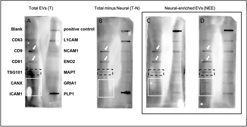
Micro RNAs (miRNAs) are short, non-coding RNAs with significant potential as diagnostic and prognostic biomarkers. However, a lack of reproducibility across studies has hindered their introduction into clinical settings. Inconsistencies between studies include a lack of consensus on the miRNAs associated with a specific disease and the direction of regulation. These differences may reflect the heterogenous nature of pathologies with multiple phenotypes, such as amyotrophic lateral sclerosis (ALS). It is also possible that discrepancies are due to different sampling, processing, and analysis protocols across labs. Using miRNA extracted from L1CAM immunoaffinity purified extracellular vesicles (neural-enriched extracellular vesicles or NEE), we thrice replicated an 8-miRNA fingerprint diagnostic of ALS, which includes the miRNA species and direction of regulation. We aimed to determine if the extra purification steps required to generate NEE created a unique extracellular vesicle (EV) fraction that might contribute to the robustness and replicability of our assay. We compared three fractions from control human plasma: 1) total heterogenous EVs (T), 2) L1CAM/neural enriched EVs (NEE), and 3) the remaining total-minus-NEE fraction (T-N). Each fraction was characterized for size, total protein content, and protein markers, then total RNA was extracted, and qPCR was run on 20 miRNAs. We report that the miRNA expression within NEE was different enough compared to T and T-N to justify the extra steps required to generate this fraction. We conclude that L1CAM immunocapture generates a unique fraction of EVs that consistently and robustly replicates a miRNA fingerprint which differentiates ALS patients from controls.
期刊介绍:
RNA has played a central role in all cellular processes since the beginning of life: decoding the genome, regulating gene expression, mediating molecular interactions, catalyzing chemical reactions. RNA Biology, as a leading journal in the field, provides a platform for presenting and discussing cutting-edge RNA research.
RNA Biology brings together a multidisciplinary community of scientists working in the areas of:
Transcription and splicing
Post-transcriptional regulation of gene expression
Non-coding RNAs
RNA localization
Translation and catalysis by RNA
Structural biology
Bioinformatics
RNA in disease and therapy
 求助内容:
求助内容: 应助结果提醒方式:


