{"title":"1例细胞形态清晰的α-胎蛋白子宫颈腺癌的免疫组织化学和分子分析。","authors":"Shu Kuriyama, Mitsutake Yano, Takahiro Kusaba, Sumika Zaitsu, Haruto Nishida, Masanori Yasuda, Kaei Nasu","doi":"10.1007/s00795-022-00336-7","DOIUrl":null,"url":null,"abstract":"<p><p>Adenocarcinomas with clear cell morphology may be associated with elevated serum alpha-fetoprotein levels in various organs. We report the case of an alpha-fetoprotein-producing cervical adenocarcinoma with clear cell morphology and compare it immunohistochemically, molecularly, and virologically with cervical clear cell carcinoma, gastric-type mucinous carcinoma, and ovarian clear cell carcinoma. A 51-year-old Japanese woman was initially diagnosed with cervical clear cell carcinoma. The tumor was resistant to standard surgery, radiotherapy, and chemotherapy. Serum carcinoembryonic antigen and alpha-fetoprotein were elevated. The tumor was immunohistochemically positive for alpha-fetoprotein, human chorionic gonadotropin, cytokeratin 20, spalt-like transcription factor 4, glypican 3, MUC6, and HIK1083. Gene panel testing revealed CCNE1 amplification, CDKN2A loss, and TP53 R282W. We compared the present case with 120 ovarian clear cell carcinoma cases using a tissue microarray. Only one case (0.8%) showed very limited immunohistochemical positivity for alpha-fetoprotein. Of the 54 cases in which serum carcinoembryonic antigen was measured, only one (1.9%) was elevated (19.9 ng/mL). We diagnosed the case as alpha-fetoprotein-producing cervical gastric-type mucinous carcinoma with enteroblastic differentiation. In conclusion, alpha-fetoprotein-producing cervical adenocarcinoma is a rare but aggressive tumor. Clinicians and pathologists should be aware of this unfamiliar tumor, its diagnostic clues, prognostic markers, and treatment strategies.</p>","PeriodicalId":18338,"journal":{"name":"Medical Molecular Morphology","volume":null,"pages":null},"PeriodicalIF":1.2000,"publicationDate":"2023-03-01","publicationTypes":"Journal Article","fieldsOfStudy":null,"isOpenAccess":false,"openAccessPdf":"","citationCount":"0","resultStr":"{\"title\":\"Immunohistochemical and molecular analysis of an α-fetoprotein-producing cervical adenocarcinoma with clear cell morphology.\",\"authors\":\"Shu Kuriyama, Mitsutake Yano, Takahiro Kusaba, Sumika Zaitsu, Haruto Nishida, Masanori Yasuda, Kaei Nasu\",\"doi\":\"10.1007/s00795-022-00336-7\",\"DOIUrl\":null,\"url\":null,\"abstract\":\"<p><p>Adenocarcinomas with clear cell morphology may be associated with elevated serum alpha-fetoprotein levels in various organs. We report the case of an alpha-fetoprotein-producing cervical adenocarcinoma with clear cell morphology and compare it immunohistochemically, molecularly, and virologically with cervical clear cell carcinoma, gastric-type mucinous carcinoma, and ovarian clear cell carcinoma. A 51-year-old Japanese woman was initially diagnosed with cervical clear cell carcinoma. The tumor was resistant to standard surgery, radiotherapy, and chemotherapy. Serum carcinoembryonic antigen and alpha-fetoprotein were elevated. The tumor was immunohistochemically positive for alpha-fetoprotein, human chorionic gonadotropin, cytokeratin 20, spalt-like transcription factor 4, glypican 3, MUC6, and HIK1083. Gene panel testing revealed CCNE1 amplification, CDKN2A loss, and TP53 R282W. We compared the present case with 120 ovarian clear cell carcinoma cases using a tissue microarray. Only one case (0.8%) showed very limited immunohistochemical positivity for alpha-fetoprotein. Of the 54 cases in which serum carcinoembryonic antigen was measured, only one (1.9%) was elevated (19.9 ng/mL). We diagnosed the case as alpha-fetoprotein-producing cervical gastric-type mucinous carcinoma with enteroblastic differentiation. In conclusion, alpha-fetoprotein-producing cervical adenocarcinoma is a rare but aggressive tumor. Clinicians and pathologists should be aware of this unfamiliar tumor, its diagnostic clues, prognostic markers, and treatment strategies.</p>\",\"PeriodicalId\":18338,\"journal\":{\"name\":\"Medical Molecular Morphology\",\"volume\":null,\"pages\":null},\"PeriodicalIF\":1.2000,\"publicationDate\":\"2023-03-01\",\"publicationTypes\":\"Journal Article\",\"fieldsOfStudy\":null,\"isOpenAccess\":false,\"openAccessPdf\":\"\",\"citationCount\":\"0\",\"resultStr\":null,\"platform\":\"Semanticscholar\",\"paperid\":null,\"PeriodicalName\":\"Medical Molecular Morphology\",\"FirstCategoryId\":\"3\",\"ListUrlMain\":\"https://doi.org/10.1007/s00795-022-00336-7\",\"RegionNum\":4,\"RegionCategory\":\"医学\",\"ArticlePicture\":[],\"TitleCN\":null,\"AbstractTextCN\":null,\"PMCID\":null,\"EPubDate\":\"\",\"PubModel\":\"\",\"JCR\":\"Q3\",\"JCRName\":\"PATHOLOGY\",\"Score\":null,\"Total\":0}","platform":"Semanticscholar","paperid":null,"PeriodicalName":"Medical Molecular Morphology","FirstCategoryId":"3","ListUrlMain":"https://doi.org/10.1007/s00795-022-00336-7","RegionNum":4,"RegionCategory":"医学","ArticlePicture":[],"TitleCN":null,"AbstractTextCN":null,"PMCID":null,"EPubDate":"","PubModel":"","JCR":"Q3","JCRName":"PATHOLOGY","Score":null,"Total":0}
Immunohistochemical and molecular analysis of an α-fetoprotein-producing cervical adenocarcinoma with clear cell morphology.
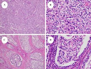
Adenocarcinomas with clear cell morphology may be associated with elevated serum alpha-fetoprotein levels in various organs. We report the case of an alpha-fetoprotein-producing cervical adenocarcinoma with clear cell morphology and compare it immunohistochemically, molecularly, and virologically with cervical clear cell carcinoma, gastric-type mucinous carcinoma, and ovarian clear cell carcinoma. A 51-year-old Japanese woman was initially diagnosed with cervical clear cell carcinoma. The tumor was resistant to standard surgery, radiotherapy, and chemotherapy. Serum carcinoembryonic antigen and alpha-fetoprotein were elevated. The tumor was immunohistochemically positive for alpha-fetoprotein, human chorionic gonadotropin, cytokeratin 20, spalt-like transcription factor 4, glypican 3, MUC6, and HIK1083. Gene panel testing revealed CCNE1 amplification, CDKN2A loss, and TP53 R282W. We compared the present case with 120 ovarian clear cell carcinoma cases using a tissue microarray. Only one case (0.8%) showed very limited immunohistochemical positivity for alpha-fetoprotein. Of the 54 cases in which serum carcinoembryonic antigen was measured, only one (1.9%) was elevated (19.9 ng/mL). We diagnosed the case as alpha-fetoprotein-producing cervical gastric-type mucinous carcinoma with enteroblastic differentiation. In conclusion, alpha-fetoprotein-producing cervical adenocarcinoma is a rare but aggressive tumor. Clinicians and pathologists should be aware of this unfamiliar tumor, its diagnostic clues, prognostic markers, and treatment strategies.
期刊介绍:
Medical Molecular Morphology is an international forum for researchers in both basic and clinical medicine to present and discuss new research on the structural mechanisms and the processes of health and disease at the molecular level. The structures of molecules, organelles, cells, tissues, and organs determine their normal function. Disease is thus best understood in terms of structural changes in these different levels of biological organization, especially in molecules and molecular interactions as well as the cellular localization of chemical components. Medical Molecular Morphology welcomes articles on basic or clinical research in the fields of cell biology, molecular biology, and medical, veterinary, and dental sciences using techniques for structural research such as electron microscopy, confocal laser scanning microscopy, enzyme histochemistry, immunohistochemistry, radioautography, X-ray microanalysis, and in situ hybridization.
Manuscripts submitted for publication must contain a statement to the effect that all human studies have been reviewed by the appropriate ethics committee and have therefore been performed in accordance with the ethical standards laid down in an appropriate version of the 1964 Declaration of Helsinki. It should also be stated clearly in the text that all persons gave their informed consent prior to their inclusion in the study. Details that might disclose the identity of the subjects under study should be omitted.
 求助内容:
求助内容: 应助结果提醒方式:


