{"title":"我国播氏分支杆菌(寡毛目:管科)腺体中的细孢子虫Jirovecia branchilis n.sp.(Microsporidia)","authors":"Xinhua Liu , Shisi Ren , Zhongyuan Chen , Qi Yin , Jianguo Xiang , Jianbo Yu , Deliang Li , Jinyong Zhang","doi":"10.1016/j.ejop.2023.125972","DOIUrl":null,"url":null,"abstract":"<div><p><em>Jirovecia</em> species primarily infect oligochaetes and are typically characterized by large rod-shaped spores with a tail-like posterior prolongation. Presently, seven <em>Jirovecia</em> spp. are reported worldwide with only one described in China. Here, a new species, <em>Jirovecia branchilis</em> n. sp. was discovered in glands of oligochaetes <em>Branchiura sowerybi</em> Beddard, 1892 in China. <em>Jirovecia branchilis</em> n. sp. elicited the formation of numerous opaque xenomas of 0.12 to 0.20 mm (n = 30) in diameter. Electron microscopic observations demonstrated that the earliest developmental stages observed were uninucleate meronts residing directly with the host cytoplasm. Mature spores were rod-shaped with blunt ends and possessed a collar-like anchoring disk, a manubrium-type polar filament, a bipartite polarplast, and a three-layered spore wall. A tail-like prolongation was distinctly observed in the posterior of spores and measured 13.2–28.6 μm long (n = 30). <em>Jirovecia branchilis</em> n. sp. showed 98.54% sequence similarity with <em>Janacekia tainunus</em> isolated from the fat body of chironomidae larvae <em>Kiefferulus tainanus</em> based on obtained partial SSU rDNA gene sequence, but was significantly different in morphology, host, and infection sites. SSU rDNA-based phylogenetic analysis indicated <em>Jirovecia branchilis</em> n. sp. clustered with <em>Janacekia tainanus</em> within the <em>Jirovecia</em>-<em>Bacillidium</em>-<em>Janacekia</em> clade. In conclusion, a new species within <em>Jirovecia</em>, <em>Jirovecia branchilis</em> n. sp. is erected herein based mainly on its morphological, ecological, and to a lesser degree on its molecular characteristics. The whole relationship between <em>Jirovecia</em> spp., <em>Janacekia</em> spp., and <em>Bacillidium</em> spp. is in need of revision and could potentially be elucidated by using additional makers and sequencing a broader diversity of the already described species.</p></div>","PeriodicalId":12042,"journal":{"name":"European journal of protistology","volume":null,"pages":null},"PeriodicalIF":1.9000,"publicationDate":"2023-04-01","publicationTypes":"Journal Article","fieldsOfStudy":null,"isOpenAccess":false,"openAccessPdf":"","citationCount":"0","resultStr":"{\"title\":\"Jirovecia branchilis n. sp. (Microsporidia) from glands of Branchiura sowerbyi (Oligochaeta: Tubificidae) in China\",\"authors\":\"Xinhua Liu , Shisi Ren , Zhongyuan Chen , Qi Yin , Jianguo Xiang , Jianbo Yu , Deliang Li , Jinyong Zhang\",\"doi\":\"10.1016/j.ejop.2023.125972\",\"DOIUrl\":null,\"url\":null,\"abstract\":\"<div><p><em>Jirovecia</em> species primarily infect oligochaetes and are typically characterized by large rod-shaped spores with a tail-like posterior prolongation. Presently, seven <em>Jirovecia</em> spp. are reported worldwide with only one described in China. Here, a new species, <em>Jirovecia branchilis</em> n. sp. was discovered in glands of oligochaetes <em>Branchiura sowerybi</em> Beddard, 1892 in China. <em>Jirovecia branchilis</em> n. sp. elicited the formation of numerous opaque xenomas of 0.12 to 0.20 mm (n = 30) in diameter. Electron microscopic observations demonstrated that the earliest developmental stages observed were uninucleate meronts residing directly with the host cytoplasm. Mature spores were rod-shaped with blunt ends and possessed a collar-like anchoring disk, a manubrium-type polar filament, a bipartite polarplast, and a three-layered spore wall. A tail-like prolongation was distinctly observed in the posterior of spores and measured 13.2–28.6 μm long (n = 30). <em>Jirovecia branchilis</em> n. sp. showed 98.54% sequence similarity with <em>Janacekia tainunus</em> isolated from the fat body of chironomidae larvae <em>Kiefferulus tainanus</em> based on obtained partial SSU rDNA gene sequence, but was significantly different in morphology, host, and infection sites. SSU rDNA-based phylogenetic analysis indicated <em>Jirovecia branchilis</em> n. sp. clustered with <em>Janacekia tainanus</em> within the <em>Jirovecia</em>-<em>Bacillidium</em>-<em>Janacekia</em> clade. In conclusion, a new species within <em>Jirovecia</em>, <em>Jirovecia branchilis</em> n. sp. is erected herein based mainly on its morphological, ecological, and to a lesser degree on its molecular characteristics. The whole relationship between <em>Jirovecia</em> spp., <em>Janacekia</em> spp., and <em>Bacillidium</em> spp. is in need of revision and could potentially be elucidated by using additional makers and sequencing a broader diversity of the already described species.</p></div>\",\"PeriodicalId\":12042,\"journal\":{\"name\":\"European journal of protistology\",\"volume\":null,\"pages\":null},\"PeriodicalIF\":1.9000,\"publicationDate\":\"2023-04-01\",\"publicationTypes\":\"Journal Article\",\"fieldsOfStudy\":null,\"isOpenAccess\":false,\"openAccessPdf\":\"\",\"citationCount\":\"0\",\"resultStr\":null,\"platform\":\"Semanticscholar\",\"paperid\":null,\"PeriodicalName\":\"European journal of protistology\",\"FirstCategoryId\":\"99\",\"ListUrlMain\":\"https://www.sciencedirect.com/science/article/pii/S0932473923000172\",\"RegionNum\":2,\"RegionCategory\":\"生物学\",\"ArticlePicture\":[],\"TitleCN\":null,\"AbstractTextCN\":null,\"PMCID\":null,\"EPubDate\":\"\",\"PubModel\":\"\",\"JCR\":\"Q4\",\"JCRName\":\"MICROBIOLOGY\",\"Score\":null,\"Total\":0}","platform":"Semanticscholar","paperid":null,"PeriodicalName":"European journal of protistology","FirstCategoryId":"99","ListUrlMain":"https://www.sciencedirect.com/science/article/pii/S0932473923000172","RegionNum":2,"RegionCategory":"生物学","ArticlePicture":[],"TitleCN":null,"AbstractTextCN":null,"PMCID":null,"EPubDate":"","PubModel":"","JCR":"Q4","JCRName":"MICROBIOLOGY","Score":null,"Total":0}
Jirovecia branchilis n. sp. (Microsporidia) from glands of Branchiura sowerbyi (Oligochaeta: Tubificidae) in China
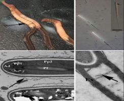
Jirovecia species primarily infect oligochaetes and are typically characterized by large rod-shaped spores with a tail-like posterior prolongation. Presently, seven Jirovecia spp. are reported worldwide with only one described in China. Here, a new species, Jirovecia branchilis n. sp. was discovered in glands of oligochaetes Branchiura sowerybi Beddard, 1892 in China. Jirovecia branchilis n. sp. elicited the formation of numerous opaque xenomas of 0.12 to 0.20 mm (n = 30) in diameter. Electron microscopic observations demonstrated that the earliest developmental stages observed were uninucleate meronts residing directly with the host cytoplasm. Mature spores were rod-shaped with blunt ends and possessed a collar-like anchoring disk, a manubrium-type polar filament, a bipartite polarplast, and a three-layered spore wall. A tail-like prolongation was distinctly observed in the posterior of spores and measured 13.2–28.6 μm long (n = 30). Jirovecia branchilis n. sp. showed 98.54% sequence similarity with Janacekia tainunus isolated from the fat body of chironomidae larvae Kiefferulus tainanus based on obtained partial SSU rDNA gene sequence, but was significantly different in morphology, host, and infection sites. SSU rDNA-based phylogenetic analysis indicated Jirovecia branchilis n. sp. clustered with Janacekia tainanus within the Jirovecia-Bacillidium-Janacekia clade. In conclusion, a new species within Jirovecia, Jirovecia branchilis n. sp. is erected herein based mainly on its morphological, ecological, and to a lesser degree on its molecular characteristics. The whole relationship between Jirovecia spp., Janacekia spp., and Bacillidium spp. is in need of revision and could potentially be elucidated by using additional makers and sequencing a broader diversity of the already described species.
期刊介绍:
Articles deal with protists, unicellular organisms encountered free-living in various habitats or as parasites or used in basic research or applications. The European Journal of Protistology covers topics such as the structure and systematics of protists, their development, ecology, molecular biology and physiology. Beside publishing original articles the journal offers a forum for announcing scientific meetings. Reviews of recently published books are included as well. With its diversity of topics, the European Journal of Protistology is an essential source of information for every active protistologist and for biologists of various fields.
 求助内容:
求助内容: 应助结果提醒方式:


