{"title":"立体定向放射手术后延髓血管母细胞瘤引起孤立的第四脑室1例。","authors":"Yuya Hama, Takahiro Sasaki, Toshikazu Yamoto, Junya Fukai, Hiroki Nishibayashi, Naoyuki Nakao","doi":"10.3892/mco.2023.2633","DOIUrl":null,"url":null,"abstract":"<p><p>Isolated fourth ventricle is a rare complication following shunt insertion of the lateral ventricles for hydrocephalus. The present report describes a rare case of a hemangioblastoma of the medulla oblongata that caused isolated fourth ventricle due to intraventricular deposition of fibrin. A 34-year-old man presented with headache a month before admission. Magnetic resonance imaging indicated multiple tumors in the medulla oblongata and the bilateral cerebellar hemisphere with surrounding edema, and the patient was diagnosed with hemangioblastoma. The patient began to develop progressive headache and nausea after stereotactic radiosurgery, and computed tomography showed obstructive hydrocephalus. Endoscopic third ventriculostomy was performed, and the intraoperative view of this showed that the walls of the lateral and third ventricles were covered with a white membrane-like substance. Endoscopic third ventriculostomy and then ventriculoperitoneal shunt did not improve the hydrocephalus. The patient's consciousness deteriorated due to isolated fourth ventricle and upward herniation. The patient underwent posterior fossa craniotomy and the tumor in the medulla oblongata was removed via a telovelar approach. Intraoperatively, the fourth ventricle was filled with a white membrane-like substance, which was surgically removed and pathologically diagnosed as fibrin. The patient's consciousness and obstructive hydrocephalus improved after surgery. The present case suggests that isolated fourth ventricle may occur after VP shunt placement for the hydrocephalus with hyperproteinorachia.</p>","PeriodicalId":18737,"journal":{"name":"Molecular and clinical oncology","volume":"18 5","pages":"37"},"PeriodicalIF":1.4000,"publicationDate":"2023-05-01","publicationTypes":"Journal Article","fieldsOfStudy":null,"isOpenAccess":false,"openAccessPdf":"https://ftp.ncbi.nlm.nih.gov/pub/pmc/oa_pdf/36/79/mco-18-05-02633.PMC10067790.pdf","citationCount":"0","resultStr":"{\"title\":\"Hemangioblastoma of the medulla oblongata that caused isolated fourth ventricle after stereotactic radiosurgery: A case report.\",\"authors\":\"Yuya Hama, Takahiro Sasaki, Toshikazu Yamoto, Junya Fukai, Hiroki Nishibayashi, Naoyuki Nakao\",\"doi\":\"10.3892/mco.2023.2633\",\"DOIUrl\":null,\"url\":null,\"abstract\":\"<p><p>Isolated fourth ventricle is a rare complication following shunt insertion of the lateral ventricles for hydrocephalus. The present report describes a rare case of a hemangioblastoma of the medulla oblongata that caused isolated fourth ventricle due to intraventricular deposition of fibrin. A 34-year-old man presented with headache a month before admission. Magnetic resonance imaging indicated multiple tumors in the medulla oblongata and the bilateral cerebellar hemisphere with surrounding edema, and the patient was diagnosed with hemangioblastoma. The patient began to develop progressive headache and nausea after stereotactic radiosurgery, and computed tomography showed obstructive hydrocephalus. Endoscopic third ventriculostomy was performed, and the intraoperative view of this showed that the walls of the lateral and third ventricles were covered with a white membrane-like substance. Endoscopic third ventriculostomy and then ventriculoperitoneal shunt did not improve the hydrocephalus. The patient's consciousness deteriorated due to isolated fourth ventricle and upward herniation. The patient underwent posterior fossa craniotomy and the tumor in the medulla oblongata was removed via a telovelar approach. Intraoperatively, the fourth ventricle was filled with a white membrane-like substance, which was surgically removed and pathologically diagnosed as fibrin. The patient's consciousness and obstructive hydrocephalus improved after surgery. The present case suggests that isolated fourth ventricle may occur after VP shunt placement for the hydrocephalus with hyperproteinorachia.</p>\",\"PeriodicalId\":18737,\"journal\":{\"name\":\"Molecular and clinical oncology\",\"volume\":\"18 5\",\"pages\":\"37\"},\"PeriodicalIF\":1.4000,\"publicationDate\":\"2023-05-01\",\"publicationTypes\":\"Journal Article\",\"fieldsOfStudy\":null,\"isOpenAccess\":false,\"openAccessPdf\":\"https://ftp.ncbi.nlm.nih.gov/pub/pmc/oa_pdf/36/79/mco-18-05-02633.PMC10067790.pdf\",\"citationCount\":\"0\",\"resultStr\":null,\"platform\":\"Semanticscholar\",\"paperid\":null,\"PeriodicalName\":\"Molecular and clinical oncology\",\"FirstCategoryId\":\"1085\",\"ListUrlMain\":\"https://doi.org/10.3892/mco.2023.2633\",\"RegionNum\":0,\"RegionCategory\":null,\"ArticlePicture\":[],\"TitleCN\":null,\"AbstractTextCN\":null,\"PMCID\":null,\"EPubDate\":\"\",\"PubModel\":\"\",\"JCR\":\"Q4\",\"JCRName\":\"ONCOLOGY\",\"Score\":null,\"Total\":0}","platform":"Semanticscholar","paperid":null,"PeriodicalName":"Molecular and clinical oncology","FirstCategoryId":"1085","ListUrlMain":"https://doi.org/10.3892/mco.2023.2633","RegionNum":0,"RegionCategory":null,"ArticlePicture":[],"TitleCN":null,"AbstractTextCN":null,"PMCID":null,"EPubDate":"","PubModel":"","JCR":"Q4","JCRName":"ONCOLOGY","Score":null,"Total":0}
Hemangioblastoma of the medulla oblongata that caused isolated fourth ventricle after stereotactic radiosurgery: A case report.
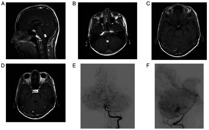
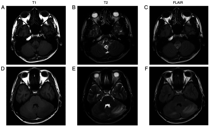
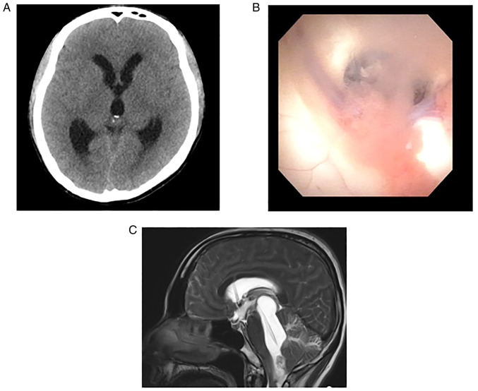
Isolated fourth ventricle is a rare complication following shunt insertion of the lateral ventricles for hydrocephalus. The present report describes a rare case of a hemangioblastoma of the medulla oblongata that caused isolated fourth ventricle due to intraventricular deposition of fibrin. A 34-year-old man presented with headache a month before admission. Magnetic resonance imaging indicated multiple tumors in the medulla oblongata and the bilateral cerebellar hemisphere with surrounding edema, and the patient was diagnosed with hemangioblastoma. The patient began to develop progressive headache and nausea after stereotactic radiosurgery, and computed tomography showed obstructive hydrocephalus. Endoscopic third ventriculostomy was performed, and the intraoperative view of this showed that the walls of the lateral and third ventricles were covered with a white membrane-like substance. Endoscopic third ventriculostomy and then ventriculoperitoneal shunt did not improve the hydrocephalus. The patient's consciousness deteriorated due to isolated fourth ventricle and upward herniation. The patient underwent posterior fossa craniotomy and the tumor in the medulla oblongata was removed via a telovelar approach. Intraoperatively, the fourth ventricle was filled with a white membrane-like substance, which was surgically removed and pathologically diagnosed as fibrin. The patient's consciousness and obstructive hydrocephalus improved after surgery. The present case suggests that isolated fourth ventricle may occur after VP shunt placement for the hydrocephalus with hyperproteinorachia.

 求助内容:
求助内容: 应助结果提醒方式:


