{"title":"广角超声与磁共振眼底形态学测量的比较。","authors":"Kohsuke Sekido, Kazuhiro Murayama, Tadashi Mizuguchi, Ryota Sakurai, Akiyoshi Iwase, Yoshiaki Shimada, Keita Suzuki, Atsuhiro Tanikawa, Masayuki Horiguchi","doi":"10.20407/fmj.2021-028","DOIUrl":null,"url":null,"abstract":"<p><strong>Objectives: </strong>To compare the eye axial length (AL), equatorial horizontal diameter (HD), and equatorial vertical diameter (VD) of normal eyes using a novel wide-angle, arc-scanning, ultrasound diagnostic device for wide-angle B-mode echography.</p><p><strong>Methods: </strong>In this cross-sectional study, wide-angle B-mode echography and magnetic resonance imaging (MRI) were conducted on 22 normal eyes; the AL, HD, and VD were measured.</p><p><strong>Results: </strong>The mean ALs were as follows: wide-angle B-mode echography, 25.22±1.47 mm and MRI, 25.24±1.46 mm; a significant correlation was observed between the two measurements (β=0.995 [0.976, 1.013]; <i>p</i><0.001; 95% R<sup>2</sup>=1.00). The mean HDs were as follows: wide-angle B-mode echography, 22.33±0.84 mm and MRI, 22.55±0.90 mm; a significant correlation was observed between the two measurements (β=0.902 [0.750, 1.179]; <i>p</i><0.001; 95% R<sup>2</sup>=0.81). The mean VDs were as follows: wide-angle B-mode echography, 22.77±0.91 mm; and MRI, 22.88±0.92 mm; a significant correlation was observed between the two measurements (β=0.966 [0.853, 1.097]; <i>p</i><0.001; 95% R<sup>2</sup>=0.93).</p><p><strong>Conclusions: </strong>There were no significant differences in the measurements for each parameter by wide-angle B-mode echography and MRI. Therefore, wide-angle B-mode echography permits accurate visualization of ocular morphology.</p>","PeriodicalId":33657,"journal":{"name":"Fujita Medical Journal","volume":"9 1","pages":"41-46"},"PeriodicalIF":0.0000,"publicationDate":"2023-02-01","publicationTypes":"Journal Article","fieldsOfStudy":null,"isOpenAccess":false,"openAccessPdf":"https://www.ncbi.nlm.nih.gov/pmc/articles/PMC9923445/pdf/","citationCount":"0","resultStr":"{\"title\":\"Comparison of ocular morphological measurement by wide-angle echography and magnetic resonance imaging.\",\"authors\":\"Kohsuke Sekido, Kazuhiro Murayama, Tadashi Mizuguchi, Ryota Sakurai, Akiyoshi Iwase, Yoshiaki Shimada, Keita Suzuki, Atsuhiro Tanikawa, Masayuki Horiguchi\",\"doi\":\"10.20407/fmj.2021-028\",\"DOIUrl\":null,\"url\":null,\"abstract\":\"<p><strong>Objectives: </strong>To compare the eye axial length (AL), equatorial horizontal diameter (HD), and equatorial vertical diameter (VD) of normal eyes using a novel wide-angle, arc-scanning, ultrasound diagnostic device for wide-angle B-mode echography.</p><p><strong>Methods: </strong>In this cross-sectional study, wide-angle B-mode echography and magnetic resonance imaging (MRI) were conducted on 22 normal eyes; the AL, HD, and VD were measured.</p><p><strong>Results: </strong>The mean ALs were as follows: wide-angle B-mode echography, 25.22±1.47 mm and MRI, 25.24±1.46 mm; a significant correlation was observed between the two measurements (β=0.995 [0.976, 1.013]; <i>p</i><0.001; 95% R<sup>2</sup>=1.00). The mean HDs were as follows: wide-angle B-mode echography, 22.33±0.84 mm and MRI, 22.55±0.90 mm; a significant correlation was observed between the two measurements (β=0.902 [0.750, 1.179]; <i>p</i><0.001; 95% R<sup>2</sup>=0.81). The mean VDs were as follows: wide-angle B-mode echography, 22.77±0.91 mm; and MRI, 22.88±0.92 mm; a significant correlation was observed between the two measurements (β=0.966 [0.853, 1.097]; <i>p</i><0.001; 95% R<sup>2</sup>=0.93).</p><p><strong>Conclusions: </strong>There were no significant differences in the measurements for each parameter by wide-angle B-mode echography and MRI. Therefore, wide-angle B-mode echography permits accurate visualization of ocular morphology.</p>\",\"PeriodicalId\":33657,\"journal\":{\"name\":\"Fujita Medical Journal\",\"volume\":\"9 1\",\"pages\":\"41-46\"},\"PeriodicalIF\":0.0000,\"publicationDate\":\"2023-02-01\",\"publicationTypes\":\"Journal Article\",\"fieldsOfStudy\":null,\"isOpenAccess\":false,\"openAccessPdf\":\"https://www.ncbi.nlm.nih.gov/pmc/articles/PMC9923445/pdf/\",\"citationCount\":\"0\",\"resultStr\":null,\"platform\":\"Semanticscholar\",\"paperid\":null,\"PeriodicalName\":\"Fujita Medical Journal\",\"FirstCategoryId\":\"1085\",\"ListUrlMain\":\"https://doi.org/10.20407/fmj.2021-028\",\"RegionNum\":0,\"RegionCategory\":null,\"ArticlePicture\":[],\"TitleCN\":null,\"AbstractTextCN\":null,\"PMCID\":null,\"EPubDate\":\"\",\"PubModel\":\"\",\"JCR\":\"\",\"JCRName\":\"\",\"Score\":null,\"Total\":0}","platform":"Semanticscholar","paperid":null,"PeriodicalName":"Fujita Medical Journal","FirstCategoryId":"1085","ListUrlMain":"https://doi.org/10.20407/fmj.2021-028","RegionNum":0,"RegionCategory":null,"ArticlePicture":[],"TitleCN":null,"AbstractTextCN":null,"PMCID":null,"EPubDate":"","PubModel":"","JCR":"","JCRName":"","Score":null,"Total":0}
Comparison of ocular morphological measurement by wide-angle echography and magnetic resonance imaging.
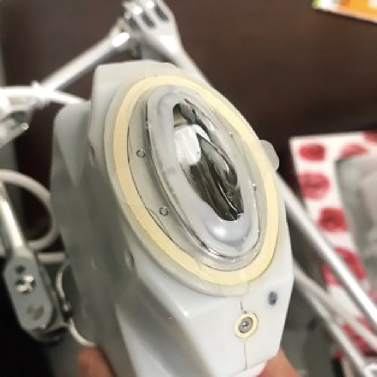
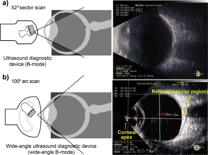
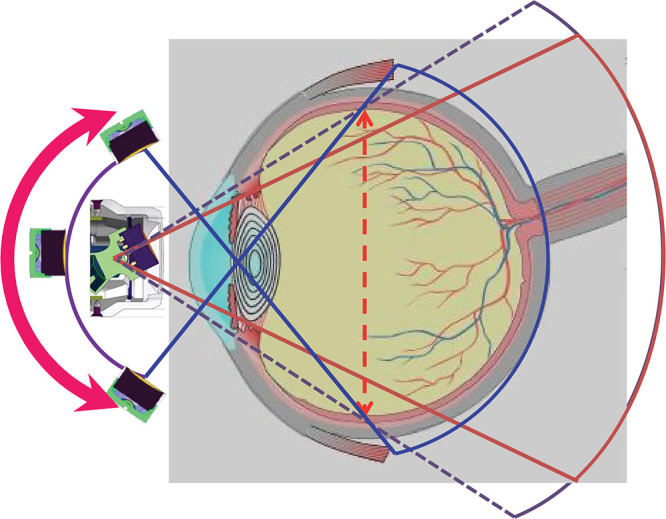
Objectives: To compare the eye axial length (AL), equatorial horizontal diameter (HD), and equatorial vertical diameter (VD) of normal eyes using a novel wide-angle, arc-scanning, ultrasound diagnostic device for wide-angle B-mode echography.
Methods: In this cross-sectional study, wide-angle B-mode echography and magnetic resonance imaging (MRI) were conducted on 22 normal eyes; the AL, HD, and VD were measured.
Results: The mean ALs were as follows: wide-angle B-mode echography, 25.22±1.47 mm and MRI, 25.24±1.46 mm; a significant correlation was observed between the two measurements (β=0.995 [0.976, 1.013]; p<0.001; 95% R2=1.00). The mean HDs were as follows: wide-angle B-mode echography, 22.33±0.84 mm and MRI, 22.55±0.90 mm; a significant correlation was observed between the two measurements (β=0.902 [0.750, 1.179]; p<0.001; 95% R2=0.81). The mean VDs were as follows: wide-angle B-mode echography, 22.77±0.91 mm; and MRI, 22.88±0.92 mm; a significant correlation was observed between the two measurements (β=0.966 [0.853, 1.097]; p<0.001; 95% R2=0.93).
Conclusions: There were no significant differences in the measurements for each parameter by wide-angle B-mode echography and MRI. Therefore, wide-angle B-mode echography permits accurate visualization of ocular morphology.

 求助内容:
求助内容: 应助结果提醒方式:


