{"title":"单次屏气时腹部弥散加权成像的同时多层回声平面成像。","authors":"Naoki Ohno, Kotaro Yoshida, Yu Ueda, Yuki Makino, Tosiaki Miyati, Toshifumi Gabata, Satoshi Kobayashi","doi":"10.2463/mrms.mp.2021-0087","DOIUrl":null,"url":null,"abstract":"<p><strong>Purpose: </strong>This multi-scanner study aimed to investigate the validity of single breath-hold (BH) diffusion-weighted imaging (DWI) using simultaneous-multislice (SMS) echo-planar imaging in multiple abdominal organs to enable faster acquisition and reliable quantification of apparent diffusion coefficient (ADC).</p><p><strong>Methods: </strong>SNR, geometric distortion (GD), and ADC in a phantom; the ADC in the liver, renal cortex, paraspinal muscle, spleen, and pancreas; and the signal intensity ratio of the portal vein-to-muscle (SIR<sub>PV-M</sub>) in healthy volunteers were compared between BH- and respiratory-triggered (RT) DWI with b-values of 0 and 800 s/mm<sup>2</sup> in two different MRI scanners.</p><p><strong>Results: </strong>The phantom study showed that the SNR of BH-DWI was significantly lower than that of the RT-DWI (P < 0.05 for both scanners), whereas the GD and ADC of BH-DWI did not differ significantly from those of the RT-DWI (P = 0.09-0.60). In the volunteer study, the scan times were 23 seconds for BH-DWI and 184±33 seconds for RT-DWI, respectively. The ADC of the liver in BH-DWI was significantly lower than that in RT-DWI (P < 0.05 for both scanners), whereas there were no significant differences in the ADCs of the renal cortex, paraspinal muscle, spleen, or pancreas between BH-DWI and RT-DWI (P = 0.07-0.86). The SIR<sub>PV-M</sub> in BH-DWI was significantly smaller than in RT-DWI (P < 0.05 for both scanners).</p><p><strong>Conclusion: </strong>The proposed method enables the acquisition of abdominal diffusion-weighted images in a single BH.</p>","PeriodicalId":18119,"journal":{"name":"Magnetic Resonance in Medical Sciences","volume":"22 2","pages":"253-262"},"PeriodicalIF":3.2000,"publicationDate":"2023-04-01","publicationTypes":"Journal Article","fieldsOfStudy":null,"isOpenAccess":false,"openAccessPdf":"https://ftp.ncbi.nlm.nih.gov/pub/pmc/oa_pdf/c9/4f/mrms-22-253.PMC10086397.pdf","citationCount":"3","resultStr":"{\"title\":\"Diffusion-weighted Imaging of the Abdomen during a Single Breath-hold Using Simultaneous-multislice Echo-planar Imaging.\",\"authors\":\"Naoki Ohno, Kotaro Yoshida, Yu Ueda, Yuki Makino, Tosiaki Miyati, Toshifumi Gabata, Satoshi Kobayashi\",\"doi\":\"10.2463/mrms.mp.2021-0087\",\"DOIUrl\":null,\"url\":null,\"abstract\":\"<p><strong>Purpose: </strong>This multi-scanner study aimed to investigate the validity of single breath-hold (BH) diffusion-weighted imaging (DWI) using simultaneous-multislice (SMS) echo-planar imaging in multiple abdominal organs to enable faster acquisition and reliable quantification of apparent diffusion coefficient (ADC).</p><p><strong>Methods: </strong>SNR, geometric distortion (GD), and ADC in a phantom; the ADC in the liver, renal cortex, paraspinal muscle, spleen, and pancreas; and the signal intensity ratio of the portal vein-to-muscle (SIR<sub>PV-M</sub>) in healthy volunteers were compared between BH- and respiratory-triggered (RT) DWI with b-values of 0 and 800 s/mm<sup>2</sup> in two different MRI scanners.</p><p><strong>Results: </strong>The phantom study showed that the SNR of BH-DWI was significantly lower than that of the RT-DWI (P < 0.05 for both scanners), whereas the GD and ADC of BH-DWI did not differ significantly from those of the RT-DWI (P = 0.09-0.60). In the volunteer study, the scan times were 23 seconds for BH-DWI and 184±33 seconds for RT-DWI, respectively. The ADC of the liver in BH-DWI was significantly lower than that in RT-DWI (P < 0.05 for both scanners), whereas there were no significant differences in the ADCs of the renal cortex, paraspinal muscle, spleen, or pancreas between BH-DWI and RT-DWI (P = 0.07-0.86). The SIR<sub>PV-M</sub> in BH-DWI was significantly smaller than in RT-DWI (P < 0.05 for both scanners).</p><p><strong>Conclusion: </strong>The proposed method enables the acquisition of abdominal diffusion-weighted images in a single BH.</p>\",\"PeriodicalId\":18119,\"journal\":{\"name\":\"Magnetic Resonance in Medical Sciences\",\"volume\":\"22 2\",\"pages\":\"253-262\"},\"PeriodicalIF\":3.2000,\"publicationDate\":\"2023-04-01\",\"publicationTypes\":\"Journal Article\",\"fieldsOfStudy\":null,\"isOpenAccess\":false,\"openAccessPdf\":\"https://ftp.ncbi.nlm.nih.gov/pub/pmc/oa_pdf/c9/4f/mrms-22-253.PMC10086397.pdf\",\"citationCount\":\"3\",\"resultStr\":null,\"platform\":\"Semanticscholar\",\"paperid\":null,\"PeriodicalName\":\"Magnetic Resonance in Medical Sciences\",\"FirstCategoryId\":\"3\",\"ListUrlMain\":\"https://doi.org/10.2463/mrms.mp.2021-0087\",\"RegionNum\":3,\"RegionCategory\":\"医学\",\"ArticlePicture\":[],\"TitleCN\":null,\"AbstractTextCN\":null,\"PMCID\":null,\"EPubDate\":\"\",\"PubModel\":\"\",\"JCR\":\"Q2\",\"JCRName\":\"RADIOLOGY, NUCLEAR MEDICINE & MEDICAL IMAGING\",\"Score\":null,\"Total\":0}","platform":"Semanticscholar","paperid":null,"PeriodicalName":"Magnetic Resonance in Medical Sciences","FirstCategoryId":"3","ListUrlMain":"https://doi.org/10.2463/mrms.mp.2021-0087","RegionNum":3,"RegionCategory":"医学","ArticlePicture":[],"TitleCN":null,"AbstractTextCN":null,"PMCID":null,"EPubDate":"","PubModel":"","JCR":"Q2","JCRName":"RADIOLOGY, NUCLEAR MEDICINE & MEDICAL IMAGING","Score":null,"Total":0}
Diffusion-weighted Imaging of the Abdomen during a Single Breath-hold Using Simultaneous-multislice Echo-planar Imaging.
Purpose: This multi-scanner study aimed to investigate the validity of single breath-hold (BH) diffusion-weighted imaging (DWI) using simultaneous-multislice (SMS) echo-planar imaging in multiple abdominal organs to enable faster acquisition and reliable quantification of apparent diffusion coefficient (ADC).
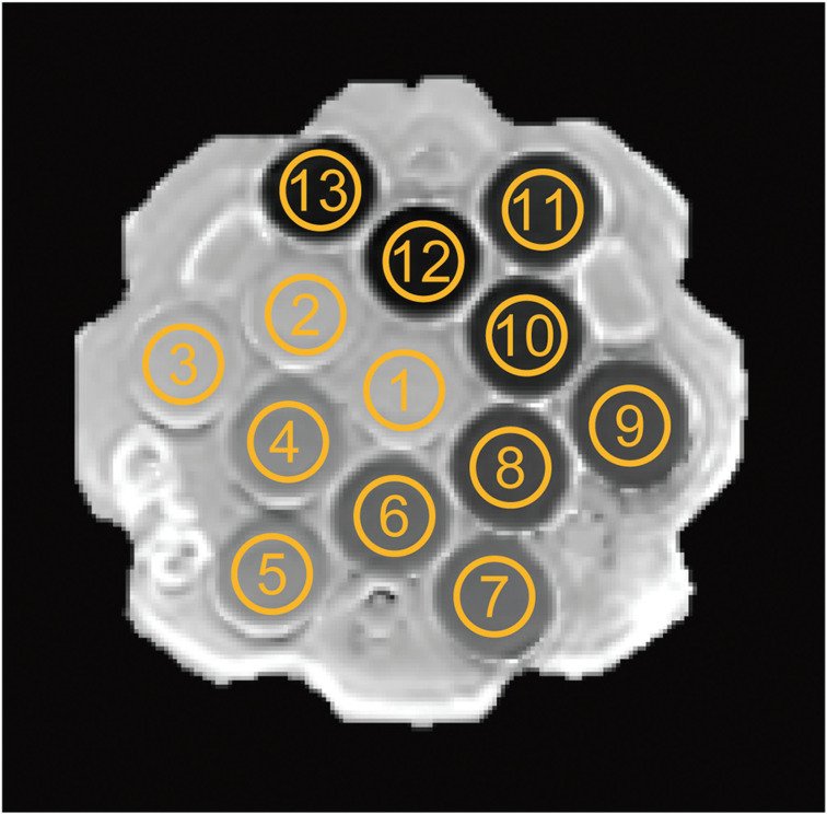
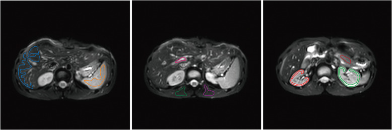
Methods: SNR, geometric distortion (GD), and ADC in a phantom; the ADC in the liver, renal cortex, paraspinal muscle, spleen, and pancreas; and the signal intensity ratio of the portal vein-to-muscle (SIRPV-M) in healthy volunteers were compared between BH- and respiratory-triggered (RT) DWI with b-values of 0 and 800 s/mm2 in two different MRI scanners.
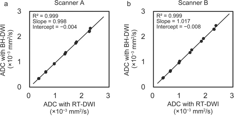
Results: The phantom study showed that the SNR of BH-DWI was significantly lower than that of the RT-DWI (P < 0.05 for both scanners), whereas the GD and ADC of BH-DWI did not differ significantly from those of the RT-DWI (P = 0.09-0.60). In the volunteer study, the scan times were 23 seconds for BH-DWI and 184±33 seconds for RT-DWI, respectively. The ADC of the liver in BH-DWI was significantly lower than that in RT-DWI (P < 0.05 for both scanners), whereas there were no significant differences in the ADCs of the renal cortex, paraspinal muscle, spleen, or pancreas between BH-DWI and RT-DWI (P = 0.07-0.86). The SIRPV-M in BH-DWI was significantly smaller than in RT-DWI (P < 0.05 for both scanners).
Conclusion: The proposed method enables the acquisition of abdominal diffusion-weighted images in a single BH.
期刊介绍:
Magnetic Resonance in Medical Sciences (MRMS or Magn
Reson Med Sci) is an international journal pursuing the
publication of original articles contributing to the progress
of magnetic resonance in the field of biomedical sciences
including technical developments and clinical applications.
MRMS is an official journal of the Japanese Society for
Magnetic Resonance in Medicine (JSMRM).
 求助内容:
求助内容: 应助结果提醒方式:


