Robert Noelken, Laura Westphal, Eik Schiegnitz, Bilal Al-Nawas
{"title":"激光辅助种植体周围缺损再生技术治疗重度种植体周围炎缺损的软硬组织再生:3年结果","authors":"Robert Noelken, Laura Westphal, Eik Schiegnitz, Bilal Al-Nawas","doi":"10.1186/s40729-023-00467-1","DOIUrl":null,"url":null,"abstract":"<p><strong>Purpose: </strong>This retrospective cohort study evaluates the regeneration of severe peri-implantitis deficiencies treated with the laser-assisted peri-implant defect regeneration (LAPIDER) approach within a 3-year follow-up.</p><p><strong>Methods: </strong>Twenty-four implants with severe peri-implantitis in 18 patients were treated according to the LAPIDER technique. In contrast to classic techniques for reconstructive peri-implantitis surgery with a marginal incision, a buccal split-flap preparation avoiding papillae separation was used. After a coronal flap elevation and a laser-assisted peri-implant defect cleaning, connective tissue and autogenous bone grafting was performed. Primary outcomes were the changes of the marginal bone levels (MBL) and the buccal bone thickness. Secondary outcomes included implant survival, peri-implant probing depths (PPD), bleeding on probing (BOP), recession, width of keratinized mucosa (KMW), thickness of keratinized mucosa (KMT), soft tissue esthetics (PES), and implant success.</p><p><strong>Results: </strong>MBL improved interproximal by 3.10 ± 2.02 mm (p < 0.001), buccal by 3.49 ± 2.89 mm (p < 0.001), and lingual by 1.46 ± 1.98 mm (p = 0.003); buccal bone thickness by 0.55 ± 0.60 mm (p = 0.005), and 1.01 ± 1.25 mm (p = 0.001) at 1 and 3 mm below reference level. Two implants were removed; 22 implants were still in function at a mean follow-up of 36 months. PPD changed from 5.05 ± 1.39 to 3.08 ± 0.71 mm (p < 0.001); recession was reduced from 2.07 ± 1.70 to 0.91 ± 1.13 mm (p = 0.001); KMW increased from 2.91 ± 1.81 to 4.18 ± 1.67 mm (p = 0.006); KMT improved from 1.73 ± 0.50 to 2.44 ± 0.43 mm (p < 0.001); PES changed from 7.7 ± 2.8 to 10.7 ± 1.9 (p < 0.001). 45.8% to 54.2% of the implants met the criteria of implant success.</p><p><strong>Conclusions: </strong>The favorable results document the proof of principle for the regeneration of severe peri-implant hard and soft tissue deficiencies by the LAPIDER treatment approach.</p>","PeriodicalId":14076,"journal":{"name":"International Journal of Implant Dentistry","volume":null,"pages":null},"PeriodicalIF":3.1000,"publicationDate":"2023-02-05","publicationTypes":"Journal Article","fieldsOfStudy":null,"isOpenAccess":false,"openAccessPdf":"https://www.ncbi.nlm.nih.gov/pmc/articles/PMC9899875/pdf/","citationCount":"0","resultStr":"{\"title\":\"Hard and soft tissue regeneration of severe peri-implantitis defects with the laser-assisted peri-implant defect regeneration technique: 3-year results.\",\"authors\":\"Robert Noelken, Laura Westphal, Eik Schiegnitz, Bilal Al-Nawas\",\"doi\":\"10.1186/s40729-023-00467-1\",\"DOIUrl\":null,\"url\":null,\"abstract\":\"<p><strong>Purpose: </strong>This retrospective cohort study evaluates the regeneration of severe peri-implantitis deficiencies treated with the laser-assisted peri-implant defect regeneration (LAPIDER) approach within a 3-year follow-up.</p><p><strong>Methods: </strong>Twenty-four implants with severe peri-implantitis in 18 patients were treated according to the LAPIDER technique. In contrast to classic techniques for reconstructive peri-implantitis surgery with a marginal incision, a buccal split-flap preparation avoiding papillae separation was used. After a coronal flap elevation and a laser-assisted peri-implant defect cleaning, connective tissue and autogenous bone grafting was performed. Primary outcomes were the changes of the marginal bone levels (MBL) and the buccal bone thickness. Secondary outcomes included implant survival, peri-implant probing depths (PPD), bleeding on probing (BOP), recession, width of keratinized mucosa (KMW), thickness of keratinized mucosa (KMT), soft tissue esthetics (PES), and implant success.</p><p><strong>Results: </strong>MBL improved interproximal by 3.10 ± 2.02 mm (p < 0.001), buccal by 3.49 ± 2.89 mm (p < 0.001), and lingual by 1.46 ± 1.98 mm (p = 0.003); buccal bone thickness by 0.55 ± 0.60 mm (p = 0.005), and 1.01 ± 1.25 mm (p = 0.001) at 1 and 3 mm below reference level. Two implants were removed; 22 implants were still in function at a mean follow-up of 36 months. PPD changed from 5.05 ± 1.39 to 3.08 ± 0.71 mm (p < 0.001); recession was reduced from 2.07 ± 1.70 to 0.91 ± 1.13 mm (p = 0.001); KMW increased from 2.91 ± 1.81 to 4.18 ± 1.67 mm (p = 0.006); KMT improved from 1.73 ± 0.50 to 2.44 ± 0.43 mm (p < 0.001); PES changed from 7.7 ± 2.8 to 10.7 ± 1.9 (p < 0.001). 45.8% to 54.2% of the implants met the criteria of implant success.</p><p><strong>Conclusions: </strong>The favorable results document the proof of principle for the regeneration of severe peri-implant hard and soft tissue deficiencies by the LAPIDER treatment approach.</p>\",\"PeriodicalId\":14076,\"journal\":{\"name\":\"International Journal of Implant Dentistry\",\"volume\":null,\"pages\":null},\"PeriodicalIF\":3.1000,\"publicationDate\":\"2023-02-05\",\"publicationTypes\":\"Journal Article\",\"fieldsOfStudy\":null,\"isOpenAccess\":false,\"openAccessPdf\":\"https://www.ncbi.nlm.nih.gov/pmc/articles/PMC9899875/pdf/\",\"citationCount\":\"0\",\"resultStr\":null,\"platform\":\"Semanticscholar\",\"paperid\":null,\"PeriodicalName\":\"International Journal of Implant Dentistry\",\"FirstCategoryId\":\"3\",\"ListUrlMain\":\"https://doi.org/10.1186/s40729-023-00467-1\",\"RegionNum\":3,\"RegionCategory\":\"医学\",\"ArticlePicture\":[],\"TitleCN\":null,\"AbstractTextCN\":null,\"PMCID\":null,\"EPubDate\":\"\",\"PubModel\":\"\",\"JCR\":\"Q1\",\"JCRName\":\"DENTISTRY, ORAL SURGERY & MEDICINE\",\"Score\":null,\"Total\":0}","platform":"Semanticscholar","paperid":null,"PeriodicalName":"International Journal of Implant Dentistry","FirstCategoryId":"3","ListUrlMain":"https://doi.org/10.1186/s40729-023-00467-1","RegionNum":3,"RegionCategory":"医学","ArticlePicture":[],"TitleCN":null,"AbstractTextCN":null,"PMCID":null,"EPubDate":"","PubModel":"","JCR":"Q1","JCRName":"DENTISTRY, ORAL SURGERY & MEDICINE","Score":null,"Total":0}
引用次数: 0
摘要
目的:本回顾性队列研究评估了激光辅助种植体周围缺损再生(LAPIDER)方法治疗严重种植体周围炎缺陷的3年随访。方法:采用LAPIDER技术对18例24例重度种植体周围炎患者进行治疗。与传统的边缘切口重建种植体周围炎手术技术相比,我们使用了一种避免乳头分离的颊裂皮瓣制备方法。冠状瓣抬高和激光辅助种植体周围缺损清理后,进行结缔组织和自体骨移植。主要结果为边缘骨水平(MBL)和颊骨厚度的变化。次要结果包括种植体存活、种植体周围探探深度(PPD)、探探出血(BOP)、退退、角化粘膜宽度(KMW)、角化粘膜厚度(KMT)、软组织美观(PES)和种植体成功。结果:MBL可使近端间缺损增加3.10±2.02 mm (p)。结论:LAPIDER治疗方法可用于种植体周围严重软组织缺损的再生。
Hard and soft tissue regeneration of severe peri-implantitis defects with the laser-assisted peri-implant defect regeneration technique: 3-year results.
Purpose: This retrospective cohort study evaluates the regeneration of severe peri-implantitis deficiencies treated with the laser-assisted peri-implant defect regeneration (LAPIDER) approach within a 3-year follow-up.
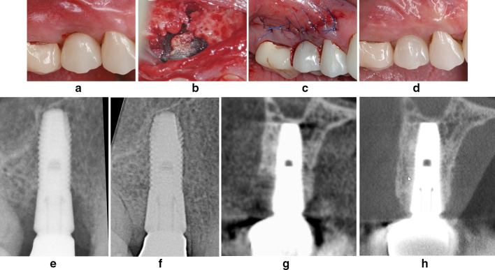
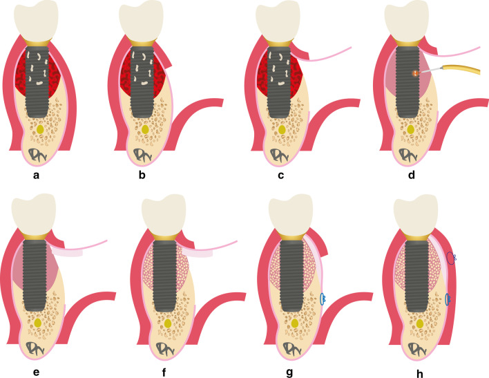
Methods: Twenty-four implants with severe peri-implantitis in 18 patients were treated according to the LAPIDER technique. In contrast to classic techniques for reconstructive peri-implantitis surgery with a marginal incision, a buccal split-flap preparation avoiding papillae separation was used. After a coronal flap elevation and a laser-assisted peri-implant defect cleaning, connective tissue and autogenous bone grafting was performed. Primary outcomes were the changes of the marginal bone levels (MBL) and the buccal bone thickness. Secondary outcomes included implant survival, peri-implant probing depths (PPD), bleeding on probing (BOP), recession, width of keratinized mucosa (KMW), thickness of keratinized mucosa (KMT), soft tissue esthetics (PES), and implant success.
Results: MBL improved interproximal by 3.10 ± 2.02 mm (p < 0.001), buccal by 3.49 ± 2.89 mm (p < 0.001), and lingual by 1.46 ± 1.98 mm (p = 0.003); buccal bone thickness by 0.55 ± 0.60 mm (p = 0.005), and 1.01 ± 1.25 mm (p = 0.001) at 1 and 3 mm below reference level. Two implants were removed; 22 implants were still in function at a mean follow-up of 36 months. PPD changed from 5.05 ± 1.39 to 3.08 ± 0.71 mm (p < 0.001); recession was reduced from 2.07 ± 1.70 to 0.91 ± 1.13 mm (p = 0.001); KMW increased from 2.91 ± 1.81 to 4.18 ± 1.67 mm (p = 0.006); KMT improved from 1.73 ± 0.50 to 2.44 ± 0.43 mm (p < 0.001); PES changed from 7.7 ± 2.8 to 10.7 ± 1.9 (p < 0.001). 45.8% to 54.2% of the implants met the criteria of implant success.
Conclusions: The favorable results document the proof of principle for the regeneration of severe peri-implant hard and soft tissue deficiencies by the LAPIDER treatment approach.
期刊介绍:
The International Journal of Implant Dentistry is a peer-reviewed open access journal published under the SpringerOpen brand. The journal is dedicated to promoting the exchange and discussion of all research areas relevant to implant dentistry in the form of systematic literature or invited reviews, prospective and retrospective clinical studies, clinical case reports, basic laboratory and animal research, and articles on material research and engineering.

 求助内容:
求助内容: 应助结果提醒方式:


