{"title":"G蛋白偶联的甲酰肽受体2促进糖尿病视网膜病变中的内皮-间充质转变。","authors":"Xueying Lou, Shuang Liu, Jian Shi, Hongliang Chen, Zichen Wang, Yingying Le, Hui Chen, Rongrong Zhu, Ying Yu","doi":"10.1159/000529578","DOIUrl":null,"url":null,"abstract":"<p><strong>Introduction: </strong>In proliferative diabetic retinopathy (PDR), retinal neovascularization is the essential pathogenic process that is linked to endothelial-to-mesenchymal transition (EndoMT) induced by high glucose (HG). This pathophysiological process may be regulated by a G-protein-coupled chemoattractant receptor FPR2 (mouse Fpr2), involved in inflammatory cell migration and proliferation. In the current study, we investigated the role of Fpr2 in regulating EndoMT and the underlying mechanisms during diabetic retinopathy progression.</p><p><strong>Methods: </strong>FPR2 agonist or inhibitor was added to human microvascular endothelial cells (HMECs) exposed to normal glucose or HG. Morphologic, phenotypic, and functional changes of HMECs as well as the formation of microvasculature related to EndoMT were assessed. EndoMT biomarkers were detected in the retinal tissues of diabetic mice and fibrovascular epiretinal membranes (FVMs) from patients with PDR.</p><p><strong>Results: </strong>HG upregulated FPR2 in HMECs, which triggered morphological changes, and the cells acquired mesenchymal phenotype, with enhanced cell migration, viability, and angiogenic process shown by tube formation and aortic ring sprouting. Inhibition of FPR2 attenuated HG-induced EndoMT and endothelial cell migration to form vessel-like tube structures. RNA sequence and protein analysis further revealed that inhibition of FPR2 decreased the expression of genes associated with EndoMT. ERK1/2 and P38 signaling pathway was activated in HMECs, promoting neovascularization in HG-induced EndoMT of HMECs. In vivo, increased expression of mesenchymal markers was detected in the retina of diabetic mice and FVMs from patients with PDR. FPR2 deficiency was associated with diminished EndoMT-related phenotypic changes in the retina of diabetic mice.</p><p><strong>Conclusions: </strong>FPR2 is actively involved in the progression of EndoMT that may contribute to the pathogenesis of PDR. Thus, FPR2 may be a potential therapeutic target for PDR.</p>","PeriodicalId":19662,"journal":{"name":"Ophthalmic Research","volume":" ","pages":"681-691"},"PeriodicalIF":2.0000,"publicationDate":"2023-01-01","publicationTypes":"Journal Article","fieldsOfStudy":null,"isOpenAccess":false,"openAccessPdf":"https://www.ncbi.nlm.nih.gov/pmc/articles/PMC10064398/pdf/","citationCount":"0","resultStr":"{\"title\":\"The G-Protein-Coupled Formyl Peptide Receptor 2 Promotes Endothelial-Mesenchymal Transition in Diabetic Retinopathy.\",\"authors\":\"Xueying Lou, Shuang Liu, Jian Shi, Hongliang Chen, Zichen Wang, Yingying Le, Hui Chen, Rongrong Zhu, Ying Yu\",\"doi\":\"10.1159/000529578\",\"DOIUrl\":null,\"url\":null,\"abstract\":\"<p><strong>Introduction: </strong>In proliferative diabetic retinopathy (PDR), retinal neovascularization is the essential pathogenic process that is linked to endothelial-to-mesenchymal transition (EndoMT) induced by high glucose (HG). This pathophysiological process may be regulated by a G-protein-coupled chemoattractant receptor FPR2 (mouse Fpr2), involved in inflammatory cell migration and proliferation. In the current study, we investigated the role of Fpr2 in regulating EndoMT and the underlying mechanisms during diabetic retinopathy progression.</p><p><strong>Methods: </strong>FPR2 agonist or inhibitor was added to human microvascular endothelial cells (HMECs) exposed to normal glucose or HG. Morphologic, phenotypic, and functional changes of HMECs as well as the formation of microvasculature related to EndoMT were assessed. EndoMT biomarkers were detected in the retinal tissues of diabetic mice and fibrovascular epiretinal membranes (FVMs) from patients with PDR.</p><p><strong>Results: </strong>HG upregulated FPR2 in HMECs, which triggered morphological changes, and the cells acquired mesenchymal phenotype, with enhanced cell migration, viability, and angiogenic process shown by tube formation and aortic ring sprouting. Inhibition of FPR2 attenuated HG-induced EndoMT and endothelial cell migration to form vessel-like tube structures. RNA sequence and protein analysis further revealed that inhibition of FPR2 decreased the expression of genes associated with EndoMT. ERK1/2 and P38 signaling pathway was activated in HMECs, promoting neovascularization in HG-induced EndoMT of HMECs. In vivo, increased expression of mesenchymal markers was detected in the retina of diabetic mice and FVMs from patients with PDR. FPR2 deficiency was associated with diminished EndoMT-related phenotypic changes in the retina of diabetic mice.</p><p><strong>Conclusions: </strong>FPR2 is actively involved in the progression of EndoMT that may contribute to the pathogenesis of PDR. Thus, FPR2 may be a potential therapeutic target for PDR.</p>\",\"PeriodicalId\":19662,\"journal\":{\"name\":\"Ophthalmic Research\",\"volume\":\" \",\"pages\":\"681-691\"},\"PeriodicalIF\":2.0000,\"publicationDate\":\"2023-01-01\",\"publicationTypes\":\"Journal Article\",\"fieldsOfStudy\":null,\"isOpenAccess\":false,\"openAccessPdf\":\"https://www.ncbi.nlm.nih.gov/pmc/articles/PMC10064398/pdf/\",\"citationCount\":\"0\",\"resultStr\":null,\"platform\":\"Semanticscholar\",\"paperid\":null,\"PeriodicalName\":\"Ophthalmic Research\",\"FirstCategoryId\":\"3\",\"ListUrlMain\":\"https://doi.org/10.1159/000529578\",\"RegionNum\":4,\"RegionCategory\":\"医学\",\"ArticlePicture\":[],\"TitleCN\":null,\"AbstractTextCN\":null,\"PMCID\":null,\"EPubDate\":\"2023/2/20 0:00:00\",\"PubModel\":\"Epub\",\"JCR\":\"Q2\",\"JCRName\":\"OPHTHALMOLOGY\",\"Score\":null,\"Total\":0}","platform":"Semanticscholar","paperid":null,"PeriodicalName":"Ophthalmic Research","FirstCategoryId":"3","ListUrlMain":"https://doi.org/10.1159/000529578","RegionNum":4,"RegionCategory":"医学","ArticlePicture":[],"TitleCN":null,"AbstractTextCN":null,"PMCID":null,"EPubDate":"2023/2/20 0:00:00","PubModel":"Epub","JCR":"Q2","JCRName":"OPHTHALMOLOGY","Score":null,"Total":0}
The G-Protein-Coupled Formyl Peptide Receptor 2 Promotes Endothelial-Mesenchymal Transition in Diabetic Retinopathy.
Introduction: In proliferative diabetic retinopathy (PDR), retinal neovascularization is the essential pathogenic process that is linked to endothelial-to-mesenchymal transition (EndoMT) induced by high glucose (HG). This pathophysiological process may be regulated by a G-protein-coupled chemoattractant receptor FPR2 (mouse Fpr2), involved in inflammatory cell migration and proliferation. In the current study, we investigated the role of Fpr2 in regulating EndoMT and the underlying mechanisms during diabetic retinopathy progression.
Methods: FPR2 agonist or inhibitor was added to human microvascular endothelial cells (HMECs) exposed to normal glucose or HG. Morphologic, phenotypic, and functional changes of HMECs as well as the formation of microvasculature related to EndoMT were assessed. EndoMT biomarkers were detected in the retinal tissues of diabetic mice and fibrovascular epiretinal membranes (FVMs) from patients with PDR.
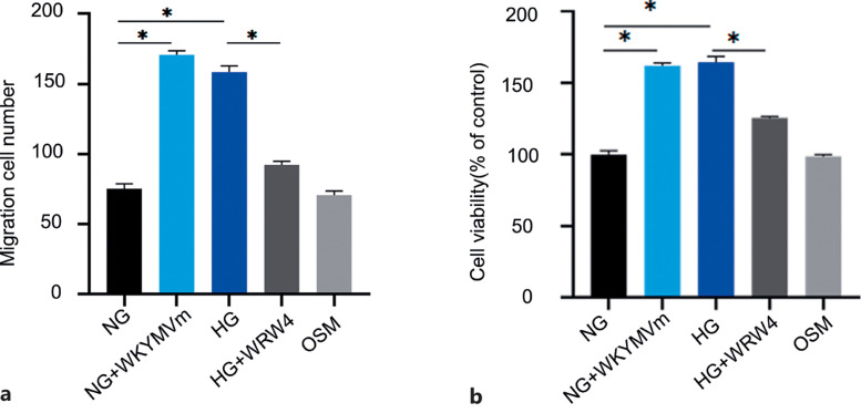
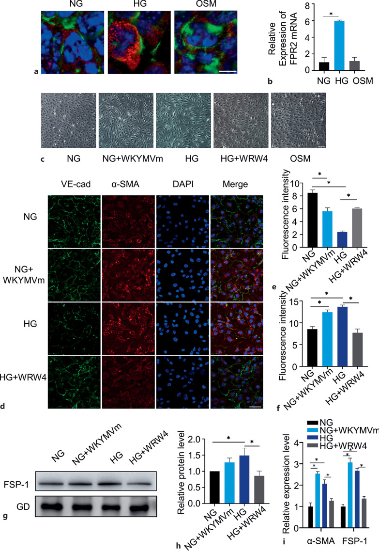
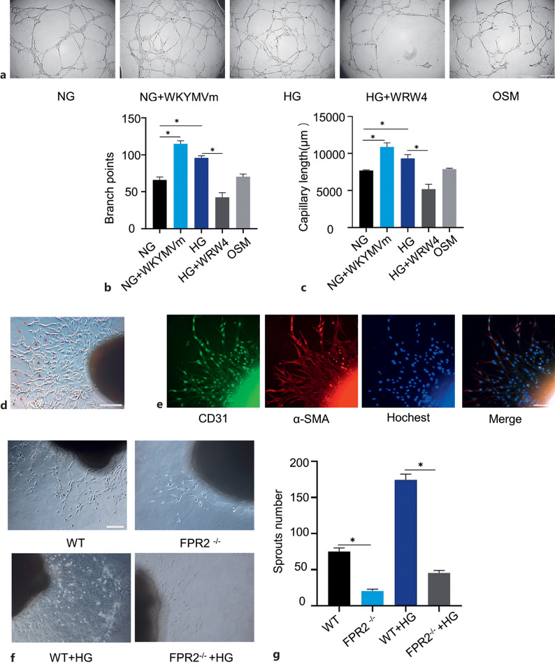
Results: HG upregulated FPR2 in HMECs, which triggered morphological changes, and the cells acquired mesenchymal phenotype, with enhanced cell migration, viability, and angiogenic process shown by tube formation and aortic ring sprouting. Inhibition of FPR2 attenuated HG-induced EndoMT and endothelial cell migration to form vessel-like tube structures. RNA sequence and protein analysis further revealed that inhibition of FPR2 decreased the expression of genes associated with EndoMT. ERK1/2 and P38 signaling pathway was activated in HMECs, promoting neovascularization in HG-induced EndoMT of HMECs. In vivo, increased expression of mesenchymal markers was detected in the retina of diabetic mice and FVMs from patients with PDR. FPR2 deficiency was associated with diminished EndoMT-related phenotypic changes in the retina of diabetic mice.
Conclusions: FPR2 is actively involved in the progression of EndoMT that may contribute to the pathogenesis of PDR. Thus, FPR2 may be a potential therapeutic target for PDR.
期刊介绍:
''Ophthalmic Research'' features original papers and reviews reporting on translational and clinical studies. Authors from throughout the world cover research topics on every field in connection with physical, physiologic, pharmacological, biochemical and molecular biological aspects of ophthalmology. This journal also aims to provide a record of international clinical research for both researchers and clinicians in ophthalmology. Finally, the transfer of information from fundamental research to clinical research and clinical practice is particularly welcome.

 求助内容:
求助内容: 应助结果提醒方式:


