Venezia G Carmona-Barrón, Inés S Fernández Del Campo, José M Delgado-García, Antonio J De la Fuente, Ignacio Plaza Lopez, Miguel A Merchán
{"title":"比较经颅交流电和颞叶干扰电刺激对全脑c-Fos免疫反应性的影响。","authors":"Venezia G Carmona-Barrón, Inés S Fernández Del Campo, José M Delgado-García, Antonio J De la Fuente, Ignacio Plaza Lopez, Miguel A Merchán","doi":"10.3389/fnana.2023.1128193","DOIUrl":null,"url":null,"abstract":"<p><p>The analysis of the topography of brain neuromodulation following transcranial alternating current (AC) stimulation is relevant for defining strategies directed to specific nuclei stimulation in patients. Among the different procedures of AC stimulation, temporal interference (tTIS) is a novel method for non-invasive neuromodulation of specific deep brain targets. However, little information is currently available about its tissue effects and its activation topography in <i>in vivo</i> animal models. After a single session (30 min, 0.12 mA) of transcranial alternate current (2,000 Hz; ES/AC group) or tTIS (2,000/2,010 Hz; Es/tTIS group) stimulation, rat brains were explored by whole-brain mapping analysis of c-Fos immunostained serial sections. For this analysis, we used two mapping methods, namely density-to-color processed channels (independent component analysis (ICA) and graphical representation (MATLAB) of morphometrical and densitometrical values obtained by density threshold segmentation. In addition, to assess tissue effects, alternate serial sections were stained for glial fibrillary acidic protein (GFAP), ionized calcium-binding adapter molecule 1 (Iba1), and Nissl. AC stimulation induced a mild superficial increase in c-Fos immunoreactivity. However, tTIS stimulation globally decreased the number of c-Fos-positive neurons and increased blood brain barrier cell immunoreactivity. tTIS also had a stronger effect around the electrode placement area and preserved neuronal activation better in restricted areas of the deep brain (directional stimulation). The enhanced activation of intramural blood vessels' cells and perivascular astrocytes suggests that low-frequency interference (10 Hz) may also have a trophic effect.</p>","PeriodicalId":12572,"journal":{"name":"Frontiers in Neuroanatomy","volume":"17 ","pages":"1128193"},"PeriodicalIF":2.3000,"publicationDate":"2023-01-01","publicationTypes":"Journal Article","fieldsOfStudy":null,"isOpenAccess":false,"openAccessPdf":"https://www.ncbi.nlm.nih.gov/pmc/articles/PMC10040600/pdf/","citationCount":"1","resultStr":"{\"title\":\"Comparing the effects of transcranial alternating current and temporal interference (tTIS) electric stimulation through whole-brain mapping of c-Fos immunoreactivity.\",\"authors\":\"Venezia G Carmona-Barrón, Inés S Fernández Del Campo, José M Delgado-García, Antonio J De la Fuente, Ignacio Plaza Lopez, Miguel A Merchán\",\"doi\":\"10.3389/fnana.2023.1128193\",\"DOIUrl\":null,\"url\":null,\"abstract\":\"<p><p>The analysis of the topography of brain neuromodulation following transcranial alternating current (AC) stimulation is relevant for defining strategies directed to specific nuclei stimulation in patients. Among the different procedures of AC stimulation, temporal interference (tTIS) is a novel method for non-invasive neuromodulation of specific deep brain targets. However, little information is currently available about its tissue effects and its activation topography in <i>in vivo</i> animal models. After a single session (30 min, 0.12 mA) of transcranial alternate current (2,000 Hz; ES/AC group) or tTIS (2,000/2,010 Hz; Es/tTIS group) stimulation, rat brains were explored by whole-brain mapping analysis of c-Fos immunostained serial sections. For this analysis, we used two mapping methods, namely density-to-color processed channels (independent component analysis (ICA) and graphical representation (MATLAB) of morphometrical and densitometrical values obtained by density threshold segmentation. In addition, to assess tissue effects, alternate serial sections were stained for glial fibrillary acidic protein (GFAP), ionized calcium-binding adapter molecule 1 (Iba1), and Nissl. AC stimulation induced a mild superficial increase in c-Fos immunoreactivity. However, tTIS stimulation globally decreased the number of c-Fos-positive neurons and increased blood brain barrier cell immunoreactivity. tTIS also had a stronger effect around the electrode placement area and preserved neuronal activation better in restricted areas of the deep brain (directional stimulation). The enhanced activation of intramural blood vessels' cells and perivascular astrocytes suggests that low-frequency interference (10 Hz) may also have a trophic effect.</p>\",\"PeriodicalId\":12572,\"journal\":{\"name\":\"Frontiers in Neuroanatomy\",\"volume\":\"17 \",\"pages\":\"1128193\"},\"PeriodicalIF\":2.3000,\"publicationDate\":\"2023-01-01\",\"publicationTypes\":\"Journal Article\",\"fieldsOfStudy\":null,\"isOpenAccess\":false,\"openAccessPdf\":\"https://www.ncbi.nlm.nih.gov/pmc/articles/PMC10040600/pdf/\",\"citationCount\":\"1\",\"resultStr\":null,\"platform\":\"Semanticscholar\",\"paperid\":null,\"PeriodicalName\":\"Frontiers in Neuroanatomy\",\"FirstCategoryId\":\"3\",\"ListUrlMain\":\"https://doi.org/10.3389/fnana.2023.1128193\",\"RegionNum\":4,\"RegionCategory\":\"医学\",\"ArticlePicture\":[],\"TitleCN\":null,\"AbstractTextCN\":null,\"PMCID\":null,\"EPubDate\":\"\",\"PubModel\":\"\",\"JCR\":\"Q1\",\"JCRName\":\"ANATOMY & MORPHOLOGY\",\"Score\":null,\"Total\":0}","platform":"Semanticscholar","paperid":null,"PeriodicalName":"Frontiers in Neuroanatomy","FirstCategoryId":"3","ListUrlMain":"https://doi.org/10.3389/fnana.2023.1128193","RegionNum":4,"RegionCategory":"医学","ArticlePicture":[],"TitleCN":null,"AbstractTextCN":null,"PMCID":null,"EPubDate":"","PubModel":"","JCR":"Q1","JCRName":"ANATOMY & MORPHOLOGY","Score":null,"Total":0}
Comparing the effects of transcranial alternating current and temporal interference (tTIS) electric stimulation through whole-brain mapping of c-Fos immunoreactivity.
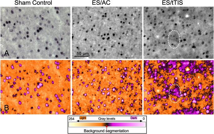
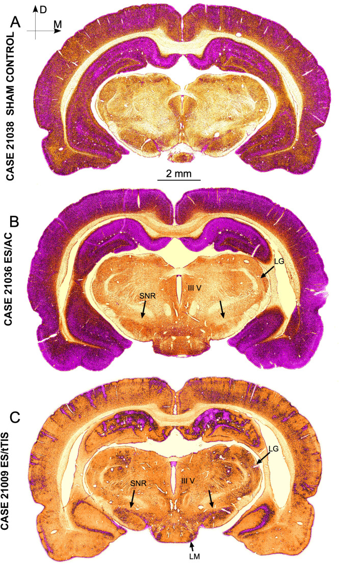
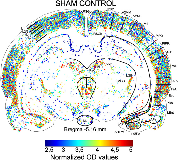
The analysis of the topography of brain neuromodulation following transcranial alternating current (AC) stimulation is relevant for defining strategies directed to specific nuclei stimulation in patients. Among the different procedures of AC stimulation, temporal interference (tTIS) is a novel method for non-invasive neuromodulation of specific deep brain targets. However, little information is currently available about its tissue effects and its activation topography in in vivo animal models. After a single session (30 min, 0.12 mA) of transcranial alternate current (2,000 Hz; ES/AC group) or tTIS (2,000/2,010 Hz; Es/tTIS group) stimulation, rat brains were explored by whole-brain mapping analysis of c-Fos immunostained serial sections. For this analysis, we used two mapping methods, namely density-to-color processed channels (independent component analysis (ICA) and graphical representation (MATLAB) of morphometrical and densitometrical values obtained by density threshold segmentation. In addition, to assess tissue effects, alternate serial sections were stained for glial fibrillary acidic protein (GFAP), ionized calcium-binding adapter molecule 1 (Iba1), and Nissl. AC stimulation induced a mild superficial increase in c-Fos immunoreactivity. However, tTIS stimulation globally decreased the number of c-Fos-positive neurons and increased blood brain barrier cell immunoreactivity. tTIS also had a stronger effect around the electrode placement area and preserved neuronal activation better in restricted areas of the deep brain (directional stimulation). The enhanced activation of intramural blood vessels' cells and perivascular astrocytes suggests that low-frequency interference (10 Hz) may also have a trophic effect.
期刊介绍:
Frontiers in Neuroanatomy publishes rigorously peer-reviewed research revealing important aspects of the anatomical organization of all nervous systems across all species. Specialty Chief Editor Javier DeFelipe at the Cajal Institute (CSIC) is supported by an outstanding Editorial Board of international experts. This multidisciplinary open-access journal is at the forefront of disseminating and communicating scientific knowledge and impactful discoveries to researchers, academics, clinicians and the public worldwide.
 求助内容:
求助内容: 应助结果提醒方式:


