Emilio S. Rivera , Andy Weiss , Lukasz G. Migas , Jeffrey A. Freiberg , Katerina V. Djambazova , Elizabeth K. Neumann , Raf Van de Plas , Jeffrey M. Spraggins , Eric P. Skaar , Richard M. Caprioli
{"title":"成像质谱显示复杂的脂质分布跨越金黄色葡萄球菌生物膜层","authors":"Emilio S. Rivera , Andy Weiss , Lukasz G. Migas , Jeffrey A. Freiberg , Katerina V. Djambazova , Elizabeth K. Neumann , Raf Van de Plas , Jeffrey M. Spraggins , Eric P. Skaar , Richard M. Caprioli","doi":"10.1016/j.jmsacl.2022.09.003","DOIUrl":null,"url":null,"abstract":"<div><h3>Introduction</h3><p>Although <em>Staphylococcus aureus</em> is the leading cause of biofilm-related infections, the lipidomic distributions within these biofilms is poorly understood. Here, lipidomic mapping of <em>S. aureus</em> biofilm cross-sections was performed to investigate heterogeneity between horizontal biofilm layers.</p></div><div><h3>Methods</h3><p><em>S. aureus</em> biofilms were grown statically, embedded in a mixture of carboxymethylcellulose/gelatin, and prepared for downstream matrix-assisted laser desorption/ionization imaging mass spectrometry (MALDI IMS). Trapped ion mobility spectrometry (TIMS) was also applied prior to mass analysis.</p></div><div><h3>Results</h3><p>Implementation of TIMS led to a ∼ threefold increase in the number of lipid species detected. Washing biofilm samples with ammonium formate (150 mM) increased signal intensity for some bacterial lipids by as much as tenfold, with minimal disruption of the biofilm structure. MALDI TIMS IMS revealed that most lipids localize primarily to a single biofilm layer, and species from the same lipid class such as cardiolipins CL(57:0) – CL(66:0) display starkly different localizations, exhibiting between 1.5 and 6.3-fold intensity differences between layers (n = 3, p < 0.03). No horizontal layers were observed within biofilms grown anaerobically, and lipids were distributed homogenously.</p></div><div><h3>Conclusions</h3><p>High spatial resolution analysis of <em>S. aureus</em> biofilm cross-sections by MALDI TIMS IMS revealed stark lipidomic heterogeneity between horizontal <em>S. aureus</em> biofilm layers demonstrating that each layer was molecularly distinct. Finally, this workflow uncovered an absence of layers in biofilms grown under anaerobic conditions, possibly indicating that oxygen contributes to the observed heterogeneity under aerobic conditions. Future applications of this workflow to study spatially localized molecular responses to antimicrobials could provide new therapeutic strategies.</p></div>","PeriodicalId":52406,"journal":{"name":"Journal of Mass Spectrometry and Advances in the Clinical Lab","volume":"26 ","pages":"Pages 36-46"},"PeriodicalIF":3.1000,"publicationDate":"2022-11-01","publicationTypes":"Journal Article","fieldsOfStudy":null,"isOpenAccess":false,"openAccessPdf":"https://www.sciencedirect.com/science/article/pii/S2667145X22000372/pdfft?md5=5cfe45d904dd71d41d3a2372d5f7f9aa&pid=1-s2.0-S2667145X22000372-main.pdf","citationCount":"2","resultStr":"{\"title\":\"Imaging mass spectrometry reveals complex lipid distributions across Staphylococcus aureus biofilm layers\",\"authors\":\"Emilio S. Rivera , Andy Weiss , Lukasz G. Migas , Jeffrey A. Freiberg , Katerina V. Djambazova , Elizabeth K. Neumann , Raf Van de Plas , Jeffrey M. Spraggins , Eric P. Skaar , Richard M. Caprioli\",\"doi\":\"10.1016/j.jmsacl.2022.09.003\",\"DOIUrl\":null,\"url\":null,\"abstract\":\"<div><h3>Introduction</h3><p>Although <em>Staphylococcus aureus</em> is the leading cause of biofilm-related infections, the lipidomic distributions within these biofilms is poorly understood. Here, lipidomic mapping of <em>S. aureus</em> biofilm cross-sections was performed to investigate heterogeneity between horizontal biofilm layers.</p></div><div><h3>Methods</h3><p><em>S. aureus</em> biofilms were grown statically, embedded in a mixture of carboxymethylcellulose/gelatin, and prepared for downstream matrix-assisted laser desorption/ionization imaging mass spectrometry (MALDI IMS). Trapped ion mobility spectrometry (TIMS) was also applied prior to mass analysis.</p></div><div><h3>Results</h3><p>Implementation of TIMS led to a ∼ threefold increase in the number of lipid species detected. Washing biofilm samples with ammonium formate (150 mM) increased signal intensity for some bacterial lipids by as much as tenfold, with minimal disruption of the biofilm structure. MALDI TIMS IMS revealed that most lipids localize primarily to a single biofilm layer, and species from the same lipid class such as cardiolipins CL(57:0) – CL(66:0) display starkly different localizations, exhibiting between 1.5 and 6.3-fold intensity differences between layers (n = 3, p < 0.03). No horizontal layers were observed within biofilms grown anaerobically, and lipids were distributed homogenously.</p></div><div><h3>Conclusions</h3><p>High spatial resolution analysis of <em>S. aureus</em> biofilm cross-sections by MALDI TIMS IMS revealed stark lipidomic heterogeneity between horizontal <em>S. aureus</em> biofilm layers demonstrating that each layer was molecularly distinct. Finally, this workflow uncovered an absence of layers in biofilms grown under anaerobic conditions, possibly indicating that oxygen contributes to the observed heterogeneity under aerobic conditions. Future applications of this workflow to study spatially localized molecular responses to antimicrobials could provide new therapeutic strategies.</p></div>\",\"PeriodicalId\":52406,\"journal\":{\"name\":\"Journal of Mass Spectrometry and Advances in the Clinical Lab\",\"volume\":\"26 \",\"pages\":\"Pages 36-46\"},\"PeriodicalIF\":3.1000,\"publicationDate\":\"2022-11-01\",\"publicationTypes\":\"Journal Article\",\"fieldsOfStudy\":null,\"isOpenAccess\":false,\"openAccessPdf\":\"https://www.sciencedirect.com/science/article/pii/S2667145X22000372/pdfft?md5=5cfe45d904dd71d41d3a2372d5f7f9aa&pid=1-s2.0-S2667145X22000372-main.pdf\",\"citationCount\":\"2\",\"resultStr\":null,\"platform\":\"Semanticscholar\",\"paperid\":null,\"PeriodicalName\":\"Journal of Mass Spectrometry and Advances in the Clinical Lab\",\"FirstCategoryId\":\"3\",\"ListUrlMain\":\"https://www.sciencedirect.com/science/article/pii/S2667145X22000372\",\"RegionNum\":4,\"RegionCategory\":\"医学\",\"ArticlePicture\":[],\"TitleCN\":null,\"AbstractTextCN\":null,\"PMCID\":null,\"EPubDate\":\"\",\"PubModel\":\"\",\"JCR\":\"Q2\",\"JCRName\":\"MEDICAL LABORATORY TECHNOLOGY\",\"Score\":null,\"Total\":0}","platform":"Semanticscholar","paperid":null,"PeriodicalName":"Journal of Mass Spectrometry and Advances in the Clinical Lab","FirstCategoryId":"3","ListUrlMain":"https://www.sciencedirect.com/science/article/pii/S2667145X22000372","RegionNum":4,"RegionCategory":"医学","ArticlePicture":[],"TitleCN":null,"AbstractTextCN":null,"PMCID":null,"EPubDate":"","PubModel":"","JCR":"Q2","JCRName":"MEDICAL LABORATORY TECHNOLOGY","Score":null,"Total":0}
Imaging mass spectrometry reveals complex lipid distributions across Staphylococcus aureus biofilm layers
Introduction
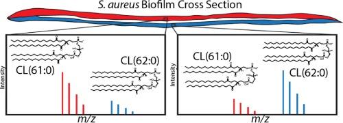
Although Staphylococcus aureus is the leading cause of biofilm-related infections, the lipidomic distributions within these biofilms is poorly understood. Here, lipidomic mapping of S. aureus biofilm cross-sections was performed to investigate heterogeneity between horizontal biofilm layers.
Methods
S. aureus biofilms were grown statically, embedded in a mixture of carboxymethylcellulose/gelatin, and prepared for downstream matrix-assisted laser desorption/ionization imaging mass spectrometry (MALDI IMS). Trapped ion mobility spectrometry (TIMS) was also applied prior to mass analysis.
Results
Implementation of TIMS led to a ∼ threefold increase in the number of lipid species detected. Washing biofilm samples with ammonium formate (150 mM) increased signal intensity for some bacterial lipids by as much as tenfold, with minimal disruption of the biofilm structure. MALDI TIMS IMS revealed that most lipids localize primarily to a single biofilm layer, and species from the same lipid class such as cardiolipins CL(57:0) – CL(66:0) display starkly different localizations, exhibiting between 1.5 and 6.3-fold intensity differences between layers (n = 3, p < 0.03). No horizontal layers were observed within biofilms grown anaerobically, and lipids were distributed homogenously.
Conclusions
High spatial resolution analysis of S. aureus biofilm cross-sections by MALDI TIMS IMS revealed stark lipidomic heterogeneity between horizontal S. aureus biofilm layers demonstrating that each layer was molecularly distinct. Finally, this workflow uncovered an absence of layers in biofilms grown under anaerobic conditions, possibly indicating that oxygen contributes to the observed heterogeneity under aerobic conditions. Future applications of this workflow to study spatially localized molecular responses to antimicrobials could provide new therapeutic strategies.
 求助内容:
求助内容: 应助结果提醒方式:


