你的诊断是什么?

IF 1.2 Q3 OBSTETRICS & GYNECOLOGY
Latika Chawla, Ankita Yadav, Mamta Sah, Shilpa Panta, Nevetha Ravichandran, Ria M, Shalini Rajaram
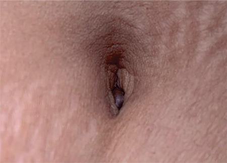
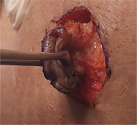
{"title":"你的诊断是什么?","authors":"Latika Chawla, Ankita Yadav, Mamta Sah, Shilpa Panta, Nevetha Ravichandran, Ria M, Shalini Rajaram","doi":"10.4274/jtgga.galenos.2022.2022-6-13","DOIUrl":null,"url":null,"abstract":"A 38-year-old lady presented with a painful swelling in the umbilicus, together with a history of increased pain and bleeding from the swelling at the time of menstruation for the last seven months. Her menstrual cycles were regular, with average flow and no dysmenorrhea. She had two living children, both were delivered vaginally. There was no history of pelvic pain, infertility, treatment for infertility, pelvic/abdominal surgery, or caesarean section. Examination revealed a 1.0x0.5 cm firm, tender, reddish-blue colored nodular swelling in the abdominal wall, located just inferior to the umbilical ring with well-defined margins and a regular surface (Figure 1). Pelvic examination was essentially normal with a multiparous-sized uterus that was anteverted, mobile, and non-tender. Both fornices were free and non-tender. The rectovaginal septum was free and there were no nodules in the pouch of Douglas. Ultrasound revealed a well-defined, hetero-echoic lesion with a peripheral rim of colour lying infra-umbilically, superficial to the rectus sheath. The same lesion appeared hyperintense on T1/T2 magnetic resonance imaging (MRI) with post-contrast enhancement. The abdomen and pelvis were found to be normal on MRI. The patient was taken up for surgical excision of the nodule. Radical omphalectomy was performed. A peri-umbilical incision was made. Umbilicus, underlying nodule, and the surrounding area of fibrosis were dissected with a 5 mm clear margin using diathermy (Figure 2). The patient is on follow-up and is free of the disease at 18 months after surgery.","PeriodicalId":17440,"journal":{"name":"Journal of the Turkish German Gynecological Association","volume":null,"pages":null},"PeriodicalIF":1.2000,"publicationDate":"2023-03-15","publicationTypes":"Journal Article","fieldsOfStudy":null,"isOpenAccess":false,"openAccessPdf":"https://ftp.ncbi.nlm.nih.gov/pub/pmc/oa_pdf/e6/51/JTGGA-24-74.PMC10019007.pdf","citationCount":"0","resultStr":"{\"title\":\"What is your diagnosis?\",\"authors\":\"Latika Chawla, Ankita Yadav, Mamta Sah, Shilpa Panta, Nevetha Ravichandran, Ria M, Shalini Rajaram\",\"doi\":\"10.4274/jtgga.galenos.2022.2022-6-13\",\"DOIUrl\":null,\"url\":null,\"abstract\":\"A 38-year-old lady presented with a painful swelling in the umbilicus, together with a history of increased pain and bleeding from the swelling at the time of menstruation for the last seven months. Her menstrual cycles were regular, with average flow and no dysmenorrhea. She had two living children, both were delivered vaginally. There was no history of pelvic pain, infertility, treatment for infertility, pelvic/abdominal surgery, or caesarean section. Examination revealed a 1.0x0.5 cm firm, tender, reddish-blue colored nodular swelling in the abdominal wall, located just inferior to the umbilical ring with well-defined margins and a regular surface (Figure 1). Pelvic examination was essentially normal with a multiparous-sized uterus that was anteverted, mobile, and non-tender. Both fornices were free and non-tender. The rectovaginal septum was free and there were no nodules in the pouch of Douglas. Ultrasound revealed a well-defined, hetero-echoic lesion with a peripheral rim of colour lying infra-umbilically, superficial to the rectus sheath. The same lesion appeared hyperintense on T1/T2 magnetic resonance imaging (MRI) with post-contrast enhancement. The abdomen and pelvis were found to be normal on MRI. The patient was taken up for surgical excision of the nodule. Radical omphalectomy was performed. A peri-umbilical incision was made. Umbilicus, underlying nodule, and the surrounding area of fibrosis were dissected with a 5 mm clear margin using diathermy (Figure 2). The patient is on follow-up and is free of the disease at 18 months after surgery.\",\"PeriodicalId\":17440,\"journal\":{\"name\":\"Journal of the Turkish German Gynecological Association\",\"volume\":null,\"pages\":null},\"PeriodicalIF\":1.2000,\"publicationDate\":\"2023-03-15\",\"publicationTypes\":\"Journal Article\",\"fieldsOfStudy\":null,\"isOpenAccess\":false,\"openAccessPdf\":\"https://ftp.ncbi.nlm.nih.gov/pub/pmc/oa_pdf/e6/51/JTGGA-24-74.PMC10019007.pdf\",\"citationCount\":\"0\",\"resultStr\":null,\"platform\":\"Semanticscholar\",\"paperid\":null,\"PeriodicalName\":\"Journal of the Turkish German Gynecological Association\",\"FirstCategoryId\":\"1085\",\"ListUrlMain\":\"https://doi.org/10.4274/jtgga.galenos.2022.2022-6-13\",\"RegionNum\":0,\"RegionCategory\":null,\"ArticlePicture\":[],\"TitleCN\":null,\"AbstractTextCN\":null,\"PMCID\":null,\"EPubDate\":\"\",\"PubModel\":\"\",\"JCR\":\"Q3\",\"JCRName\":\"OBSTETRICS & GYNECOLOGY\",\"Score\":null,\"Total\":0}","platform":"Semanticscholar","paperid":null,"PeriodicalName":"Journal of the Turkish German Gynecological Association","FirstCategoryId":"1085","ListUrlMain":"https://doi.org/10.4274/jtgga.galenos.2022.2022-6-13","RegionNum":0,"RegionCategory":null,"ArticlePicture":[],"TitleCN":null,"AbstractTextCN":null,"PMCID":null,"EPubDate":"","PubModel":"","JCR":"Q3","JCRName":"OBSTETRICS & GYNECOLOGY","Score":null,"Total":0}
引用次数: 0

摘要

本文章由计算机程序翻译,如有差异,请以英文原文为准。
What is your diagnosis?

What is your diagnosis?

What is your diagnosis?

What is your diagnosis?
A 38-year-old lady presented with a painful swelling in the umbilicus, together with a history of increased pain and bleeding from the swelling at the time of menstruation for the last seven months. Her menstrual cycles were regular, with average flow and no dysmenorrhea. She had two living children, both were delivered vaginally. There was no history of pelvic pain, infertility, treatment for infertility, pelvic/abdominal surgery, or caesarean section. Examination revealed a 1.0x0.5 cm firm, tender, reddish-blue colored nodular swelling in the abdominal wall, located just inferior to the umbilical ring with well-defined margins and a regular surface (Figure 1). Pelvic examination was essentially normal with a multiparous-sized uterus that was anteverted, mobile, and non-tender. Both fornices were free and non-tender. The rectovaginal septum was free and there were no nodules in the pouch of Douglas. Ultrasound revealed a well-defined, hetero-echoic lesion with a peripheral rim of colour lying infra-umbilically, superficial to the rectus sheath. The same lesion appeared hyperintense on T1/T2 magnetic resonance imaging (MRI) with post-contrast enhancement. The abdomen and pelvis were found to be normal on MRI. The patient was taken up for surgical excision of the nodule. Radical omphalectomy was performed. A peri-umbilical incision was made. Umbilicus, underlying nodule, and the surrounding area of fibrosis were dissected with a 5 mm clear margin using diathermy (Figure 2). The patient is on follow-up and is free of the disease at 18 months after surgery.
求助全文
通过发布文献求助,成功后即可免费获取论文全文。 去求助
来源期刊
CiteScore
2.40
自引率
7.10%
发文量
56
期刊介绍: Journal of the Turkish-German Gynecological Association is the official, open access publication of the Turkish-German Gynecological Education and Research Foundation and Turkish-German Gynecological Association and is published quarterly on March, June, September and December. It is an independent peer-reviewed international journal printed in English language. Manuscripts are reviewed in accordance with “double-blind peer review” process for both reviewers and authors. The target audience of Journal of the Turkish-German Gynecological Association includes gynecologists and primary care physicians interested in gynecology practice. It publishes original works on all aspects of obstertrics and gynecology. The aim of Journal of the Turkish-German Gynecological Association is to publish high quality original research articles. In addition to research articles, reviews, editorials, letters to the editor, diagnostic puzzle are also published. Suggestions for new books are also welcomed. Journal of the Turkish-German Gynecological Association does not charge any fee for article submission or processing.
×
引用
GB/T 7714-2015
复制
MLA
复制
APA
复制
导出至
BibTeX EndNote RefMan NoteFirst NoteExpress
×
提示
您的信息不完整,为了账户安全,请先补充。
现在去补充
×
提示
您因"违规操作"
具体请查看互助需知
我知道了
×
提示
确定
请完成安全验证×
copy
已复制链接
快去分享给好友吧!
我知道了
右上角分享
点击右上角分享
0
联系我们:info@booksci.cn Book学术提供免费学术资源搜索服务,方便国内外学者检索中英文文献。致力于提供最便捷和优质的服务体验。 Copyright © 2023 布克学术 All rights reserved.
京ICP备2023020795号-1
ghs 京公网安备 11010802042870号
Book学术文献互助
Book学术文献互助群
群 号:481959085
Book学术官方微信