Pia Iben Pietersen, Janni Lynggård Bo Madsen, Jon Asmussen, Lars Lund, Tommy Kjærgaard Nielsen, Michael Pedersen, Birte Engvad, Ole Graumann
{"title":"多参数磁共振成像表征肾脏肿瘤:Cornelis等人提出的算法的验证研究。","authors":"Pia Iben Pietersen, Janni Lynggård Bo Madsen, Jon Asmussen, Lars Lund, Tommy Kjærgaard Nielsen, Michael Pedersen, Birte Engvad, Ole Graumann","doi":"10.25259/JCIS_124_2022","DOIUrl":null,"url":null,"abstract":"<p><strong>Objectives: </strong>In the last decade, the incidence of renal cell carcinoma (RCC) has been rising, with the greatest increase observed for solid tumors. Magnetic resonance imaging (MRI) protocols and algorithms have recently been available for classifying RCC subtypes and benign subtypes. The objective of this study was to prospectively validate the MRI algorithm presented by Cornelis <i>et al</i>. for RCC classification.</p><p><strong>Material and methods: </strong>Over a 7-month period, 38 patients with 44 renal tumors were prospectively included in the study and received an MRI examination in addition to the conventional investigation program. The MRI sequences were: T2-weighted, dual chemical shift MRI, diffusion-weighted imaging (DWI), and dynamic contrast-enhanced T1-weighted in wash-in and wash-out phases. The images were evaluated according to the algorithm by two experienced, blinded radiologists, and the histopathological diagnosis served as the gold standard.</p><p><strong>Results: </strong>Of 44 tumors in 38 patients, only 8 tumors (18.2%) received the same MRI diagnosis according to the algorithm as the histopathological diagnosis. MRI diagnosed 16 angiomyolipoma, 14 clear cell RCC (ccRCC), 12 chromophobe RCC (chRCC), and two papillary RCC (pRCC), while histopathological examination diagnosed 24 ccRCC, four pRCC, one chRCC, and one mixed tumor of both pRCC and chRCC. Malignant tumors were statistically significantly larger than the benign (3.16 ± 1.34 cm vs. 2.00 ± 1.04 cm, <i>P</i> = 0.006).</p><p><strong>Conclusion: </strong>This prospective study could not reproduce Cornelis <i>et al</i>.'s results and does not support differentiating renal masses using multiparametric MRI without percutaneous biopsy in the future. The MRI algorithm showed few promising results to categorize renal tumors, indicating histopathology for clinical decisions and follow-up regimes of renal masses are still required.</p>","PeriodicalId":15512,"journal":{"name":"Journal of Clinical Imaging Science","volume":"13 ","pages":"7"},"PeriodicalIF":1.3000,"publicationDate":"2023-01-01","publicationTypes":"Journal Article","fieldsOfStudy":null,"isOpenAccess":false,"openAccessPdf":"https://ftp.ncbi.nlm.nih.gov/pub/pmc/oa_pdf/3d/96/JCIS-13-7.PMC9992978.pdf","citationCount":"0","resultStr":"{\"title\":\"Multiparametric magnetic resonance imaging for characterizing renal tumors: A validation study of the algorithm presented by Cornelis <i>et al</i>.\",\"authors\":\"Pia Iben Pietersen, Janni Lynggård Bo Madsen, Jon Asmussen, Lars Lund, Tommy Kjærgaard Nielsen, Michael Pedersen, Birte Engvad, Ole Graumann\",\"doi\":\"10.25259/JCIS_124_2022\",\"DOIUrl\":null,\"url\":null,\"abstract\":\"<p><strong>Objectives: </strong>In the last decade, the incidence of renal cell carcinoma (RCC) has been rising, with the greatest increase observed for solid tumors. Magnetic resonance imaging (MRI) protocols and algorithms have recently been available for classifying RCC subtypes and benign subtypes. The objective of this study was to prospectively validate the MRI algorithm presented by Cornelis <i>et al</i>. for RCC classification.</p><p><strong>Material and methods: </strong>Over a 7-month period, 38 patients with 44 renal tumors were prospectively included in the study and received an MRI examination in addition to the conventional investigation program. The MRI sequences were: T2-weighted, dual chemical shift MRI, diffusion-weighted imaging (DWI), and dynamic contrast-enhanced T1-weighted in wash-in and wash-out phases. The images were evaluated according to the algorithm by two experienced, blinded radiologists, and the histopathological diagnosis served as the gold standard.</p><p><strong>Results: </strong>Of 44 tumors in 38 patients, only 8 tumors (18.2%) received the same MRI diagnosis according to the algorithm as the histopathological diagnosis. MRI diagnosed 16 angiomyolipoma, 14 clear cell RCC (ccRCC), 12 chromophobe RCC (chRCC), and two papillary RCC (pRCC), while histopathological examination diagnosed 24 ccRCC, four pRCC, one chRCC, and one mixed tumor of both pRCC and chRCC. Malignant tumors were statistically significantly larger than the benign (3.16 ± 1.34 cm vs. 2.00 ± 1.04 cm, <i>P</i> = 0.006).</p><p><strong>Conclusion: </strong>This prospective study could not reproduce Cornelis <i>et al</i>.'s results and does not support differentiating renal masses using multiparametric MRI without percutaneous biopsy in the future. The MRI algorithm showed few promising results to categorize renal tumors, indicating histopathology for clinical decisions and follow-up regimes of renal masses are still required.</p>\",\"PeriodicalId\":15512,\"journal\":{\"name\":\"Journal of Clinical Imaging Science\",\"volume\":\"13 \",\"pages\":\"7\"},\"PeriodicalIF\":1.3000,\"publicationDate\":\"2023-01-01\",\"publicationTypes\":\"Journal Article\",\"fieldsOfStudy\":null,\"isOpenAccess\":false,\"openAccessPdf\":\"https://ftp.ncbi.nlm.nih.gov/pub/pmc/oa_pdf/3d/96/JCIS-13-7.PMC9992978.pdf\",\"citationCount\":\"0\",\"resultStr\":null,\"platform\":\"Semanticscholar\",\"paperid\":null,\"PeriodicalName\":\"Journal of Clinical Imaging Science\",\"FirstCategoryId\":\"1085\",\"ListUrlMain\":\"https://doi.org/10.25259/JCIS_124_2022\",\"RegionNum\":0,\"RegionCategory\":null,\"ArticlePicture\":[],\"TitleCN\":null,\"AbstractTextCN\":null,\"PMCID\":null,\"EPubDate\":\"\",\"PubModel\":\"\",\"JCR\":\"Q3\",\"JCRName\":\"RADIOLOGY, NUCLEAR MEDICINE & MEDICAL IMAGING\",\"Score\":null,\"Total\":0}","platform":"Semanticscholar","paperid":null,"PeriodicalName":"Journal of Clinical Imaging Science","FirstCategoryId":"1085","ListUrlMain":"https://doi.org/10.25259/JCIS_124_2022","RegionNum":0,"RegionCategory":null,"ArticlePicture":[],"TitleCN":null,"AbstractTextCN":null,"PMCID":null,"EPubDate":"","PubModel":"","JCR":"Q3","JCRName":"RADIOLOGY, NUCLEAR MEDICINE & MEDICAL IMAGING","Score":null,"Total":0}
引用次数: 0
摘要
目的:近十年来,肾细胞癌(RCC)的发病率呈上升趋势,其中实体瘤的发病率增幅最大。磁共振成像(MRI)协议和算法最近可用于分类RCC亚型和良性亚型。本研究的目的是前瞻性地验证Cornelis等人提出的用于RCC分类的MRI算法。材料和方法:在7个月的时间里,38例44例肾脏肿瘤患者被前瞻性纳入研究,并在常规调查方案的基础上接受MRI检查。MRI序列为:t2加权,双化学移位MRI,弥散加权成像(DWI)和动态对比增强t1加权冲洗和冲洗期。图像由两名经验丰富的盲法放射科医生根据算法进行评估,组织病理学诊断作为金标准。结果:38例患者44个肿瘤中,仅8个肿瘤(18.2%)的MRI诊断与组织病理学诊断一致。MRI诊断血管平滑肌脂肪瘤16例,透明细胞RCC (ccRCC) 14例,疏色RCC (chRCC) 12例,乳头状RCC (pRCC) 2例,组织病理学诊断ccRCC 24例,pRCC 4例,chRCC 1例,pRCC和chRCC混合肿瘤1例。恶性肿瘤大于良性肿瘤(3.16±1.34 cm∶2.00±1.04 cm, P = 0.006)。结论:这项前瞻性研究不能重现Cornelis等人的结果,也不支持未来在不经皮活检的情况下使用多参数MRI来鉴别肾肿块。MRI算法对肾脏肿瘤的分类显示出很少有希望的结果,这表明仍然需要临床决策的组织病理学和肾脏肿块的随访制度。
Multiparametric magnetic resonance imaging for characterizing renal tumors: A validation study of the algorithm presented by Cornelis et al.
Objectives: In the last decade, the incidence of renal cell carcinoma (RCC) has been rising, with the greatest increase observed for solid tumors. Magnetic resonance imaging (MRI) protocols and algorithms have recently been available for classifying RCC subtypes and benign subtypes. The objective of this study was to prospectively validate the MRI algorithm presented by Cornelis et al. for RCC classification.
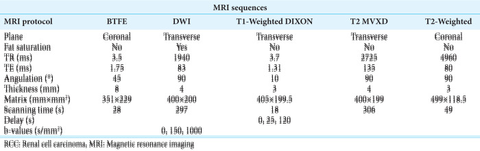
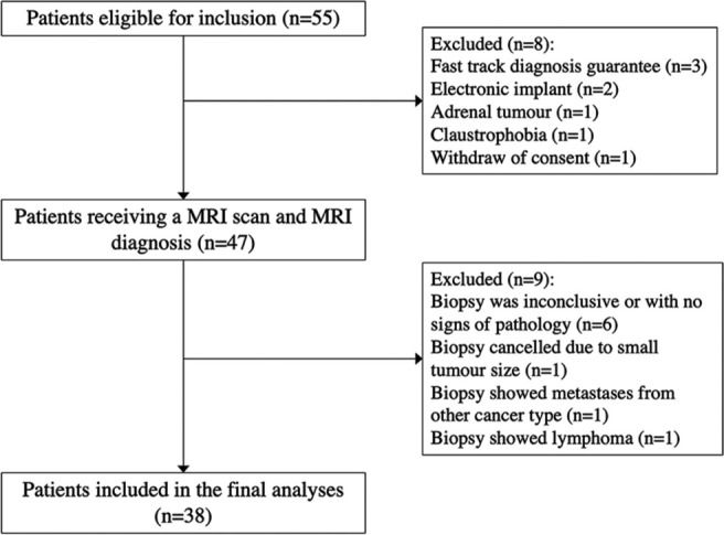
Material and methods: Over a 7-month period, 38 patients with 44 renal tumors were prospectively included in the study and received an MRI examination in addition to the conventional investigation program. The MRI sequences were: T2-weighted, dual chemical shift MRI, diffusion-weighted imaging (DWI), and dynamic contrast-enhanced T1-weighted in wash-in and wash-out phases. The images were evaluated according to the algorithm by two experienced, blinded radiologists, and the histopathological diagnosis served as the gold standard.
Results: Of 44 tumors in 38 patients, only 8 tumors (18.2%) received the same MRI diagnosis according to the algorithm as the histopathological diagnosis. MRI diagnosed 16 angiomyolipoma, 14 clear cell RCC (ccRCC), 12 chromophobe RCC (chRCC), and two papillary RCC (pRCC), while histopathological examination diagnosed 24 ccRCC, four pRCC, one chRCC, and one mixed tumor of both pRCC and chRCC. Malignant tumors were statistically significantly larger than the benign (3.16 ± 1.34 cm vs. 2.00 ± 1.04 cm, P = 0.006).
Conclusion: This prospective study could not reproduce Cornelis et al.'s results and does not support differentiating renal masses using multiparametric MRI without percutaneous biopsy in the future. The MRI algorithm showed few promising results to categorize renal tumors, indicating histopathology for clinical decisions and follow-up regimes of renal masses are still required.
期刊介绍:
The Journal of Clinical Imaging Science (JCIS) is an open access peer-reviewed journal committed to publishing high-quality articles in the field of Imaging Science. The journal aims to present Imaging Science and relevant clinical information in an understandable and useful format. The journal is owned and published by the Scientific Scholar. Audience Our audience includes Radiologists, Researchers, Clinicians, medical professionals and students. Review process JCIS has a highly rigorous peer-review process that makes sure that manuscripts are scientifically accurate, relevant, novel and important. Authors disclose all conflicts, affiliations and financial associations such that the published content is not biased.

 求助内容:
求助内容: 应助结果提醒方式:


