{"title":"非活动性甲状腺相关性眼病脉络膜血管的评价。","authors":"Seren Pehlivanoglu, Funda Ebru Aksoy, Gamze Ozturk Karabulut, Ugur Tunc, Korhan Fazil, Ozgur Artunay, Muhittin Taskapili","doi":"10.14744/bej.2022.04900","DOIUrl":null,"url":null,"abstract":"<p><strong>Objectives: </strong>The objectives of the study were to evaluate the vascular and stromal structure of the choroid in patients with inactive thyroid associated orbitopathy (TAO) by measuring choroidal vascularity index (CVI) and choroidal thickness (CT) using enhanced depth imaging (EDI) optical coherence tomography (OCT).</p><p><strong>Methods: </strong>The choroidal image was taken with EDI mode spectral domain (SD)-OCT. All scans were taken between 9.30 am and 11.30 am to avoid the diurnal variation of CT and CVI. To calculate CVI, macular SD-OCT scans were binarized using the publicly available software ImageJ and luminal area and total choroidal area (TCA) were measured. CVI was calculated as the proportion of LA to TCA. Furthermore, the relation between CVI and axial length, gender, and age was evaluated.</p><p><strong>Results: </strong>This study included 78 individuals with a mean age of 51.4±7.3 years. Group 1 consisted of 44 patients with inactive stage TAO, and Group 2 consisted of 34 healthy controls. Subfoveal CT was 338.92±73.93 µm in Group 1 and 303.97±40.35 µm in Group 2 (p=0.174). The CVI significantly differed between the two groups, which was higher in group 1 (p=0.000).</p><p><strong>Conclusion: </strong>Although CT was not different between groups, CVI which is the indicator of the vascular status of the choroid, was higher in patients with TAO in the inactive stage compared with healthy control subjects.</p>","PeriodicalId":8740,"journal":{"name":"Beyoglu Eye Journal","volume":"8 1","pages":"38-44"},"PeriodicalIF":0.0000,"publicationDate":"2023-01-01","publicationTypes":"Journal Article","fieldsOfStudy":null,"isOpenAccess":false,"openAccessPdf":"https://ftp.ncbi.nlm.nih.gov/pub/pmc/oa_pdf/3f/69/BEJ-8-38.PMC9993418.pdf","citationCount":"0","resultStr":"{\"title\":\"Assessment of Choroidal Vascularity in Inactive Thyroid Associated Orbitopathy.\",\"authors\":\"Seren Pehlivanoglu, Funda Ebru Aksoy, Gamze Ozturk Karabulut, Ugur Tunc, Korhan Fazil, Ozgur Artunay, Muhittin Taskapili\",\"doi\":\"10.14744/bej.2022.04900\",\"DOIUrl\":null,\"url\":null,\"abstract\":\"<p><strong>Objectives: </strong>The objectives of the study were to evaluate the vascular and stromal structure of the choroid in patients with inactive thyroid associated orbitopathy (TAO) by measuring choroidal vascularity index (CVI) and choroidal thickness (CT) using enhanced depth imaging (EDI) optical coherence tomography (OCT).</p><p><strong>Methods: </strong>The choroidal image was taken with EDI mode spectral domain (SD)-OCT. All scans were taken between 9.30 am and 11.30 am to avoid the diurnal variation of CT and CVI. To calculate CVI, macular SD-OCT scans were binarized using the publicly available software ImageJ and luminal area and total choroidal area (TCA) were measured. CVI was calculated as the proportion of LA to TCA. Furthermore, the relation between CVI and axial length, gender, and age was evaluated.</p><p><strong>Results: </strong>This study included 78 individuals with a mean age of 51.4±7.3 years. Group 1 consisted of 44 patients with inactive stage TAO, and Group 2 consisted of 34 healthy controls. Subfoveal CT was 338.92±73.93 µm in Group 1 and 303.97±40.35 µm in Group 2 (p=0.174). The CVI significantly differed between the two groups, which was higher in group 1 (p=0.000).</p><p><strong>Conclusion: </strong>Although CT was not different between groups, CVI which is the indicator of the vascular status of the choroid, was higher in patients with TAO in the inactive stage compared with healthy control subjects.</p>\",\"PeriodicalId\":8740,\"journal\":{\"name\":\"Beyoglu Eye Journal\",\"volume\":\"8 1\",\"pages\":\"38-44\"},\"PeriodicalIF\":0.0000,\"publicationDate\":\"2023-01-01\",\"publicationTypes\":\"Journal Article\",\"fieldsOfStudy\":null,\"isOpenAccess\":false,\"openAccessPdf\":\"https://ftp.ncbi.nlm.nih.gov/pub/pmc/oa_pdf/3f/69/BEJ-8-38.PMC9993418.pdf\",\"citationCount\":\"0\",\"resultStr\":null,\"platform\":\"Semanticscholar\",\"paperid\":null,\"PeriodicalName\":\"Beyoglu Eye Journal\",\"FirstCategoryId\":\"1085\",\"ListUrlMain\":\"https://doi.org/10.14744/bej.2022.04900\",\"RegionNum\":0,\"RegionCategory\":null,\"ArticlePicture\":[],\"TitleCN\":null,\"AbstractTextCN\":null,\"PMCID\":null,\"EPubDate\":\"\",\"PubModel\":\"\",\"JCR\":\"\",\"JCRName\":\"\",\"Score\":null,\"Total\":0}","platform":"Semanticscholar","paperid":null,"PeriodicalName":"Beyoglu Eye Journal","FirstCategoryId":"1085","ListUrlMain":"https://doi.org/10.14744/bej.2022.04900","RegionNum":0,"RegionCategory":null,"ArticlePicture":[],"TitleCN":null,"AbstractTextCN":null,"PMCID":null,"EPubDate":"","PubModel":"","JCR":"","JCRName":"","Score":null,"Total":0}
Assessment of Choroidal Vascularity in Inactive Thyroid Associated Orbitopathy.
Objectives: The objectives of the study were to evaluate the vascular and stromal structure of the choroid in patients with inactive thyroid associated orbitopathy (TAO) by measuring choroidal vascularity index (CVI) and choroidal thickness (CT) using enhanced depth imaging (EDI) optical coherence tomography (OCT).
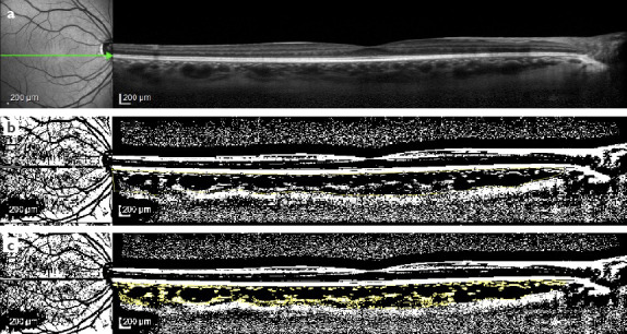
Methods: The choroidal image was taken with EDI mode spectral domain (SD)-OCT. All scans were taken between 9.30 am and 11.30 am to avoid the diurnal variation of CT and CVI. To calculate CVI, macular SD-OCT scans were binarized using the publicly available software ImageJ and luminal area and total choroidal area (TCA) were measured. CVI was calculated as the proportion of LA to TCA. Furthermore, the relation between CVI and axial length, gender, and age was evaluated.
Results: This study included 78 individuals with a mean age of 51.4±7.3 years. Group 1 consisted of 44 patients with inactive stage TAO, and Group 2 consisted of 34 healthy controls. Subfoveal CT was 338.92±73.93 µm in Group 1 and 303.97±40.35 µm in Group 2 (p=0.174). The CVI significantly differed between the two groups, which was higher in group 1 (p=0.000).
Conclusion: Although CT was not different between groups, CVI which is the indicator of the vascular status of the choroid, was higher in patients with TAO in the inactive stage compared with healthy control subjects.

 求助内容:
求助内容: 应助结果提醒方式:


