F Suárez-Cabrera, M Encinoso, A Artiles, I Castellano, C Melián, J R Jaber
{"title":"猫的一种引起布-恰里样综合征的纵隔肿块。","authors":"F Suárez-Cabrera, M Encinoso, A Artiles, I Castellano, C Melián, J R Jaber","doi":"10.22099/IJVR.2022.42203.6136","DOIUrl":null,"url":null,"abstract":"<p><strong>Background: </strong>Budd-Chiari syndrome (BCS) is considered a rare condition in cats that is characterized by the obstruction of the hepatic venous outflow tract from the level of the small hepatic veins to the level of the termination of the inferior vena cava into the right atrium in the absence of cardiac or pericardial disease, or sinusoidal obstruction syndrome.</p><p><strong>Case description: </strong>This report presents a 13-year-old cat with a two-week history of progressive lethargy, inappetence, weight loss, and abdominal distension.</p><p><strong>Findings/treatment and outcome: </strong>The radiological study was consistent with pleural effusion, as well as alveolar and interstitial pulmonary patterns. Ultrasonography confirmed hepatic venin congestion and ascites. Abdominocentesis revealed a modified transudate. A computed tomography (CT) angiography showed a mass at the level of the caudal mediastinum that compressed the caudal vena cava (CVC). Mediastinal lymphoma was considered the most likely differential diagnosis. These findings were interpreted as Budd-Chiari-like syndrome (BCLS) secondary to a mediastinal mass although, unfortunately, no further diagnostic or treatment procedures were accepted by the owners. BCLS is a rare condition in cats, where most of the reported cases occurred as a result of obstruction of the caudal vena cava. In this report, BCLS was caused by a mass located in the caudal mediastinum oppressing the caudal vena cava.</p><p><strong>Conclusion: </strong>This is the first report of BCLS in cats diagnosed by CT angiography, and it shows the value of this technique to define the origin and extent of the mass and to evaluate the presence or absence of metastatic lesions.</p>","PeriodicalId":14629,"journal":{"name":"Iranian journal of veterinary research","volume":"23 4","pages":"380-384"},"PeriodicalIF":1.0000,"publicationDate":"2022-01-01","publicationTypes":"Journal Article","fieldsOfStudy":null,"isOpenAccess":false,"openAccessPdf":"https://ftp.ncbi.nlm.nih.gov/pub/pmc/oa_pdf/e2/64/ijvr-23-380.PMC9984135.pdf","citationCount":"0","resultStr":"{\"title\":\"A mediastinal mass causing Budd-Chiari-like syndrome in a cat.\",\"authors\":\"F Suárez-Cabrera, M Encinoso, A Artiles, I Castellano, C Melián, J R Jaber\",\"doi\":\"10.22099/IJVR.2022.42203.6136\",\"DOIUrl\":null,\"url\":null,\"abstract\":\"<p><strong>Background: </strong>Budd-Chiari syndrome (BCS) is considered a rare condition in cats that is characterized by the obstruction of the hepatic venous outflow tract from the level of the small hepatic veins to the level of the termination of the inferior vena cava into the right atrium in the absence of cardiac or pericardial disease, or sinusoidal obstruction syndrome.</p><p><strong>Case description: </strong>This report presents a 13-year-old cat with a two-week history of progressive lethargy, inappetence, weight loss, and abdominal distension.</p><p><strong>Findings/treatment and outcome: </strong>The radiological study was consistent with pleural effusion, as well as alveolar and interstitial pulmonary patterns. Ultrasonography confirmed hepatic venin congestion and ascites. Abdominocentesis revealed a modified transudate. A computed tomography (CT) angiography showed a mass at the level of the caudal mediastinum that compressed the caudal vena cava (CVC). Mediastinal lymphoma was considered the most likely differential diagnosis. These findings were interpreted as Budd-Chiari-like syndrome (BCLS) secondary to a mediastinal mass although, unfortunately, no further diagnostic or treatment procedures were accepted by the owners. BCLS is a rare condition in cats, where most of the reported cases occurred as a result of obstruction of the caudal vena cava. In this report, BCLS was caused by a mass located in the caudal mediastinum oppressing the caudal vena cava.</p><p><strong>Conclusion: </strong>This is the first report of BCLS in cats diagnosed by CT angiography, and it shows the value of this technique to define the origin and extent of the mass and to evaluate the presence or absence of metastatic lesions.</p>\",\"PeriodicalId\":14629,\"journal\":{\"name\":\"Iranian journal of veterinary research\",\"volume\":\"23 4\",\"pages\":\"380-384\"},\"PeriodicalIF\":1.0000,\"publicationDate\":\"2022-01-01\",\"publicationTypes\":\"Journal Article\",\"fieldsOfStudy\":null,\"isOpenAccess\":false,\"openAccessPdf\":\"https://ftp.ncbi.nlm.nih.gov/pub/pmc/oa_pdf/e2/64/ijvr-23-380.PMC9984135.pdf\",\"citationCount\":\"0\",\"resultStr\":null,\"platform\":\"Semanticscholar\",\"paperid\":null,\"PeriodicalName\":\"Iranian journal of veterinary research\",\"FirstCategoryId\":\"97\",\"ListUrlMain\":\"https://doi.org/10.22099/IJVR.2022.42203.6136\",\"RegionNum\":4,\"RegionCategory\":\"农林科学\",\"ArticlePicture\":[],\"TitleCN\":null,\"AbstractTextCN\":null,\"PMCID\":null,\"EPubDate\":\"\",\"PubModel\":\"\",\"JCR\":\"Q3\",\"JCRName\":\"VETERINARY SCIENCES\",\"Score\":null,\"Total\":0}","platform":"Semanticscholar","paperid":null,"PeriodicalName":"Iranian journal of veterinary research","FirstCategoryId":"97","ListUrlMain":"https://doi.org/10.22099/IJVR.2022.42203.6136","RegionNum":4,"RegionCategory":"农林科学","ArticlePicture":[],"TitleCN":null,"AbstractTextCN":null,"PMCID":null,"EPubDate":"","PubModel":"","JCR":"Q3","JCRName":"VETERINARY SCIENCES","Score":null,"Total":0}
A mediastinal mass causing Budd-Chiari-like syndrome in a cat.
Background: Budd-Chiari syndrome (BCS) is considered a rare condition in cats that is characterized by the obstruction of the hepatic venous outflow tract from the level of the small hepatic veins to the level of the termination of the inferior vena cava into the right atrium in the absence of cardiac or pericardial disease, or sinusoidal obstruction syndrome.
Case description: This report presents a 13-year-old cat with a two-week history of progressive lethargy, inappetence, weight loss, and abdominal distension.
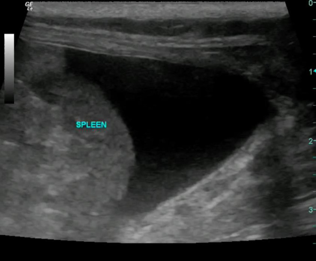
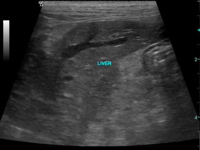
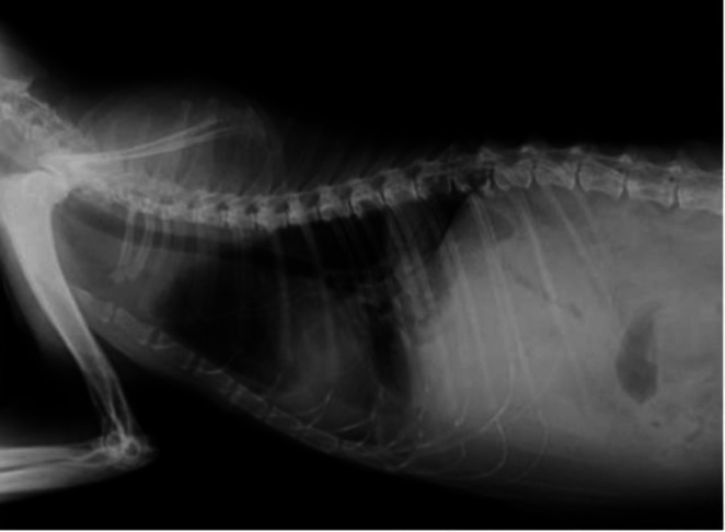
Findings/treatment and outcome: The radiological study was consistent with pleural effusion, as well as alveolar and interstitial pulmonary patterns. Ultrasonography confirmed hepatic venin congestion and ascites. Abdominocentesis revealed a modified transudate. A computed tomography (CT) angiography showed a mass at the level of the caudal mediastinum that compressed the caudal vena cava (CVC). Mediastinal lymphoma was considered the most likely differential diagnosis. These findings were interpreted as Budd-Chiari-like syndrome (BCLS) secondary to a mediastinal mass although, unfortunately, no further diagnostic or treatment procedures were accepted by the owners. BCLS is a rare condition in cats, where most of the reported cases occurred as a result of obstruction of the caudal vena cava. In this report, BCLS was caused by a mass located in the caudal mediastinum oppressing the caudal vena cava.
Conclusion: This is the first report of BCLS in cats diagnosed by CT angiography, and it shows the value of this technique to define the origin and extent of the mass and to evaluate the presence or absence of metastatic lesions.
期刊介绍:
The Iranian Journal of Veterinary Research(IJVR) is published quarterly in 4 issues. The aims of this journal are to improve and expand knowledge in all veterinary fields. It is an international journal indexed by the Thomson Institute for Scientific Information (ISI), Elsevier, Scopus, CAB International, Veterinary Bulletin and several other international databases. Research papers and reports on a wide range of veterinary topics are published in the journal after being evaluated by expert reviewers.The Editor-in-Chief is responsible for the editorial content of the journal—including peer-reviewed manuscripts—and the timing of its publication.
 求助内容:
求助内容: 应助结果提醒方式:


