Hang-Nga Mai, Thaw Thaw Win, Minh Son Tong, Cheong-Hee Lee, Kyu-Bok Lee, So-Yeun Kim, Hyun-Woo Lee, Du-Hyeong Lee
{"title":"不同微笑表情的虚拟笑脸图像中面部单元的三维形态计量分析。","authors":"Hang-Nga Mai, Thaw Thaw Win, Minh Son Tong, Cheong-Hee Lee, Kyu-Bok Lee, So-Yeun Kim, Hyun-Woo Lee, Du-Hyeong Lee","doi":"10.4047/jap.2023.15.1.1","DOIUrl":null,"url":null,"abstract":"<p><strong>Purpose: </strong>Accuracy of image matching between resting and smiling facial models is affected by the stability of the reference surfaces. This study aimed to investigate the morphometric variations in subdivided facial units during resting, posed and spontaneous smiling.</p><p><strong>Materials and methods: </strong>The posed and spontaneous smiling faces of 33 adults were digitized and registered to the resting faces. The morphological changes of subdivided facial units at the forehead (upper and lower central, upper and lower lateral, and temple), nasal (dorsum, tip, lateral wall, and alar lobules), and chin (central and lateral) regions were assessed by measuring the 3D mesh deviations between the smiling and resting facial models. The one-way analysis of variance, Duncan post hoc tests, and Student's t-test were used to determine the differences among the groups (α = .05).</p><p><strong>Results: </strong>The smallest morphometric changes were observed at the upper and central forehead and nasal dorsum; meanwhile, the largest deviation was found at the nasal alar lobules in both the posed and spontaneous smiles (<i>P</i> < .001). The spontaneous smile generally resulted in larger facial unit changes than the posed smile, and significant difference was observed at the alar lobules, central chin, and lateral chin units (<i>P</i> < .001).</p><p><strong>Conclusion: </strong>The upper and central forehead and nasal dorsum are reliable areas for image matching between resting and smiling 3D facial images. The central chin area can be considered an additional reference area for posed smiles; however, special cautions should be taken when selecting this area as references for spontaneous smiles.</p>","PeriodicalId":51291,"journal":{"name":"Journal of Advanced Prosthodontics","volume":"15 1","pages":"1-10"},"PeriodicalIF":2.5000,"publicationDate":"2023-02-01","publicationTypes":"Journal Article","fieldsOfStudy":null,"isOpenAccess":false,"openAccessPdf":"https://ftp.ncbi.nlm.nih.gov/pub/pmc/oa_pdf/4a/73/jap-15-1.PMC9992697.pdf","citationCount":"1","resultStr":"{\"title\":\"Three-dimensional morphometric analysis of facial units in virtual smiling facial images with different smile expressions.\",\"authors\":\"Hang-Nga Mai, Thaw Thaw Win, Minh Son Tong, Cheong-Hee Lee, Kyu-Bok Lee, So-Yeun Kim, Hyun-Woo Lee, Du-Hyeong Lee\",\"doi\":\"10.4047/jap.2023.15.1.1\",\"DOIUrl\":null,\"url\":null,\"abstract\":\"<p><strong>Purpose: </strong>Accuracy of image matching between resting and smiling facial models is affected by the stability of the reference surfaces. This study aimed to investigate the morphometric variations in subdivided facial units during resting, posed and spontaneous smiling.</p><p><strong>Materials and methods: </strong>The posed and spontaneous smiling faces of 33 adults were digitized and registered to the resting faces. The morphological changes of subdivided facial units at the forehead (upper and lower central, upper and lower lateral, and temple), nasal (dorsum, tip, lateral wall, and alar lobules), and chin (central and lateral) regions were assessed by measuring the 3D mesh deviations between the smiling and resting facial models. The one-way analysis of variance, Duncan post hoc tests, and Student's t-test were used to determine the differences among the groups (α = .05).</p><p><strong>Results: </strong>The smallest morphometric changes were observed at the upper and central forehead and nasal dorsum; meanwhile, the largest deviation was found at the nasal alar lobules in both the posed and spontaneous smiles (<i>P</i> < .001). The spontaneous smile generally resulted in larger facial unit changes than the posed smile, and significant difference was observed at the alar lobules, central chin, and lateral chin units (<i>P</i> < .001).</p><p><strong>Conclusion: </strong>The upper and central forehead and nasal dorsum are reliable areas for image matching between resting and smiling 3D facial images. The central chin area can be considered an additional reference area for posed smiles; however, special cautions should be taken when selecting this area as references for spontaneous smiles.</p>\",\"PeriodicalId\":51291,\"journal\":{\"name\":\"Journal of Advanced Prosthodontics\",\"volume\":\"15 1\",\"pages\":\"1-10\"},\"PeriodicalIF\":2.5000,\"publicationDate\":\"2023-02-01\",\"publicationTypes\":\"Journal Article\",\"fieldsOfStudy\":null,\"isOpenAccess\":false,\"openAccessPdf\":\"https://ftp.ncbi.nlm.nih.gov/pub/pmc/oa_pdf/4a/73/jap-15-1.PMC9992697.pdf\",\"citationCount\":\"1\",\"resultStr\":null,\"platform\":\"Semanticscholar\",\"paperid\":null,\"PeriodicalName\":\"Journal of Advanced Prosthodontics\",\"FirstCategoryId\":\"3\",\"ListUrlMain\":\"https://doi.org/10.4047/jap.2023.15.1.1\",\"RegionNum\":3,\"RegionCategory\":\"医学\",\"ArticlePicture\":[],\"TitleCN\":null,\"AbstractTextCN\":null,\"PMCID\":null,\"EPubDate\":\"\",\"PubModel\":\"\",\"JCR\":\"Q1\",\"JCRName\":\"DENTISTRY, ORAL SURGERY & MEDICINE\",\"Score\":null,\"Total\":0}","platform":"Semanticscholar","paperid":null,"PeriodicalName":"Journal of Advanced Prosthodontics","FirstCategoryId":"3","ListUrlMain":"https://doi.org/10.4047/jap.2023.15.1.1","RegionNum":3,"RegionCategory":"医学","ArticlePicture":[],"TitleCN":null,"AbstractTextCN":null,"PMCID":null,"EPubDate":"","PubModel":"","JCR":"Q1","JCRName":"DENTISTRY, ORAL SURGERY & MEDICINE","Score":null,"Total":0}
Three-dimensional morphometric analysis of facial units in virtual smiling facial images with different smile expressions.
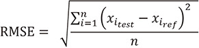
Purpose: Accuracy of image matching between resting and smiling facial models is affected by the stability of the reference surfaces. This study aimed to investigate the morphometric variations in subdivided facial units during resting, posed and spontaneous smiling.
Materials and methods: The posed and spontaneous smiling faces of 33 adults were digitized and registered to the resting faces. The morphological changes of subdivided facial units at the forehead (upper and lower central, upper and lower lateral, and temple), nasal (dorsum, tip, lateral wall, and alar lobules), and chin (central and lateral) regions were assessed by measuring the 3D mesh deviations between the smiling and resting facial models. The one-way analysis of variance, Duncan post hoc tests, and Student's t-test were used to determine the differences among the groups (α = .05).
Results: The smallest morphometric changes were observed at the upper and central forehead and nasal dorsum; meanwhile, the largest deviation was found at the nasal alar lobules in both the posed and spontaneous smiles (P < .001). The spontaneous smile generally resulted in larger facial unit changes than the posed smile, and significant difference was observed at the alar lobules, central chin, and lateral chin units (P < .001).
Conclusion: The upper and central forehead and nasal dorsum are reliable areas for image matching between resting and smiling 3D facial images. The central chin area can be considered an additional reference area for posed smiles; however, special cautions should be taken when selecting this area as references for spontaneous smiles.
期刊介绍:
This journal aims to convey scientific and clinical progress in the field of prosthodontics and its related areas to many dental communities concerned with esthetic and functional restorations, occlusion, implants, prostheses, and biomaterials related to prosthodontics.
This journal publishes
• Original research data of high scientific merit in the field of diagnosis, function, esthetics and stomatognathic physiology related to prosthodontic rehabilitation, physiology and mechanics of occlusion, mechanical and biologic aspects of prosthodontic materials including dental implants.
• Review articles by experts on controversies and new developments in prosthodontics.
• Case reports if they provide or document new fundamental knowledge.

 求助内容:
求助内容: 应助结果提醒方式:


