{"title":"从正常认知衰老到阿尔茨海默病的眼部生物标志物的变化:一项初步研究","authors":"Pareena Chaitanuwong, Supharat Jariyakosol, Supanut Apinyawasisuk, Parima Hirunwiwatkul, Hathairat Lawanlattanagul, Solaphat Hemrungrojn, Yuda Chongpison","doi":"10.2147/EB.S391608","DOIUrl":null,"url":null,"abstract":"<p><strong>Purpose: </strong>To identify ophthalmic findings in Alzheimer's type dementia (ATD) compared to normal subjects.</p><p><strong>Patients and methods: </strong>This comparative descriptive study included participants from the institution's cognitive fitness center. Complete ophthalmic examinations were performed. Optical coherence tomography (OCT) and OCT angiography (OCTA) were used to analyze retinal thickness and vascular density. The Ocular Surface Disease Index (OSDI) score and tear breakup time (TBUT) were used to assess dry eye. The blink rate was counted by a well-trained observer. Cognitive function was evaluated using the Thai Mental State Examination (TMSE) score. Correlation analysis was performed to compare OCT, OCTA parameters, and TMSE.</p><p><strong>Results: </strong>We included 24 ATD patients and 39 normal participants as a control group by age and sex-matched. The prevalence of dry eye using the Asia Dry Eye Society criteria was 15% and 13% in normal and ATD patients, respectively. The differences in OSDI scores, TBUT, and blink rate between the two groups were not statistically significant. The parafoveal and perifoveal macular thickness of the ATD group were significantly lower than that of the control group (p<0.01). All parameters of the vessel density of the ATD group were significantly lower than in the control group, including the whole macular vessel density (p<0.01), optic disc vessel density at the nerve head level (p<0.01), and optic disc vessel density at the radial peripapillary capillary level (p<0.05). After age adjustment, there were no statistically significant differences in all the OCT and OCTA parameters. There was a positive correlation between retinal thickness and vessel density in the macular and optic disc region and TMSE scores.</p><p><strong>Conclusion: </strong>Perifoveal and parafoveal retinal thickness might be more sensitive than peripapillary RNFL thickness to detect neurodegenerative changes in patients with ATD. Macular thickness and vessel density reduction were also positively correlated with cognitive decline.</p>","PeriodicalId":51844,"journal":{"name":"Eye and Brain","volume":"15 ","pages":"15-23"},"PeriodicalIF":2.4000,"publicationDate":"2023-01-01","publicationTypes":"Journal Article","fieldsOfStudy":null,"isOpenAccess":false,"openAccessPdf":"https://ftp.ncbi.nlm.nih.gov/pub/pmc/oa_pdf/1e/df/eb-15-15.PMC9986468.pdf","citationCount":"1","resultStr":"{\"title\":\"Changes in Ocular Biomarkers from Normal Cognitive Aging to Alzheimer's Disease: A Pilot Study.\",\"authors\":\"Pareena Chaitanuwong, Supharat Jariyakosol, Supanut Apinyawasisuk, Parima Hirunwiwatkul, Hathairat Lawanlattanagul, Solaphat Hemrungrojn, Yuda Chongpison\",\"doi\":\"10.2147/EB.S391608\",\"DOIUrl\":null,\"url\":null,\"abstract\":\"<p><strong>Purpose: </strong>To identify ophthalmic findings in Alzheimer's type dementia (ATD) compared to normal subjects.</p><p><strong>Patients and methods: </strong>This comparative descriptive study included participants from the institution's cognitive fitness center. Complete ophthalmic examinations were performed. Optical coherence tomography (OCT) and OCT angiography (OCTA) were used to analyze retinal thickness and vascular density. The Ocular Surface Disease Index (OSDI) score and tear breakup time (TBUT) were used to assess dry eye. The blink rate was counted by a well-trained observer. Cognitive function was evaluated using the Thai Mental State Examination (TMSE) score. Correlation analysis was performed to compare OCT, OCTA parameters, and TMSE.</p><p><strong>Results: </strong>We included 24 ATD patients and 39 normal participants as a control group by age and sex-matched. The prevalence of dry eye using the Asia Dry Eye Society criteria was 15% and 13% in normal and ATD patients, respectively. The differences in OSDI scores, TBUT, and blink rate between the two groups were not statistically significant. The parafoveal and perifoveal macular thickness of the ATD group were significantly lower than that of the control group (p<0.01). All parameters of the vessel density of the ATD group were significantly lower than in the control group, including the whole macular vessel density (p<0.01), optic disc vessel density at the nerve head level (p<0.01), and optic disc vessel density at the radial peripapillary capillary level (p<0.05). After age adjustment, there were no statistically significant differences in all the OCT and OCTA parameters. There was a positive correlation between retinal thickness and vessel density in the macular and optic disc region and TMSE scores.</p><p><strong>Conclusion: </strong>Perifoveal and parafoveal retinal thickness might be more sensitive than peripapillary RNFL thickness to detect neurodegenerative changes in patients with ATD. Macular thickness and vessel density reduction were also positively correlated with cognitive decline.</p>\",\"PeriodicalId\":51844,\"journal\":{\"name\":\"Eye and Brain\",\"volume\":\"15 \",\"pages\":\"15-23\"},\"PeriodicalIF\":2.4000,\"publicationDate\":\"2023-01-01\",\"publicationTypes\":\"Journal Article\",\"fieldsOfStudy\":null,\"isOpenAccess\":false,\"openAccessPdf\":\"https://ftp.ncbi.nlm.nih.gov/pub/pmc/oa_pdf/1e/df/eb-15-15.PMC9986468.pdf\",\"citationCount\":\"1\",\"resultStr\":null,\"platform\":\"Semanticscholar\",\"paperid\":null,\"PeriodicalName\":\"Eye and Brain\",\"FirstCategoryId\":\"1085\",\"ListUrlMain\":\"https://doi.org/10.2147/EB.S391608\",\"RegionNum\":0,\"RegionCategory\":null,\"ArticlePicture\":[],\"TitleCN\":null,\"AbstractTextCN\":null,\"PMCID\":null,\"EPubDate\":\"\",\"PubModel\":\"\",\"JCR\":\"Q1\",\"JCRName\":\"OPHTHALMOLOGY\",\"Score\":null,\"Total\":0}","platform":"Semanticscholar","paperid":null,"PeriodicalName":"Eye and Brain","FirstCategoryId":"1085","ListUrlMain":"https://doi.org/10.2147/EB.S391608","RegionNum":0,"RegionCategory":null,"ArticlePicture":[],"TitleCN":null,"AbstractTextCN":null,"PMCID":null,"EPubDate":"","PubModel":"","JCR":"Q1","JCRName":"OPHTHALMOLOGY","Score":null,"Total":0}
Changes in Ocular Biomarkers from Normal Cognitive Aging to Alzheimer's Disease: A Pilot Study.
Purpose: To identify ophthalmic findings in Alzheimer's type dementia (ATD) compared to normal subjects.
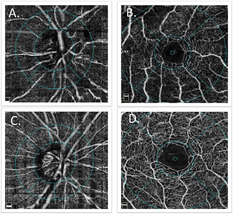
Patients and methods: This comparative descriptive study included participants from the institution's cognitive fitness center. Complete ophthalmic examinations were performed. Optical coherence tomography (OCT) and OCT angiography (OCTA) were used to analyze retinal thickness and vascular density. The Ocular Surface Disease Index (OSDI) score and tear breakup time (TBUT) were used to assess dry eye. The blink rate was counted by a well-trained observer. Cognitive function was evaluated using the Thai Mental State Examination (TMSE) score. Correlation analysis was performed to compare OCT, OCTA parameters, and TMSE.
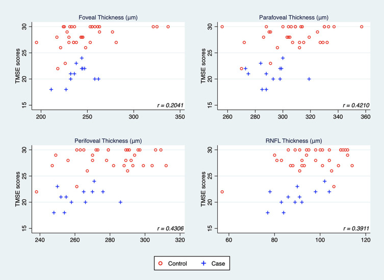
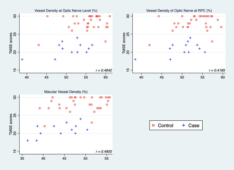
Results: We included 24 ATD patients and 39 normal participants as a control group by age and sex-matched. The prevalence of dry eye using the Asia Dry Eye Society criteria was 15% and 13% in normal and ATD patients, respectively. The differences in OSDI scores, TBUT, and blink rate between the two groups were not statistically significant. The parafoveal and perifoveal macular thickness of the ATD group were significantly lower than that of the control group (p<0.01). All parameters of the vessel density of the ATD group were significantly lower than in the control group, including the whole macular vessel density (p<0.01), optic disc vessel density at the nerve head level (p<0.01), and optic disc vessel density at the radial peripapillary capillary level (p<0.05). After age adjustment, there were no statistically significant differences in all the OCT and OCTA parameters. There was a positive correlation between retinal thickness and vessel density in the macular and optic disc region and TMSE scores.
Conclusion: Perifoveal and parafoveal retinal thickness might be more sensitive than peripapillary RNFL thickness to detect neurodegenerative changes in patients with ATD. Macular thickness and vessel density reduction were also positively correlated with cognitive decline.
期刊介绍:
Eye and Brain is an international, peer-reviewed, open access journal focusing on basic research, clinical findings, and expert reviews in the field of visual science and neuro-ophthalmology. The journal’s unique focus is the link between two well-known visual centres, the eye and the brain, with an emphasis on the importance of such connections. All aspects of clinical and especially basic research on the visual system are addressed within the journal as well as significant future directions in vision research and therapeutic measures. This unique journal focuses on neurological aspects of vision – both physiological and pathological. The scope of the journal spans from the cornea to the associational visual cortex and all the visual centers in between. Topics range from basic biological mechanisms to therapeutic treatment, from simple organisms to humans, and utilizing techniques from molecular biology to behavior. The journal especially welcomes primary research articles or review papers that make the connection between the eye and the brain. Specific areas covered in the journal include: Physiology and pathophysiology of visual centers, Eye movement disorders and strabismus, Cellular, biochemical, and molecular features of the visual system, Structural and functional organization of the eye and of the visual cortex, Metabolic demands of the visual system, Diseases and disorders with neuro-ophthalmic manifestations, Clinical and experimental neuro-ophthalmology and visual system pathologies, Epidemiological studies.

 求助内容:
求助内容: 应助结果提醒方式:


