Siddhartha Dhiman , Stephanie Fountain-Zaragoza , Jens H. Jensen , Maria Fatima Falangola , Emilie T. McKinnon , Hunter G. Moss , Kathryn E. Thorn , William J. Rieter , Maria Vittoria Spampinato , Paul J. Nietert , Joseph A. Helpern , Andreana Benitez
{"title":"纤维球白质模型揭示了健康大脑衰老过程中的微结构变化","authors":"Siddhartha Dhiman , Stephanie Fountain-Zaragoza , Jens H. Jensen , Maria Fatima Falangola , Emilie T. McKinnon , Hunter G. Moss , Kathryn E. Thorn , William J. Rieter , Maria Vittoria Spampinato , Paul J. Nietert , Joseph A. Helpern , Andreana Benitez","doi":"10.1016/j.nbas.2022.100037","DOIUrl":null,"url":null,"abstract":"<div><p>Age-related white matter degeneration is characterized by myelin breakdown and neuronal fiber loss that preferentially occur in regions that myelinate later in development. Conventional diffusion MRI (dMRI) has demonstrated age-related increases in diffusivity but provides limited information regarding the tissue-specific changes driving these effects. A recently developed dMRI biophysical modeling technique, Fiber Ball White Matter (FBWM) modeling, offers enhanced biological interpretability by estimating microstructural properties specific to the intra-axonal and extra-axonal spaces. We used FBWM to illustrate the biological mechanisms underlying changes throughout white matter in healthy aging using data from 63 cognitively unimpaired adults ages 45–85 with no radiological evidence of neurodegeneration or incipient Alzheimer’s disease. Conventional dMRI and FBWM metrics were computed for two late-myelinating (genu of the corpus callosum and association tracts) and two early-myelinating regions (splenium of the corpus callosum and projection tracts). We examined the associations between age and these metrics in each region and tested whether age was differentially associated with these metrics in late- vs. early-myelinating regions. We found that conventional metrics replicated patterns of age-related increases in diffusivity in late-myelinating regions. FBWM additionally revealed specific intra- and extra-axonal changes suggestive of myelin breakdown and preferential loss of smaller-diameter axons, yielding <em>in vivo</em> corroboration of findings from histopathological studies of aged brains. These results demonstrate that advanced biophysical modeling approaches, such as FBWM, offer novel information about the microstructure-specific alterations contributing to white matter changes in healthy aging. These tools hold promise as sensitive indicators of early pathological changes related to neurodegenerative disease.</p></div>","PeriodicalId":72131,"journal":{"name":"Aging brain","volume":"2 ","pages":"Article 100037"},"PeriodicalIF":1.7000,"publicationDate":"2022-01-01","publicationTypes":"Journal Article","fieldsOfStudy":null,"isOpenAccess":false,"openAccessPdf":"https://ftp.ncbi.nlm.nih.gov/pub/pmc/oa_pdf/d6/fe/main.PMC9624504.pdf","citationCount":"3","resultStr":"{\"title\":\"Fiber Ball white matter modeling reveals microstructural alterations in healthy brain aging\",\"authors\":\"Siddhartha Dhiman , Stephanie Fountain-Zaragoza , Jens H. Jensen , Maria Fatima Falangola , Emilie T. McKinnon , Hunter G. Moss , Kathryn E. Thorn , William J. Rieter , Maria Vittoria Spampinato , Paul J. Nietert , Joseph A. Helpern , Andreana Benitez\",\"doi\":\"10.1016/j.nbas.2022.100037\",\"DOIUrl\":null,\"url\":null,\"abstract\":\"<div><p>Age-related white matter degeneration is characterized by myelin breakdown and neuronal fiber loss that preferentially occur in regions that myelinate later in development. Conventional diffusion MRI (dMRI) has demonstrated age-related increases in diffusivity but provides limited information regarding the tissue-specific changes driving these effects. A recently developed dMRI biophysical modeling technique, Fiber Ball White Matter (FBWM) modeling, offers enhanced biological interpretability by estimating microstructural properties specific to the intra-axonal and extra-axonal spaces. We used FBWM to illustrate the biological mechanisms underlying changes throughout white matter in healthy aging using data from 63 cognitively unimpaired adults ages 45–85 with no radiological evidence of neurodegeneration or incipient Alzheimer’s disease. Conventional dMRI and FBWM metrics were computed for two late-myelinating (genu of the corpus callosum and association tracts) and two early-myelinating regions (splenium of the corpus callosum and projection tracts). We examined the associations between age and these metrics in each region and tested whether age was differentially associated with these metrics in late- vs. early-myelinating regions. We found that conventional metrics replicated patterns of age-related increases in diffusivity in late-myelinating regions. FBWM additionally revealed specific intra- and extra-axonal changes suggestive of myelin breakdown and preferential loss of smaller-diameter axons, yielding <em>in vivo</em> corroboration of findings from histopathological studies of aged brains. These results demonstrate that advanced biophysical modeling approaches, such as FBWM, offer novel information about the microstructure-specific alterations contributing to white matter changes in healthy aging. These tools hold promise as sensitive indicators of early pathological changes related to neurodegenerative disease.</p></div>\",\"PeriodicalId\":72131,\"journal\":{\"name\":\"Aging brain\",\"volume\":\"2 \",\"pages\":\"Article 100037\"},\"PeriodicalIF\":1.7000,\"publicationDate\":\"2022-01-01\",\"publicationTypes\":\"Journal Article\",\"fieldsOfStudy\":null,\"isOpenAccess\":false,\"openAccessPdf\":\"https://ftp.ncbi.nlm.nih.gov/pub/pmc/oa_pdf/d6/fe/main.PMC9624504.pdf\",\"citationCount\":\"3\",\"resultStr\":null,\"platform\":\"Semanticscholar\",\"paperid\":null,\"PeriodicalName\":\"Aging brain\",\"FirstCategoryId\":\"1085\",\"ListUrlMain\":\"https://www.sciencedirect.com/science/article/pii/S2589958922000093\",\"RegionNum\":0,\"RegionCategory\":null,\"ArticlePicture\":[],\"TitleCN\":null,\"AbstractTextCN\":null,\"PMCID\":null,\"EPubDate\":\"\",\"PubModel\":\"\",\"JCR\":\"Q3\",\"JCRName\":\"CLINICAL NEUROLOGY\",\"Score\":null,\"Total\":0}","platform":"Semanticscholar","paperid":null,"PeriodicalName":"Aging brain","FirstCategoryId":"1085","ListUrlMain":"https://www.sciencedirect.com/science/article/pii/S2589958922000093","RegionNum":0,"RegionCategory":null,"ArticlePicture":[],"TitleCN":null,"AbstractTextCN":null,"PMCID":null,"EPubDate":"","PubModel":"","JCR":"Q3","JCRName":"CLINICAL NEUROLOGY","Score":null,"Total":0}
Fiber Ball white matter modeling reveals microstructural alterations in healthy brain aging
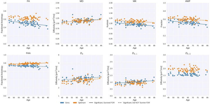
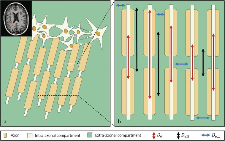
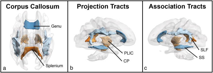
Age-related white matter degeneration is characterized by myelin breakdown and neuronal fiber loss that preferentially occur in regions that myelinate later in development. Conventional diffusion MRI (dMRI) has demonstrated age-related increases in diffusivity but provides limited information regarding the tissue-specific changes driving these effects. A recently developed dMRI biophysical modeling technique, Fiber Ball White Matter (FBWM) modeling, offers enhanced biological interpretability by estimating microstructural properties specific to the intra-axonal and extra-axonal spaces. We used FBWM to illustrate the biological mechanisms underlying changes throughout white matter in healthy aging using data from 63 cognitively unimpaired adults ages 45–85 with no radiological evidence of neurodegeneration or incipient Alzheimer’s disease. Conventional dMRI and FBWM metrics were computed for two late-myelinating (genu of the corpus callosum and association tracts) and two early-myelinating regions (splenium of the corpus callosum and projection tracts). We examined the associations between age and these metrics in each region and tested whether age was differentially associated with these metrics in late- vs. early-myelinating regions. We found that conventional metrics replicated patterns of age-related increases in diffusivity in late-myelinating regions. FBWM additionally revealed specific intra- and extra-axonal changes suggestive of myelin breakdown and preferential loss of smaller-diameter axons, yielding in vivo corroboration of findings from histopathological studies of aged brains. These results demonstrate that advanced biophysical modeling approaches, such as FBWM, offer novel information about the microstructure-specific alterations contributing to white matter changes in healthy aging. These tools hold promise as sensitive indicators of early pathological changes related to neurodegenerative disease.

 求助内容:
求助内容: 应助结果提醒方式:


