Ju In Park, Chang Kyu Park, Bong Jin Park, Seok Keun Choi
{"title":"大量肥厚性脑膜瘤斑块模拟纤维发育不良。","authors":"Ju In Park, Chang Kyu Park, Bong Jin Park, Seok Keun Choi","doi":"10.14791/btrt.2023.0027","DOIUrl":null,"url":null,"abstract":"<p><p>The authors report an extremely rare case of a massive hyperostotic meningioma en plaque, which had characteristics of unique bony growth. A 34-year-old man presented with a palpable solid mass in the left cranial region that had gradually grown in size with a broad base on the calvarium for 8 years. Radiologically, the area involved by the mass ranged from the sphenoid bone to the frontal, parietal, temporal, and occipital bones. Three-dimensional CT revealed multiple growing spiculate features on the inner and outer cranial surface. Even though the radiologic features resembled fibrous dysplasia, it was histologically found to be a type of meningioma.</p>","PeriodicalId":72453,"journal":{"name":"Brain tumor research and treatment","volume":"11 4","pages":"271-273"},"PeriodicalIF":0.0000,"publicationDate":"2023-10-01","publicationTypes":"Journal Article","fieldsOfStudy":null,"isOpenAccess":false,"openAccessPdf":"https://www.ncbi.nlm.nih.gov/pmc/articles/PMC10641317/pdf/","citationCount":"0","resultStr":"{\"title\":\"Massive Hyperostotic Meningioma En Plaque Mimicking Fibrous Dysplasia.\",\"authors\":\"Ju In Park, Chang Kyu Park, Bong Jin Park, Seok Keun Choi\",\"doi\":\"10.14791/btrt.2023.0027\",\"DOIUrl\":null,\"url\":null,\"abstract\":\"<p><p>The authors report an extremely rare case of a massive hyperostotic meningioma en plaque, which had characteristics of unique bony growth. A 34-year-old man presented with a palpable solid mass in the left cranial region that had gradually grown in size with a broad base on the calvarium for 8 years. Radiologically, the area involved by the mass ranged from the sphenoid bone to the frontal, parietal, temporal, and occipital bones. Three-dimensional CT revealed multiple growing spiculate features on the inner and outer cranial surface. Even though the radiologic features resembled fibrous dysplasia, it was histologically found to be a type of meningioma.</p>\",\"PeriodicalId\":72453,\"journal\":{\"name\":\"Brain tumor research and treatment\",\"volume\":\"11 4\",\"pages\":\"271-273\"},\"PeriodicalIF\":0.0000,\"publicationDate\":\"2023-10-01\",\"publicationTypes\":\"Journal Article\",\"fieldsOfStudy\":null,\"isOpenAccess\":false,\"openAccessPdf\":\"https://www.ncbi.nlm.nih.gov/pmc/articles/PMC10641317/pdf/\",\"citationCount\":\"0\",\"resultStr\":null,\"platform\":\"Semanticscholar\",\"paperid\":null,\"PeriodicalName\":\"Brain tumor research and treatment\",\"FirstCategoryId\":\"1085\",\"ListUrlMain\":\"https://doi.org/10.14791/btrt.2023.0027\",\"RegionNum\":0,\"RegionCategory\":null,\"ArticlePicture\":[],\"TitleCN\":null,\"AbstractTextCN\":null,\"PMCID\":null,\"EPubDate\":\"\",\"PubModel\":\"\",\"JCR\":\"\",\"JCRName\":\"\",\"Score\":null,\"Total\":0}","platform":"Semanticscholar","paperid":null,"PeriodicalName":"Brain tumor research and treatment","FirstCategoryId":"1085","ListUrlMain":"https://doi.org/10.14791/btrt.2023.0027","RegionNum":0,"RegionCategory":null,"ArticlePicture":[],"TitleCN":null,"AbstractTextCN":null,"PMCID":null,"EPubDate":"","PubModel":"","JCR":"","JCRName":"","Score":null,"Total":0}
Massive Hyperostotic Meningioma En Plaque Mimicking Fibrous Dysplasia.
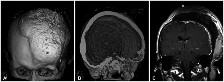
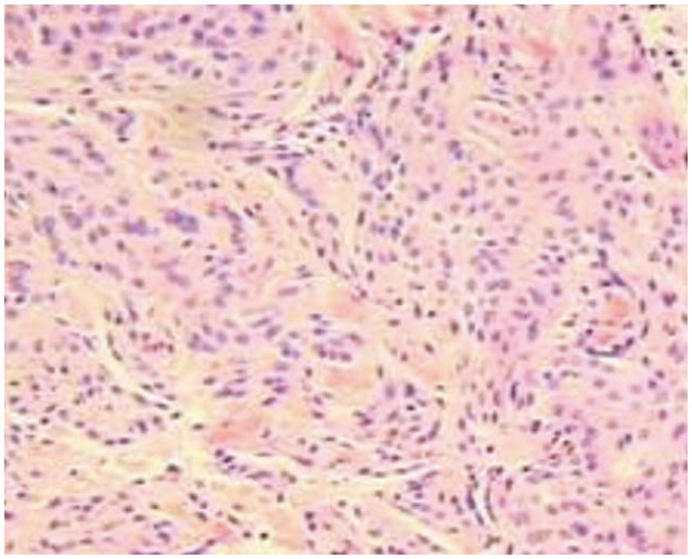
The authors report an extremely rare case of a massive hyperostotic meningioma en plaque, which had characteristics of unique bony growth. A 34-year-old man presented with a palpable solid mass in the left cranial region that had gradually grown in size with a broad base on the calvarium for 8 years. Radiologically, the area involved by the mass ranged from the sphenoid bone to the frontal, parietal, temporal, and occipital bones. Three-dimensional CT revealed multiple growing spiculate features on the inner and outer cranial surface. Even though the radiologic features resembled fibrous dysplasia, it was histologically found to be a type of meningioma.

 求助内容:
求助内容: 应助结果提醒方式:


