氧致视网膜病变小鼠模型右旋糖酐灌注与GSI-B4分离素染色的比较。

Shaofen Huang, Jiajian Liang, G. Yam, Zhihao Lu, C. Pang, H. Chen
{"title":"氧致视网膜病变小鼠模型右旋糖酐灌注与GSI-B4分离素染色的比较。","authors":"Shaofen Huang, Jiajian Liang, G. Yam, Zhihao Lu, C. Pang, H. Chen","doi":"10.3969/J.ISSN.1000-4432.2015.02.006","DOIUrl":null,"url":null,"abstract":"PURPOSE Oxygen-induced retinopathy (OIR) is a robust and widely used animal model for the study of retinal neovascularization (NV). Dextran perfusion and Griffonia simplicifolia isolectin B4 (GSI-B4) staining are two common methods for examining the occurrence and extent of OIR. This study provides a quantitative comparison of the two for OIR detection. METHODS At postnatal day 7 (PN7), fifteen C57BL/6J mice were exposed to a 75% hyperoxic condition for 5 days and then returned to room air conditions. At PN17, the mice received intravitreal injection of GSI-B4 Alexa Fluor 568 conjugate. After 10 hours, they were infused with FITC-dextran conjugate via the left ventricle. Retinal flat mounts were photographed by confocal microscopy. Areas with fluorescent signals and the total retinal areas were quantified by Image J software. RESULTS Both GSI-B4 and dextran detected the peripheral neovascular area. The mean hyper fluorescence area was 0.33 ± 0.14% of whole retinal area determined by GSI-B4 staining and 0.25 ± 0.28% determined by dextran perfusion. The difference between the two measures was 0.08% (95% CI:-0.59%, 0.43%). The Pearson correlation coefficient between the two methods was 0.386,P =0.035. The mean coincidence rates were 14.3 ± 13.4% and 24.9 ± 18.5% for GSI-B4 and dextran staining, respectively. CONCLUSION Both methods can complement each other in demonstrating and quantitatively evaluating retinal NV. A poor agreement was found between the two methods; GSI-B4 isolectin was more effective than FITC-dextran perfusion in evaluating the extent of retinal NV in a mouse model of OIR.","PeriodicalId":12096,"journal":{"name":"眼科学报","volume":"63 1","pages":"70-4"},"PeriodicalIF":0.0000,"publicationDate":"2015-06-11","publicationTypes":"Journal Article","fieldsOfStudy":null,"isOpenAccess":false,"openAccessPdf":"","citationCount":"1","resultStr":"{\"title\":\"Comparison of Dextran Perfusion and GSI-B4 Isolectin Staining in a Mouse Model of Oxygen-induced Retinopathy.\",\"authors\":\"Shaofen Huang, Jiajian Liang, G. Yam, Zhihao Lu, C. Pang, H. Chen\",\"doi\":\"10.3969/J.ISSN.1000-4432.2015.02.006\",\"DOIUrl\":null,\"url\":null,\"abstract\":\"PURPOSE Oxygen-induced retinopathy (OIR) is a robust and widely used animal model for the study of retinal neovascularization (NV). Dextran perfusion and Griffonia simplicifolia isolectin B4 (GSI-B4) staining are two common methods for examining the occurrence and extent of OIR. This study provides a quantitative comparison of the two for OIR detection. METHODS At postnatal day 7 (PN7), fifteen C57BL/6J mice were exposed to a 75% hyperoxic condition for 5 days and then returned to room air conditions. At PN17, the mice received intravitreal injection of GSI-B4 Alexa Fluor 568 conjugate. After 10 hours, they were infused with FITC-dextran conjugate via the left ventricle. Retinal flat mounts were photographed by confocal microscopy. Areas with fluorescent signals and the total retinal areas were quantified by Image J software. RESULTS Both GSI-B4 and dextran detected the peripheral neovascular area. The mean hyper fluorescence area was 0.33 ± 0.14% of whole retinal area determined by GSI-B4 staining and 0.25 ± 0.28% determined by dextran perfusion. The difference between the two measures was 0.08% (95% CI:-0.59%, 0.43%). The Pearson correlation coefficient between the two methods was 0.386,P =0.035. The mean coincidence rates were 14.3 ± 13.4% and 24.9 ± 18.5% for GSI-B4 and dextran staining, respectively. CONCLUSION Both methods can complement each other in demonstrating and quantitatively evaluating retinal NV. A poor agreement was found between the two methods; GSI-B4 isolectin was more effective than FITC-dextran perfusion in evaluating the extent of retinal NV in a mouse model of OIR.\",\"PeriodicalId\":12096,\"journal\":{\"name\":\"眼科学报\",\"volume\":\"63 1\",\"pages\":\"70-4\"},\"PeriodicalIF\":0.0000,\"publicationDate\":\"2015-06-11\",\"publicationTypes\":\"Journal Article\",\"fieldsOfStudy\":null,\"isOpenAccess\":false,\"openAccessPdf\":\"\",\"citationCount\":\"1\",\"resultStr\":null,\"platform\":\"Semanticscholar\",\"paperid\":null,\"PeriodicalName\":\"眼科学报\",\"FirstCategoryId\":\"3\",\"ListUrlMain\":\"https://doi.org/10.3969/J.ISSN.1000-4432.2015.02.006\",\"RegionNum\":0,\"RegionCategory\":null,\"ArticlePicture\":[],\"TitleCN\":null,\"AbstractTextCN\":null,\"PMCID\":null,\"EPubDate\":\"\",\"PubModel\":\"\",\"JCR\":\"\",\"JCRName\":\"\",\"Score\":null,\"Total\":0}","platform":"Semanticscholar","paperid":null,"PeriodicalName":"眼科学报","FirstCategoryId":"3","ListUrlMain":"https://doi.org/10.3969/J.ISSN.1000-4432.2015.02.006","RegionNum":0,"RegionCategory":null,"ArticlePicture":[],"TitleCN":null,"AbstractTextCN":null,"PMCID":null,"EPubDate":"","PubModel":"","JCR":"","JCRName":"","Score":null,"Total":0}
引用次数: 1

摘要

目的氧诱导视网膜病变(OIR)是研究视网膜新生血管(NV)的一种强大且广泛应用的动物模型。右旋糖酐灌注和单叶Griffonia simplicifolia isolectin B4 (GSI-B4)染色是检测OIR发生和程度的两种常用方法。本研究为红外光谱检测提供了一种定量比较方法。方法在出生后第7天(PN7), 15只C57BL/6J小鼠暴露于75%高氧条件下5 d,然后返回室温环境。在PN17时,小鼠接受玻璃体内注射GSI-B4 Alexa Fluor 568偶联物。10小时后,经左心室注入fitc -葡聚糖缀合物。用共聚焦显微镜拍摄视网膜平面支架。用Image J软件对有荧光信号的区域和视网膜总面积进行量化。结果GSI-B4和葡聚糖均能检测外周新生血管区。GSI-B4染色法测定的平均高荧光面积为视网膜全面积的0.33±0.14%,葡聚糖灌注法测定的平均高荧光面积为0.25±0.28%。两种测量方法的差异为0.08% (95% CI:-0.59%, 0.43%)。两种方法的Pearson相关系数为0.386,P =0.035。GSI-B4染色和葡聚糖染色的平均符合率分别为14.3±13.4%和24.9±18.5%。结论两种方法在显示和定量评价视网膜NV方面可以互补,但两种方法的一致性较差;GSI-B4分离素比fitc -葡聚糖灌注更有效地评估小鼠视网膜NV的程度。
本文章由计算机程序翻译,如有差异,请以英文原文为准。
Comparison of Dextran Perfusion and GSI-B4 Isolectin Staining in a Mouse Model of Oxygen-induced Retinopathy.
PURPOSE Oxygen-induced retinopathy (OIR) is a robust and widely used animal model for the study of retinal neovascularization (NV). Dextran perfusion and Griffonia simplicifolia isolectin B4 (GSI-B4) staining are two common methods for examining the occurrence and extent of OIR. This study provides a quantitative comparison of the two for OIR detection. METHODS At postnatal day 7 (PN7), fifteen C57BL/6J mice were exposed to a 75% hyperoxic condition for 5 days and then returned to room air conditions. At PN17, the mice received intravitreal injection of GSI-B4 Alexa Fluor 568 conjugate. After 10 hours, they were infused with FITC-dextran conjugate via the left ventricle. Retinal flat mounts were photographed by confocal microscopy. Areas with fluorescent signals and the total retinal areas were quantified by Image J software. RESULTS Both GSI-B4 and dextran detected the peripheral neovascular area. The mean hyper fluorescence area was 0.33 ± 0.14% of whole retinal area determined by GSI-B4 staining and 0.25 ± 0.28% determined by dextran perfusion. The difference between the two measures was 0.08% (95% CI:-0.59%, 0.43%). The Pearson correlation coefficient between the two methods was 0.386,P =0.035. The mean coincidence rates were 14.3 ± 13.4% and 24.9 ± 18.5% for GSI-B4 and dextran staining, respectively. CONCLUSION Both methods can complement each other in demonstrating and quantitatively evaluating retinal NV. A poor agreement was found between the two methods; GSI-B4 isolectin was more effective than FITC-dextran perfusion in evaluating the extent of retinal NV in a mouse model of OIR.
求助全文
通过发布文献求助,成功后即可免费获取论文全文。 去求助
来源期刊
自引率
0.00%
发文量
1312
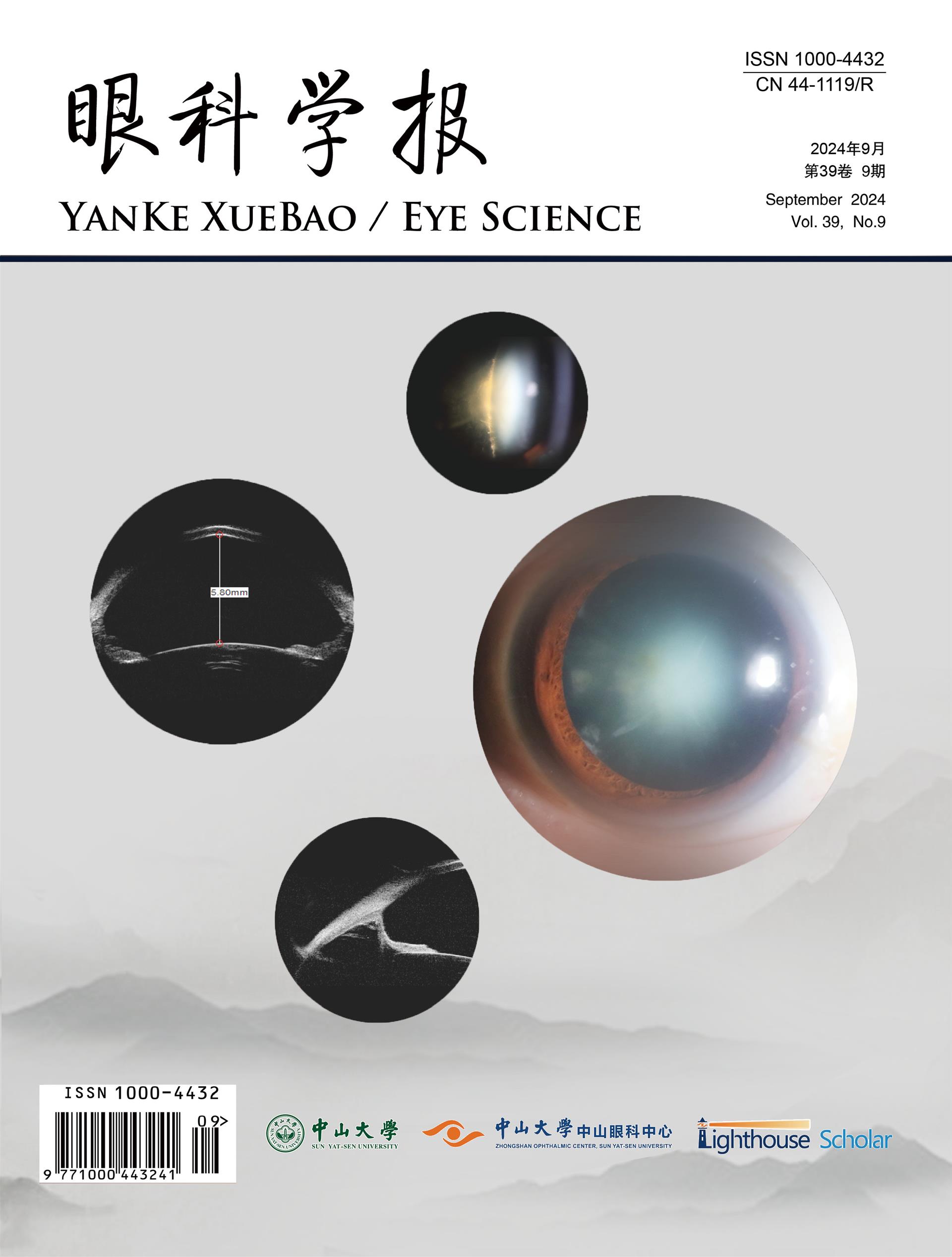
期刊介绍: Eye science was founded in 1985. It is a national medical journal supervised by the Ministry of Education of the People's Republic of China, sponsored by Sun Yat-sen University, and hosted by Sun Yat-sen University Zhongshan Eye Center (in October 2020, it was changed from a quarterly to a monthly, with the publication number: ISSN: 1000-4432; CN: 44-1119/R). It is edited by Ge Jian, former dean of Sun Yat-sen University Zhongshan Eye Center, Liu Yizhi, director and dean of Sun Yat-sen University Zhongshan Eye Center, and Lin Haotian, deputy director of Sun Yat-sen University Zhongshan Eye Center, as executive editor. It mainly reports on new developments and trends in the field of ophthalmology at home and abroad, focusing on basic research in ophthalmology, clinical experience, and theoretical knowledge and technical operations related to epidemiology. It has been included in important databases at home and abroad, such as Chemical Abstract (CA), China Journal Full-text Database (CNKI), China Core Journals (Selection) Database (Wanfang), and Chinese Science and Technology Journal Database (VIP).
×
引用
GB/T 7714-2015
复制
MLA
复制
APA
复制
导出至
BibTeX EndNote RefMan NoteFirst NoteExpress
×
提示
您的信息不完整,为了账户安全,请先补充。
现在去补充
×
提示
您因"违规操作"
具体请查看互助需知
我知道了
×
提示
确定
请完成安全验证×
copy
已复制链接
快去分享给好友吧!
我知道了
右上角分享
点击右上角分享
0
联系我们:info@booksci.cn Book学术提供免费学术资源搜索服务,方便国内外学者检索中英文文献。致力于提供最便捷和优质的服务体验。 Copyright © 2023 布克学术 All rights reserved.
京ICP备2023020795号-1
ghs 京公网安备 11010802042870号
Book学术文献互助
Book学术文献互助群
群 号:481959085
Book学术官方微信