{"title":"混合表型急性白血病(T 型/巨核细胞型):真的存在吗?","authors":"Neelum Mansoor, Omer Javed, Naila Rafiq, Anila Aali, Fatima Meraj","doi":"10.1007/s12308-023-00535-w","DOIUrl":null,"url":null,"abstract":"<p><p>Mixed-phenotype acute leukemias (MPAL) account for < 4% of all cases of acute leukemias. These are a heterogeneous group of leukemias grouped together by the WHO classification as \"rare subtypes.\" The diagnosis and treatment of MPAL is extremely challenging particularly for low middle income countries. Of these, B/myeloid and T/myeloid combinations are relatively common subtypes. However, megakaryoblastic and erythroid lineages in combination with other lineages are still rare enough to not even be addressed in the WHO classification. To date, there have been only a few reports of mixed B or T cell and megakaryocytic or mixed B or T cell and erythroid leukemias. We report the clinical presentation, diagnostic profile, and disease course of MPAL cases with a biphenotypic pattern consistent with T/megakaryoblastic lineage which is not yet defined in WHO classification. These cases were phenotyped using 8-color flow cytometry (BD FACS CANTO-II) using an extensive panel of markers. Interphase fluorescence in situ hybridization (FISH) was done using dual color dual fusion probes for BCR::ABL1, RUNX1::RUNX1T1, and ETV6::RUNX1, while MLL and CBFB gene rearrangement was tested by break-apart probes. Karyotyping was performed using the conventional GTG-banding technique. Both FISH and karyotyping were analyzed by the automated cell imaging system Leica Biosystems, using Cytovision MB8. The cases presented here satisfy the criteria for both T-lineage assignment (cyCD3 intensity reaches that of normal T-lymphocytes) and acute megakaryoblastic leukemia (≥ 1 megakaryocytic marker in > 50% blasts) and thus represent the first documented examples of this unusual entity from Pakistan. It is crucial to report these cases to gather more data about clinical presentation, diagnostic profile, and disease course. Additionally, the reported cases highlight the limitations of existing classifications which do not address rare subtypes. More importantly, T/megakaryoblastic MPAL needs to be included in the WHO classification as a separate entity.</p>","PeriodicalId":51320,"journal":{"name":"Journal of Hematopathology","volume":null,"pages":null},"PeriodicalIF":0.6000,"publicationDate":"2023-03-01","publicationTypes":"Journal Article","fieldsOfStudy":null,"isOpenAccess":false,"openAccessPdf":"","citationCount":"0","resultStr":"{\"title\":\"Mixed-phenotype acute leukemia, T/megakaryoblastic: does it really exist?\",\"authors\":\"Neelum Mansoor, Omer Javed, Naila Rafiq, Anila Aali, Fatima Meraj\",\"doi\":\"10.1007/s12308-023-00535-w\",\"DOIUrl\":null,\"url\":null,\"abstract\":\"<p><p>Mixed-phenotype acute leukemias (MPAL) account for < 4% of all cases of acute leukemias. These are a heterogeneous group of leukemias grouped together by the WHO classification as \\\"rare subtypes.\\\" The diagnosis and treatment of MPAL is extremely challenging particularly for low middle income countries. Of these, B/myeloid and T/myeloid combinations are relatively common subtypes. However, megakaryoblastic and erythroid lineages in combination with other lineages are still rare enough to not even be addressed in the WHO classification. To date, there have been only a few reports of mixed B or T cell and megakaryocytic or mixed B or T cell and erythroid leukemias. We report the clinical presentation, diagnostic profile, and disease course of MPAL cases with a biphenotypic pattern consistent with T/megakaryoblastic lineage which is not yet defined in WHO classification. These cases were phenotyped using 8-color flow cytometry (BD FACS CANTO-II) using an extensive panel of markers. Interphase fluorescence in situ hybridization (FISH) was done using dual color dual fusion probes for BCR::ABL1, RUNX1::RUNX1T1, and ETV6::RUNX1, while MLL and CBFB gene rearrangement was tested by break-apart probes. Karyotyping was performed using the conventional GTG-banding technique. Both FISH and karyotyping were analyzed by the automated cell imaging system Leica Biosystems, using Cytovision MB8. The cases presented here satisfy the criteria for both T-lineage assignment (cyCD3 intensity reaches that of normal T-lymphocytes) and acute megakaryoblastic leukemia (≥ 1 megakaryocytic marker in > 50% blasts) and thus represent the first documented examples of this unusual entity from Pakistan. It is crucial to report these cases to gather more data about clinical presentation, diagnostic profile, and disease course. Additionally, the reported cases highlight the limitations of existing classifications which do not address rare subtypes. More importantly, T/megakaryoblastic MPAL needs to be included in the WHO classification as a separate entity.</p>\",\"PeriodicalId\":51320,\"journal\":{\"name\":\"Journal of Hematopathology\",\"volume\":null,\"pages\":null},\"PeriodicalIF\":0.6000,\"publicationDate\":\"2023-03-01\",\"publicationTypes\":\"Journal Article\",\"fieldsOfStudy\":null,\"isOpenAccess\":false,\"openAccessPdf\":\"\",\"citationCount\":\"0\",\"resultStr\":null,\"platform\":\"Semanticscholar\",\"paperid\":null,\"PeriodicalName\":\"Journal of Hematopathology\",\"FirstCategoryId\":\"3\",\"ListUrlMain\":\"https://doi.org/10.1007/s12308-023-00535-w\",\"RegionNum\":4,\"RegionCategory\":\"医学\",\"ArticlePicture\":[],\"TitleCN\":null,\"AbstractTextCN\":null,\"PMCID\":null,\"EPubDate\":\"\",\"PubModel\":\"\",\"JCR\":\"Q4\",\"JCRName\":\"HEMATOLOGY\",\"Score\":null,\"Total\":0}","platform":"Semanticscholar","paperid":null,"PeriodicalName":"Journal of Hematopathology","FirstCategoryId":"3","ListUrlMain":"https://doi.org/10.1007/s12308-023-00535-w","RegionNum":4,"RegionCategory":"医学","ArticlePicture":[],"TitleCN":null,"AbstractTextCN":null,"PMCID":null,"EPubDate":"","PubModel":"","JCR":"Q4","JCRName":"HEMATOLOGY","Score":null,"Total":0}
Mixed-phenotype acute leukemia, T/megakaryoblastic: does it really exist?
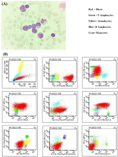
Mixed-phenotype acute leukemias (MPAL) account for < 4% of all cases of acute leukemias. These are a heterogeneous group of leukemias grouped together by the WHO classification as "rare subtypes." The diagnosis and treatment of MPAL is extremely challenging particularly for low middle income countries. Of these, B/myeloid and T/myeloid combinations are relatively common subtypes. However, megakaryoblastic and erythroid lineages in combination with other lineages are still rare enough to not even be addressed in the WHO classification. To date, there have been only a few reports of mixed B or T cell and megakaryocytic or mixed B or T cell and erythroid leukemias. We report the clinical presentation, diagnostic profile, and disease course of MPAL cases with a biphenotypic pattern consistent with T/megakaryoblastic lineage which is not yet defined in WHO classification. These cases were phenotyped using 8-color flow cytometry (BD FACS CANTO-II) using an extensive panel of markers. Interphase fluorescence in situ hybridization (FISH) was done using dual color dual fusion probes for BCR::ABL1, RUNX1::RUNX1T1, and ETV6::RUNX1, while MLL and CBFB gene rearrangement was tested by break-apart probes. Karyotyping was performed using the conventional GTG-banding technique. Both FISH and karyotyping were analyzed by the automated cell imaging system Leica Biosystems, using Cytovision MB8. The cases presented here satisfy the criteria for both T-lineage assignment (cyCD3 intensity reaches that of normal T-lymphocytes) and acute megakaryoblastic leukemia (≥ 1 megakaryocytic marker in > 50% blasts) and thus represent the first documented examples of this unusual entity from Pakistan. It is crucial to report these cases to gather more data about clinical presentation, diagnostic profile, and disease course. Additionally, the reported cases highlight the limitations of existing classifications which do not address rare subtypes. More importantly, T/megakaryoblastic MPAL needs to be included in the WHO classification as a separate entity.
期刊介绍:
The Journal of Hematopathology aims at providing pathologists with a special interest in hematopathology with all the information needed to perform modern pathology in evaluating lymphoid tissues and bone marrow. To this end the journal publishes reviews, editorials, comments, original papers, guidelines and protocols, papers on ancillary techniques, and occasional case reports in the fields of the pathology, molecular biology, and clinical features of diseases of the hematopoietic system.
The journal is the unique reference point for all pathologists with an interest in hematopathology. Molecular biologists involved in the expanding field of molecular diagnostics and research on lymphomas and leukemia benefit from the journal, too. Furthermore, the journal is of major interest for hematologists dealing with patients suffering from lymphomas, leukemias, and other diseases.
The journal is unique in its true international character. Especially in the field of hematopathology it is clear that there are huge geographical variations in incidence of diseases. This is not only locally relevant, but due to globalization, relevant for all those involved in the management of patients.

 求助内容:
求助内容: 应助结果提醒方式:


