Renato Ambrósio, Marcella Q Salomão, Lorena Barros, João Batista R da Fonseca Filho, Jaime Guedes, Alexandre Neto, Aydano P Machado, Bernardo T Lopes, Nelson Sena, Louise Pellegrino Gomes Esporcatte
{"title":"圆锥角膜和扩张性角膜疾病的多模式诊断:范式转变。","authors":"Renato Ambrósio, Marcella Q Salomão, Lorena Barros, João Batista R da Fonseca Filho, Jaime Guedes, Alexandre Neto, Aydano P Machado, Bernardo T Lopes, Nelson Sena, Louise Pellegrino Gomes Esporcatte","doi":"10.1186/s40662-023-00363-0","DOIUrl":null,"url":null,"abstract":"<p><p>Different diagnostic approaches for ectatic corneal diseases (ECD) include screening, diagnosis confirmation, classification of the ECD type, severity staging, prognostic evaluation, and clinical follow-up. The comprehensive assessment must start with a directed clinical history. However, multimodal imaging tools, including Placido-disk topography, Scheimpflug three-dimensional (3D) tomography, corneal biomechanical evaluations, and layered (or segmental) tomography with epithelial thickness by optical coherence tomography (OCT), or digital very high-frequency ultrasound (dVHF-US) serve as fundamental complementary exams for measuring different characteristics of the cornea. Also, ocular wavefront analysis, axial length measurements, corneal specular or confocal microscopy, and genetic or molecular biology tests are relevant for clinical decisions. Artificial intelligence enhances interpretation and enables combining such a plethora of data, boosting accuracy and facilitating clinical decisions. The applications of diagnostic information for individualized treatments became relevant concerning the therapeutic refractive procedures that emerged as alternatives to keratoplasty. The first paradigm shift concerns the surgical management of patients with ECD with different techniques, such as crosslinking and intrastromal corneal ring segments. A second paradigm shift involved the quest for identifying patients at higher risk of progressive iatrogenic ectasia after elective refractive corrections on the cornea. Beyond augmenting the sensitivity to detect very mild (subclinical or fruste) forms of ECD, ectasia risk assessment evolved to characterize the inherent susceptibility for ectasia development and progression. Furthermore, ectasia risk is also related to environmental factors, including eye rubbing and the relational impact of the surgical procedure on the cornea.</p>","PeriodicalId":12194,"journal":{"name":"Eye and Vision","volume":"10 1","pages":"45"},"PeriodicalIF":4.0000,"publicationDate":"2023-11-03","publicationTypes":"Journal Article","fieldsOfStudy":null,"isOpenAccess":false,"openAccessPdf":"https://www.ncbi.nlm.nih.gov/pmc/articles/PMC10623885/pdf/","citationCount":"0","resultStr":"{\"title\":\"Multimodal diagnostics for keratoconus and ectatic corneal diseases: a paradigm shift.\",\"authors\":\"Renato Ambrósio, Marcella Q Salomão, Lorena Barros, João Batista R da Fonseca Filho, Jaime Guedes, Alexandre Neto, Aydano P Machado, Bernardo T Lopes, Nelson Sena, Louise Pellegrino Gomes Esporcatte\",\"doi\":\"10.1186/s40662-023-00363-0\",\"DOIUrl\":null,\"url\":null,\"abstract\":\"<p><p>Different diagnostic approaches for ectatic corneal diseases (ECD) include screening, diagnosis confirmation, classification of the ECD type, severity staging, prognostic evaluation, and clinical follow-up. The comprehensive assessment must start with a directed clinical history. However, multimodal imaging tools, including Placido-disk topography, Scheimpflug three-dimensional (3D) tomography, corneal biomechanical evaluations, and layered (or segmental) tomography with epithelial thickness by optical coherence tomography (OCT), or digital very high-frequency ultrasound (dVHF-US) serve as fundamental complementary exams for measuring different characteristics of the cornea. Also, ocular wavefront analysis, axial length measurements, corneal specular or confocal microscopy, and genetic or molecular biology tests are relevant for clinical decisions. Artificial intelligence enhances interpretation and enables combining such a plethora of data, boosting accuracy and facilitating clinical decisions. The applications of diagnostic information for individualized treatments became relevant concerning the therapeutic refractive procedures that emerged as alternatives to keratoplasty. The first paradigm shift concerns the surgical management of patients with ECD with different techniques, such as crosslinking and intrastromal corneal ring segments. A second paradigm shift involved the quest for identifying patients at higher risk of progressive iatrogenic ectasia after elective refractive corrections on the cornea. Beyond augmenting the sensitivity to detect very mild (subclinical or fruste) forms of ECD, ectasia risk assessment evolved to characterize the inherent susceptibility for ectasia development and progression. Furthermore, ectasia risk is also related to environmental factors, including eye rubbing and the relational impact of the surgical procedure on the cornea.</p>\",\"PeriodicalId\":12194,\"journal\":{\"name\":\"Eye and Vision\",\"volume\":\"10 1\",\"pages\":\"45\"},\"PeriodicalIF\":4.0000,\"publicationDate\":\"2023-11-03\",\"publicationTypes\":\"Journal Article\",\"fieldsOfStudy\":null,\"isOpenAccess\":false,\"openAccessPdf\":\"https://www.ncbi.nlm.nih.gov/pmc/articles/PMC10623885/pdf/\",\"citationCount\":\"0\",\"resultStr\":null,\"platform\":\"Semanticscholar\",\"paperid\":null,\"PeriodicalName\":\"Eye and Vision\",\"FirstCategoryId\":\"3\",\"ListUrlMain\":\"https://doi.org/10.1186/s40662-023-00363-0\",\"RegionNum\":1,\"RegionCategory\":\"医学\",\"ArticlePicture\":[],\"TitleCN\":null,\"AbstractTextCN\":null,\"PMCID\":null,\"EPubDate\":\"\",\"PubModel\":\"\",\"JCR\":\"Q1\",\"JCRName\":\"OPHTHALMOLOGY\",\"Score\":null,\"Total\":0}","platform":"Semanticscholar","paperid":null,"PeriodicalName":"Eye and Vision","FirstCategoryId":"3","ListUrlMain":"https://doi.org/10.1186/s40662-023-00363-0","RegionNum":1,"RegionCategory":"医学","ArticlePicture":[],"TitleCN":null,"AbstractTextCN":null,"PMCID":null,"EPubDate":"","PubModel":"","JCR":"Q1","JCRName":"OPHTHALMOLOGY","Score":null,"Total":0}
Multimodal diagnostics for keratoconus and ectatic corneal diseases: a paradigm shift.
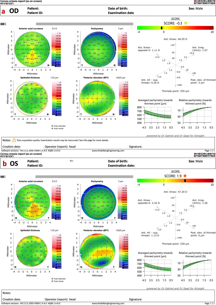
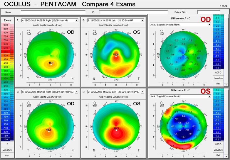
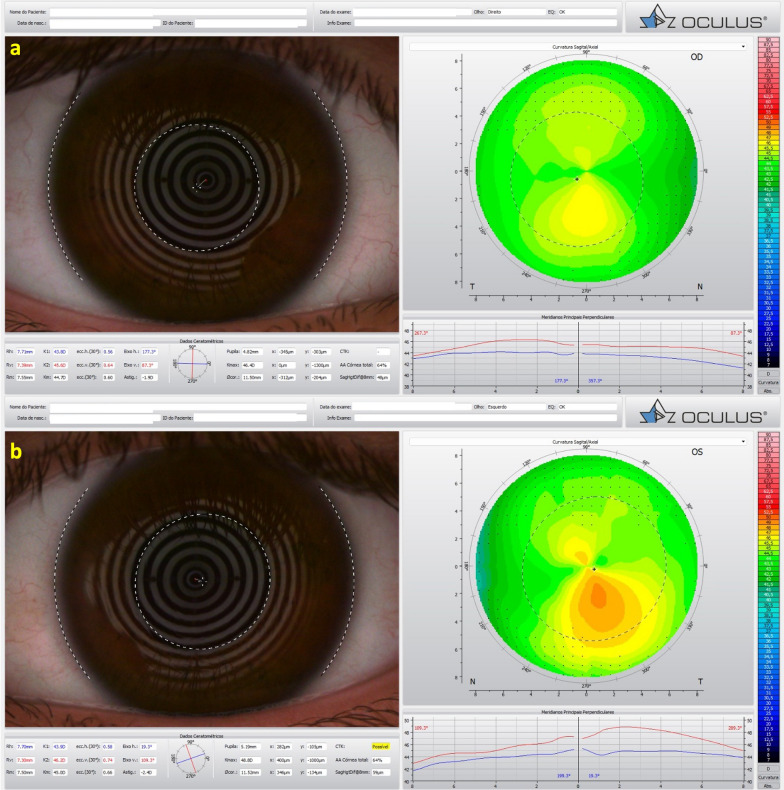
Different diagnostic approaches for ectatic corneal diseases (ECD) include screening, diagnosis confirmation, classification of the ECD type, severity staging, prognostic evaluation, and clinical follow-up. The comprehensive assessment must start with a directed clinical history. However, multimodal imaging tools, including Placido-disk topography, Scheimpflug three-dimensional (3D) tomography, corneal biomechanical evaluations, and layered (or segmental) tomography with epithelial thickness by optical coherence tomography (OCT), or digital very high-frequency ultrasound (dVHF-US) serve as fundamental complementary exams for measuring different characteristics of the cornea. Also, ocular wavefront analysis, axial length measurements, corneal specular or confocal microscopy, and genetic or molecular biology tests are relevant for clinical decisions. Artificial intelligence enhances interpretation and enables combining such a plethora of data, boosting accuracy and facilitating clinical decisions. The applications of diagnostic information for individualized treatments became relevant concerning the therapeutic refractive procedures that emerged as alternatives to keratoplasty. The first paradigm shift concerns the surgical management of patients with ECD with different techniques, such as crosslinking and intrastromal corneal ring segments. A second paradigm shift involved the quest for identifying patients at higher risk of progressive iatrogenic ectasia after elective refractive corrections on the cornea. Beyond augmenting the sensitivity to detect very mild (subclinical or fruste) forms of ECD, ectasia risk assessment evolved to characterize the inherent susceptibility for ectasia development and progression. Furthermore, ectasia risk is also related to environmental factors, including eye rubbing and the relational impact of the surgical procedure on the cornea.
期刊介绍:
Eye and Vision is an open access, peer-reviewed journal for ophthalmologists and visual science specialists. It welcomes research articles, reviews, methodologies, commentaries, case reports, perspectives and short reports encompassing all aspects of eye and vision. Topics of interest include but are not limited to: current developments of theoretical, experimental and clinical investigations in ophthalmology, optometry and vision science which focus on novel and high-impact findings on central issues pertaining to biology, pathophysiology and etiology of eye diseases as well as advances in diagnostic techniques, surgical treatment, instrument updates, the latest drug findings, results of clinical trials and research findings. It aims to provide ophthalmologists and visual science specialists with the latest developments in theoretical, experimental and clinical investigations in eye and vision.
 求助内容:
求助内容: 应助结果提醒方式:


