{"title":"使用多重灵敏度编码的前列腺高分辨率扩散加权成像:与传统和缩小视野技术的比较。","authors":"Atsushi Nakamoto, Hiromitsu Onishi, Takahiro Tsuboyama, Hideyuki Fukui, Takashi Ota, Keigo Yano, Kengo Kiso, Toru Honda, Hiroyuki Tarewaki, Yoshihiro Koyama, Mitsuaki Tatsumi, Noriyuki Tomiyama","doi":"10.2463/mrms.mp.2023-0039","DOIUrl":null,"url":null,"abstract":"<p><strong>Purpose: </strong>To compare objective and subjective image quality, lesion conspicuity, and apparent diffusion coefficient (ADC) of high-resolution multiplexed sensitivity-encoding diffusion-weighted imaging (MUSE-DWI) with conventional DWI (c-DWI) and reduced FOV DWI (rFOV-DWI) in prostate MRI.</p><p><strong>Methods: </strong>Forty-seven patients who underwent prostate MRI, including c-DWI, rFOV-DWI, and MUSE-DWI, were retrospectively evaluated. SNR and ADC of normal prostate tissue and contrast-to-noise ratio (CNR) and ADC of prostate cancer (PCa) were measured and compared between the three sequences. Image quality and lesion conspicuity were independently graded by two radiologists using a 5-point scale and compared between the three sequences.</p><p><strong>Results: </strong>The SNR of normal prostate tissue was significantly higher with rFOV-DWI than with the other two DWI techniques (P ≤ 0.01). The CNR of the PCa was significantly higher with rFOV-DWI than with MUSE-DWI (P < 0.05). The ADC of normal prostate tissue measured by rFOV-DWI was lower than that measured by MUSE-DWI and c-DWI (P < 0.01), while there was no difference in the ADC of cancers. In the qualitative analysis, MUSE-DWI showed significantly higher scores than rFOV-DWI and c-DWI for visibility of anatomy and overall image quality in both readers, and significantly higher scores for distortion in one of the two readers (P < 0.001). There was no difference in lesion conspicuity between the three sequences.</p><p><strong>Conclusion: </strong>High-resolution MUSE-DWI showed higher image quality and reduced distortion compared to c-DWI, while maintaining a wide FOV and similar ADC quantification, although no difference in lesion conspicuity was observed.</p>","PeriodicalId":94126,"journal":{"name":"Magnetic resonance in medical sciences : MRMS : an official journal of Japan Society of Magnetic Resonance in Medicine","volume":" ","pages":"58-65"},"PeriodicalIF":3.2000,"publicationDate":"2025-01-01","publicationTypes":"Journal Article","fieldsOfStudy":null,"isOpenAccess":false,"openAccessPdf":"https://www.ncbi.nlm.nih.gov/pmc/articles/PMC11733513/pdf/","citationCount":"0","resultStr":"{\"title\":\"High-resolution Diffusion-weighted Imaging of the Prostate Using Multiplexed Sensitivity-encoding: Comparison with the Conventional and Reduced Field-of-view Techniques.\",\"authors\":\"Atsushi Nakamoto, Hiromitsu Onishi, Takahiro Tsuboyama, Hideyuki Fukui, Takashi Ota, Keigo Yano, Kengo Kiso, Toru Honda, Hiroyuki Tarewaki, Yoshihiro Koyama, Mitsuaki Tatsumi, Noriyuki Tomiyama\",\"doi\":\"10.2463/mrms.mp.2023-0039\",\"DOIUrl\":null,\"url\":null,\"abstract\":\"<p><strong>Purpose: </strong>To compare objective and subjective image quality, lesion conspicuity, and apparent diffusion coefficient (ADC) of high-resolution multiplexed sensitivity-encoding diffusion-weighted imaging (MUSE-DWI) with conventional DWI (c-DWI) and reduced FOV DWI (rFOV-DWI) in prostate MRI.</p><p><strong>Methods: </strong>Forty-seven patients who underwent prostate MRI, including c-DWI, rFOV-DWI, and MUSE-DWI, were retrospectively evaluated. SNR and ADC of normal prostate tissue and contrast-to-noise ratio (CNR) and ADC of prostate cancer (PCa) were measured and compared between the three sequences. Image quality and lesion conspicuity were independently graded by two radiologists using a 5-point scale and compared between the three sequences.</p><p><strong>Results: </strong>The SNR of normal prostate tissue was significantly higher with rFOV-DWI than with the other two DWI techniques (P ≤ 0.01). The CNR of the PCa was significantly higher with rFOV-DWI than with MUSE-DWI (P < 0.05). The ADC of normal prostate tissue measured by rFOV-DWI was lower than that measured by MUSE-DWI and c-DWI (P < 0.01), while there was no difference in the ADC of cancers. In the qualitative analysis, MUSE-DWI showed significantly higher scores than rFOV-DWI and c-DWI for visibility of anatomy and overall image quality in both readers, and significantly higher scores for distortion in one of the two readers (P < 0.001). There was no difference in lesion conspicuity between the three sequences.</p><p><strong>Conclusion: </strong>High-resolution MUSE-DWI showed higher image quality and reduced distortion compared to c-DWI, while maintaining a wide FOV and similar ADC quantification, although no difference in lesion conspicuity was observed.</p>\",\"PeriodicalId\":94126,\"journal\":{\"name\":\"Magnetic resonance in medical sciences : MRMS : an official journal of Japan Society of Magnetic Resonance in Medicine\",\"volume\":\" \",\"pages\":\"58-65\"},\"PeriodicalIF\":3.2000,\"publicationDate\":\"2025-01-01\",\"publicationTypes\":\"Journal Article\",\"fieldsOfStudy\":null,\"isOpenAccess\":false,\"openAccessPdf\":\"https://www.ncbi.nlm.nih.gov/pmc/articles/PMC11733513/pdf/\",\"citationCount\":\"0\",\"resultStr\":null,\"platform\":\"Semanticscholar\",\"paperid\":null,\"PeriodicalName\":\"Magnetic resonance in medical sciences : MRMS : an official journal of Japan Society of Magnetic Resonance in Medicine\",\"FirstCategoryId\":\"1085\",\"ListUrlMain\":\"https://doi.org/10.2463/mrms.mp.2023-0039\",\"RegionNum\":0,\"RegionCategory\":null,\"ArticlePicture\":[],\"TitleCN\":null,\"AbstractTextCN\":null,\"PMCID\":null,\"EPubDate\":\"2023/10/28 0:00:00\",\"PubModel\":\"Epub\",\"JCR\":\"\",\"JCRName\":\"\",\"Score\":null,\"Total\":0}","platform":"Semanticscholar","paperid":null,"PeriodicalName":"Magnetic resonance in medical sciences : MRMS : an official journal of Japan Society of Magnetic Resonance in Medicine","FirstCategoryId":"1085","ListUrlMain":"https://doi.org/10.2463/mrms.mp.2023-0039","RegionNum":0,"RegionCategory":null,"ArticlePicture":[],"TitleCN":null,"AbstractTextCN":null,"PMCID":null,"EPubDate":"2023/10/28 0:00:00","PubModel":"Epub","JCR":"","JCRName":"","Score":null,"Total":0}
High-resolution Diffusion-weighted Imaging of the Prostate Using Multiplexed Sensitivity-encoding: Comparison with the Conventional and Reduced Field-of-view Techniques.
Purpose: To compare objective and subjective image quality, lesion conspicuity, and apparent diffusion coefficient (ADC) of high-resolution multiplexed sensitivity-encoding diffusion-weighted imaging (MUSE-DWI) with conventional DWI (c-DWI) and reduced FOV DWI (rFOV-DWI) in prostate MRI.
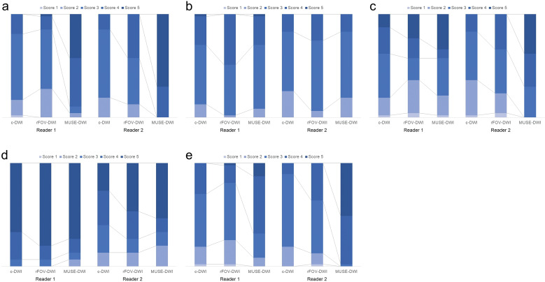
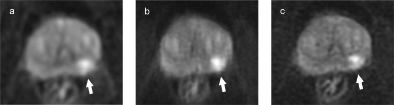
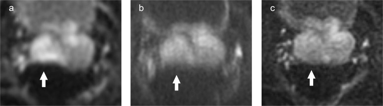
Methods: Forty-seven patients who underwent prostate MRI, including c-DWI, rFOV-DWI, and MUSE-DWI, were retrospectively evaluated. SNR and ADC of normal prostate tissue and contrast-to-noise ratio (CNR) and ADC of prostate cancer (PCa) were measured and compared between the three sequences. Image quality and lesion conspicuity were independently graded by two radiologists using a 5-point scale and compared between the three sequences.
Results: The SNR of normal prostate tissue was significantly higher with rFOV-DWI than with the other two DWI techniques (P ≤ 0.01). The CNR of the PCa was significantly higher with rFOV-DWI than with MUSE-DWI (P < 0.05). The ADC of normal prostate tissue measured by rFOV-DWI was lower than that measured by MUSE-DWI and c-DWI (P < 0.01), while there was no difference in the ADC of cancers. In the qualitative analysis, MUSE-DWI showed significantly higher scores than rFOV-DWI and c-DWI for visibility of anatomy and overall image quality in both readers, and significantly higher scores for distortion in one of the two readers (P < 0.001). There was no difference in lesion conspicuity between the three sequences.
Conclusion: High-resolution MUSE-DWI showed higher image quality and reduced distortion compared to c-DWI, while maintaining a wide FOV and similar ADC quantification, although no difference in lesion conspicuity was observed.

 求助内容:
求助内容: 应助结果提醒方式:


