阑尾粘液囊肿:特别强调影像研究

Q4 Medicine
C. Chiang, Kuang-Yang Lin, Chaur‐Shine Wang, Yi-Chang Chen, Meng‐Dar Lee, Hsueh-Chin Huang, Chin-Tai Huang, H. Tu, Chung‐kwe Wang, Jean-Dean Li
{"title":"阑尾粘液囊肿:特别强调影像研究","authors":"C. Chiang, Kuang-Yang Lin, Chaur‐Shine Wang, Yi-Chang Chen, Meng‐Dar Lee, Hsueh-Chin Huang, Chin-Tai Huang, H. Tu, Chung‐kwe Wang, Jean-Dean Li","doi":"10.6557/GJTA.199009_7(3).0003","DOIUrl":null,"url":null,"abstract":"In a review of 1,268 appendectomies from January 1984 to April 1989, ten cases were diagnosed as ”mucocele” by pathology. All these cases presented with a right lower quadrant (RLQ) mass or pain of variable duration except one whose mucocele was found incidentally during an operation for gastric cancer. One case revealed curvilinear and mottled calcification outlining a large mucocele on plain film of the abdomen. Abdominal ultrasonography in 7 patients showed negative finding in one case, a hypoechoic or anechoic tubular lesion with good transmission in two, an ovoid cystic lesion with variable internal echoes in dependent portion in two, intussusception in one and a periappendiceal abscess in one. Barium enema of 4 patients showed nonvisualization of the appendix but either filling defects with round smooth shape or external compression to the cecum were found. Fibercolonoscopy was performed in 4 cases, all were disclosed intrusions of mucocele from the appendiceal orifice. In one of them, the mucocele invaginated deeply and caused chronic appendico-cecal intussusception. Computed tomography was performed in one case, the mucocele showed a near-water-density tubular lesion with good enhancement of the wall after contrast injection. Though appendiceal mucocele is a rare clinical entity, it should be in the list of differential diagnosis of RLQ pain. With the disease in mind and advanced imaging modalities available, preoperative diagnosis is probable.","PeriodicalId":10003,"journal":{"name":"胃肠病学","volume":"7 1","pages":"122-131"},"PeriodicalIF":0.0000,"publicationDate":"1990-09-01","publicationTypes":"Journal Article","fieldsOfStudy":null,"isOpenAccess":false,"openAccessPdf":"","citationCount":"0","resultStr":"{\"title\":\"Mucocele of the Appendix: With Special Emphasis on Image Studies\",\"authors\":\"C. Chiang, Kuang-Yang Lin, Chaur‐Shine Wang, Yi-Chang Chen, Meng‐Dar Lee, Hsueh-Chin Huang, Chin-Tai Huang, H. Tu, Chung‐kwe Wang, Jean-Dean Li\",\"doi\":\"10.6557/GJTA.199009_7(3).0003\",\"DOIUrl\":null,\"url\":null,\"abstract\":\"In a review of 1,268 appendectomies from January 1984 to April 1989, ten cases were diagnosed as ”mucocele” by pathology. All these cases presented with a right lower quadrant (RLQ) mass or pain of variable duration except one whose mucocele was found incidentally during an operation for gastric cancer. One case revealed curvilinear and mottled calcification outlining a large mucocele on plain film of the abdomen. Abdominal ultrasonography in 7 patients showed negative finding in one case, a hypoechoic or anechoic tubular lesion with good transmission in two, an ovoid cystic lesion with variable internal echoes in dependent portion in two, intussusception in one and a periappendiceal abscess in one. Barium enema of 4 patients showed nonvisualization of the appendix but either filling defects with round smooth shape or external compression to the cecum were found. Fibercolonoscopy was performed in 4 cases, all were disclosed intrusions of mucocele from the appendiceal orifice. In one of them, the mucocele invaginated deeply and caused chronic appendico-cecal intussusception. Computed tomography was performed in one case, the mucocele showed a near-water-density tubular lesion with good enhancement of the wall after contrast injection. Though appendiceal mucocele is a rare clinical entity, it should be in the list of differential diagnosis of RLQ pain. With the disease in mind and advanced imaging modalities available, preoperative diagnosis is probable.\",\"PeriodicalId\":10003,\"journal\":{\"name\":\"胃肠病学\",\"volume\":\"7 1\",\"pages\":\"122-131\"},\"PeriodicalIF\":0.0000,\"publicationDate\":\"1990-09-01\",\"publicationTypes\":\"Journal Article\",\"fieldsOfStudy\":null,\"isOpenAccess\":false,\"openAccessPdf\":\"\",\"citationCount\":\"0\",\"resultStr\":null,\"platform\":\"Semanticscholar\",\"paperid\":null,\"PeriodicalName\":\"胃肠病学\",\"FirstCategoryId\":\"3\",\"ListUrlMain\":\"https://doi.org/10.6557/GJTA.199009_7(3).0003\",\"RegionNum\":0,\"RegionCategory\":null,\"ArticlePicture\":[],\"TitleCN\":null,\"AbstractTextCN\":null,\"PMCID\":null,\"EPubDate\":\"\",\"PubModel\":\"\",\"JCR\":\"Q4\",\"JCRName\":\"Medicine\",\"Score\":null,\"Total\":0}","platform":"Semanticscholar","paperid":null,"PeriodicalName":"胃肠病学","FirstCategoryId":"3","ListUrlMain":"https://doi.org/10.6557/GJTA.199009_7(3).0003","RegionNum":0,"RegionCategory":null,"ArticlePicture":[],"TitleCN":null,"AbstractTextCN":null,"PMCID":null,"EPubDate":"","PubModel":"","JCR":"Q4","JCRName":"Medicine","Score":null,"Total":0}
引用次数: 0

摘要

本文回顾了1984年1月至1989年4月的1268例阑尾切除术,其中10例经病理诊断为粘液囊肿。所有病例均表现为右下腹(RLQ)肿块或持续时间不等的疼痛,除了一例在胃癌手术中偶然发现粘液囊肿。1例腹部平片示曲线状斑驳钙化,示大粘液囊肿。7例患者腹部超声1例为阴性,2例为低回声或无回声管状病变,透射良好,2例为卵球形囊状病变,依赖部分内回声变化,1例为肠套叠,1例为阑尾周围脓肿。4例患者行钡剂灌肠均未见阑尾,但均可见圆形平滑填充缺损或外压迫盲肠。4例患者行纤维结肠镜检查,均发现阑尾口粘液囊肿侵入。其中一例粘液囊肿深度内陷,引起慢性阑尾-盲肠套叠。1例行计算机断层扫描,粘液囊肿显示近水密度管状病变,注射造影剂后壁增强良好。虽然阑尾粘液囊肿是一种罕见的临床疾病,但它应该被列入RLQ疼痛的鉴别诊断清单。考虑到疾病和先进的影像学手段,术前诊断是可能的。
本文章由计算机程序翻译,如有差异,请以英文原文为准。
Mucocele of the Appendix: With Special Emphasis on Image Studies
In a review of 1,268 appendectomies from January 1984 to April 1989, ten cases were diagnosed as ”mucocele” by pathology. All these cases presented with a right lower quadrant (RLQ) mass or pain of variable duration except one whose mucocele was found incidentally during an operation for gastric cancer. One case revealed curvilinear and mottled calcification outlining a large mucocele on plain film of the abdomen. Abdominal ultrasonography in 7 patients showed negative finding in one case, a hypoechoic or anechoic tubular lesion with good transmission in two, an ovoid cystic lesion with variable internal echoes in dependent portion in two, intussusception in one and a periappendiceal abscess in one. Barium enema of 4 patients showed nonvisualization of the appendix but either filling defects with round smooth shape or external compression to the cecum were found. Fibercolonoscopy was performed in 4 cases, all were disclosed intrusions of mucocele from the appendiceal orifice. In one of them, the mucocele invaginated deeply and caused chronic appendico-cecal intussusception. Computed tomography was performed in one case, the mucocele showed a near-water-density tubular lesion with good enhancement of the wall after contrast injection. Though appendiceal mucocele is a rare clinical entity, it should be in the list of differential diagnosis of RLQ pain. With the disease in mind and advanced imaging modalities available, preoperative diagnosis is probable.
求助全文
通过发布文献求助,成功后即可免费获取论文全文。 去求助
来源期刊
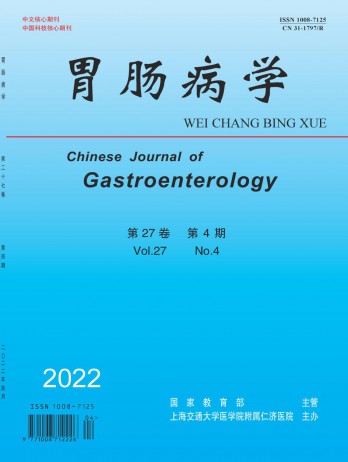
胃肠病学
胃肠病学 Medicine-Gastroenterology
CiteScore
0.10
自引率
0.00%
发文量
4567
期刊介绍: Gastroenterology is an academic journal sponsored by Shanghai Jiao Tong University School of Medicine. It mainly publishes original research papers, reviews and comments in this field. The journal was founded in 1996 and is included in well-known databases such as Peking University Journal (Chinese Journal of Humanities and Social Sciences) and Statistical Source Journal (China's Excellent Science and Technology Papers Journal). It is one of the national key academic journals under the jurisdiction of the Ministry of Education. Gastroenterology enjoys a high reputation and influence in the academic community. The articles published in this journal have a high academic level and practical value, providing readers with more actual cases and industry information, and have received widespread attention and citations from readers.
×
引用
GB/T 7714-2015
复制
MLA
复制
APA
复制
导出至
BibTeX EndNote RefMan NoteFirst NoteExpress
×
提示
您的信息不完整,为了账户安全,请先补充。
现在去补充
×
提示
您因"违规操作"
具体请查看互助需知
我知道了
×
提示
确定
请完成安全验证×
copy
已复制链接
快去分享给好友吧!
我知道了
右上角分享
点击右上角分享
0
联系我们:info@booksci.cn Book学术提供免费学术资源搜索服务,方便国内外学者检索中英文文献。致力于提供最便捷和优质的服务体验。 Copyright © 2023 布克学术 All rights reserved.
京ICP备2023020795号-1
ghs 京公网安备 11010802042870号
Book学术文献互助
Book学术文献互助群
群 号:481959085
Book学术官方微信