{"title":"年轻女性乳房错构瘤:放射病理相关性。","authors":"Ravikanth Reddy","doi":"10.4103/jmau.jmau_142_20","DOIUrl":null,"url":null,"abstract":"<p><p>Hamartoma of the breast is a rare benign lesion that leads to unilateral breast enlargement with evidence of a localized palpable mass. Ultrasonography findings are typical and include a well-defined mass lesion of heterogeneous echotexture consisting of mixed echogenic and sonolucent areas. This case report describes a hamartoma of the breast in a 17-year-old female.</p>","PeriodicalId":16340,"journal":{"name":"Journal of Microscopy and Ultrastructure","volume":"1 1","pages":"52-53"},"PeriodicalIF":0.0000,"publicationDate":"2023-02-07","publicationTypes":"Journal Article","fieldsOfStudy":null,"isOpenAccess":false,"openAccessPdf":"https://www.ncbi.nlm.nih.gov/pmc/articles/PMC12063920/pdf/","citationCount":"0","resultStr":"{\"title\":\"Hamartoma of the Breast in a Young Female: Radio-Pathological Correlation.\",\"authors\":\"Ravikanth Reddy\",\"doi\":\"10.4103/jmau.jmau_142_20\",\"DOIUrl\":null,\"url\":null,\"abstract\":\"<p><p>Hamartoma of the breast is a rare benign lesion that leads to unilateral breast enlargement with evidence of a localized palpable mass. Ultrasonography findings are typical and include a well-defined mass lesion of heterogeneous echotexture consisting of mixed echogenic and sonolucent areas. This case report describes a hamartoma of the breast in a 17-year-old female.</p>\",\"PeriodicalId\":16340,\"journal\":{\"name\":\"Journal of Microscopy and Ultrastructure\",\"volume\":\"1 1\",\"pages\":\"52-53\"},\"PeriodicalIF\":0.0000,\"publicationDate\":\"2023-02-07\",\"publicationTypes\":\"Journal Article\",\"fieldsOfStudy\":null,\"isOpenAccess\":false,\"openAccessPdf\":\"https://www.ncbi.nlm.nih.gov/pmc/articles/PMC12063920/pdf/\",\"citationCount\":\"0\",\"resultStr\":null,\"platform\":\"Semanticscholar\",\"paperid\":null,\"PeriodicalName\":\"Journal of Microscopy and Ultrastructure\",\"FirstCategoryId\":\"1085\",\"ListUrlMain\":\"https://doi.org/10.4103/jmau.jmau_142_20\",\"RegionNum\":0,\"RegionCategory\":null,\"ArticlePicture\":[],\"TitleCN\":null,\"AbstractTextCN\":null,\"PMCID\":null,\"EPubDate\":\"2025/1/1 0:00:00\",\"PubModel\":\"eCollection\",\"JCR\":\"Q3\",\"JCRName\":\"Medicine\",\"Score\":null,\"Total\":0}","platform":"Semanticscholar","paperid":null,"PeriodicalName":"Journal of Microscopy and Ultrastructure","FirstCategoryId":"1085","ListUrlMain":"https://doi.org/10.4103/jmau.jmau_142_20","RegionNum":0,"RegionCategory":null,"ArticlePicture":[],"TitleCN":null,"AbstractTextCN":null,"PMCID":null,"EPubDate":"2025/1/1 0:00:00","PubModel":"eCollection","JCR":"Q3","JCRName":"Medicine","Score":null,"Total":0}
Hamartoma of the Breast in a Young Female: Radio-Pathological Correlation.
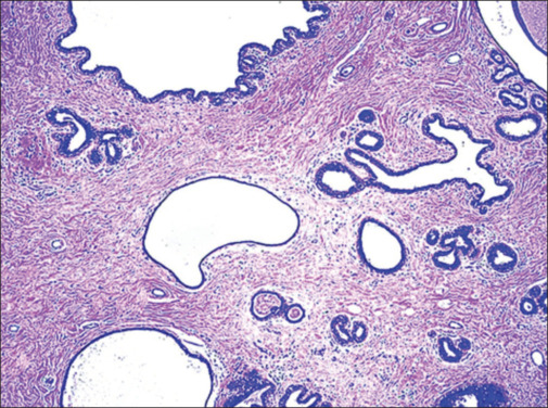
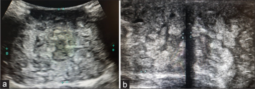
Hamartoma of the breast is a rare benign lesion that leads to unilateral breast enlargement with evidence of a localized palpable mass. Ultrasonography findings are typical and include a well-defined mass lesion of heterogeneous echotexture consisting of mixed echogenic and sonolucent areas. This case report describes a hamartoma of the breast in a 17-year-old female.

 求助内容:
求助内容: 应助结果提醒方式:


