{"title":"大口黑鲈(Micropterus salmoides, Centrarchidae)嗅器官解剖、超微结构和组织学","authors":"Hyun Tae Kim, Seung Woon Yun, Jong Young Park","doi":"10.1186/s42649-019-0023-3","DOIUrl":null,"url":null,"abstract":"<p>The detailed anatomy, ultrastructure and histology of the olfactory organ of <i>Micropterus salmoides</i> were investigated by a stereo microscope, a light microscope, and a scanning electron microscope. Its external structure shows a tube-like anterior nostril to stick out and a posterior nostril flat to the skin surface. Meanwhile, its internal structure, the olfactory chamber, contains a fan-shaped rosette structure with 9 to 11 lamellae in adult fish over 35?cm in standard length (SL) and two accessory nasal sacs (ethmoidal and lacrimal sacs) were found. Interestingly, the rosette in young fish under 15?cm in SL was a longitudinal structure in parallel with each of 4–5 lamellae. Histologically, the sensory epithelium (SE) on the olfactory chamber consists of 5 types of cells: olfactory receptor neurons, supporting cells, basal cells, lymphatic cells and mucous cells. In contrast, the non-sensory epithelium (NSE) has stratified epithelial cells, lymphatic cells and mucous cells. The mucous cells of the SE are abundant and distributed densely in one row on the outermost superficial surface, but the one of the NSE are less than the SE. From these results, the olfactory characters of <i>M. salmoides</i> may be related with its ecological habit spending in the middle layer of stagnant water contaminated, more or less.</p>","PeriodicalId":470,"journal":{"name":"Applied Microscopy","volume":"49 1","pages":""},"PeriodicalIF":0.0000,"publicationDate":"2019-12-23","publicationTypes":"Journal Article","fieldsOfStudy":null,"isOpenAccess":false,"openAccessPdf":"https://sci-hub-pdf.com/10.1186/s42649-019-0023-3","citationCount":"2","resultStr":"{\"title\":\"Anatomy, ultrastructure and histology of the olfactory organ of the largemouth bass Micropterus salmoides, Centrarchidae\",\"authors\":\"Hyun Tae Kim, Seung Woon Yun, Jong Young Park\",\"doi\":\"10.1186/s42649-019-0023-3\",\"DOIUrl\":null,\"url\":null,\"abstract\":\"<p>The detailed anatomy, ultrastructure and histology of the olfactory organ of <i>Micropterus salmoides</i> were investigated by a stereo microscope, a light microscope, and a scanning electron microscope. Its external structure shows a tube-like anterior nostril to stick out and a posterior nostril flat to the skin surface. Meanwhile, its internal structure, the olfactory chamber, contains a fan-shaped rosette structure with 9 to 11 lamellae in adult fish over 35?cm in standard length (SL) and two accessory nasal sacs (ethmoidal and lacrimal sacs) were found. Interestingly, the rosette in young fish under 15?cm in SL was a longitudinal structure in parallel with each of 4–5 lamellae. Histologically, the sensory epithelium (SE) on the olfactory chamber consists of 5 types of cells: olfactory receptor neurons, supporting cells, basal cells, lymphatic cells and mucous cells. In contrast, the non-sensory epithelium (NSE) has stratified epithelial cells, lymphatic cells and mucous cells. The mucous cells of the SE are abundant and distributed densely in one row on the outermost superficial surface, but the one of the NSE are less than the SE. From these results, the olfactory characters of <i>M. salmoides</i> may be related with its ecological habit spending in the middle layer of stagnant water contaminated, more or less.</p>\",\"PeriodicalId\":470,\"journal\":{\"name\":\"Applied Microscopy\",\"volume\":\"49 1\",\"pages\":\"\"},\"PeriodicalIF\":0.0000,\"publicationDate\":\"2019-12-23\",\"publicationTypes\":\"Journal Article\",\"fieldsOfStudy\":null,\"isOpenAccess\":false,\"openAccessPdf\":\"https://sci-hub-pdf.com/10.1186/s42649-019-0023-3\",\"citationCount\":\"2\",\"resultStr\":null,\"platform\":\"Semanticscholar\",\"paperid\":null,\"PeriodicalName\":\"Applied Microscopy\",\"FirstCategoryId\":\"1085\",\"ListUrlMain\":\"https://link.springer.com/article/10.1186/s42649-019-0023-3\",\"RegionNum\":0,\"RegionCategory\":null,\"ArticlePicture\":[],\"TitleCN\":null,\"AbstractTextCN\":null,\"PMCID\":null,\"EPubDate\":\"\",\"PubModel\":\"\",\"JCR\":\"Q3\",\"JCRName\":\"Immunology and Microbiology\",\"Score\":null,\"Total\":0}","platform":"Semanticscholar","paperid":null,"PeriodicalName":"Applied Microscopy","FirstCategoryId":"1085","ListUrlMain":"https://link.springer.com/article/10.1186/s42649-019-0023-3","RegionNum":0,"RegionCategory":null,"ArticlePicture":[],"TitleCN":null,"AbstractTextCN":null,"PMCID":null,"EPubDate":"","PubModel":"","JCR":"Q3","JCRName":"Immunology and Microbiology","Score":null,"Total":0}
Anatomy, ultrastructure and histology of the olfactory organ of the largemouth bass Micropterus salmoides, Centrarchidae
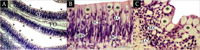
The detailed anatomy, ultrastructure and histology of the olfactory organ of Micropterus salmoides were investigated by a stereo microscope, a light microscope, and a scanning electron microscope. Its external structure shows a tube-like anterior nostril to stick out and a posterior nostril flat to the skin surface. Meanwhile, its internal structure, the olfactory chamber, contains a fan-shaped rosette structure with 9 to 11 lamellae in adult fish over 35?cm in standard length (SL) and two accessory nasal sacs (ethmoidal and lacrimal sacs) were found. Interestingly, the rosette in young fish under 15?cm in SL was a longitudinal structure in parallel with each of 4–5 lamellae. Histologically, the sensory epithelium (SE) on the olfactory chamber consists of 5 types of cells: olfactory receptor neurons, supporting cells, basal cells, lymphatic cells and mucous cells. In contrast, the non-sensory epithelium (NSE) has stratified epithelial cells, lymphatic cells and mucous cells. The mucous cells of the SE are abundant and distributed densely in one row on the outermost superficial surface, but the one of the NSE are less than the SE. From these results, the olfactory characters of M. salmoides may be related with its ecological habit spending in the middle layer of stagnant water contaminated, more or less.
Applied MicroscopyImmunology and Microbiology-Applied Microbiology and Biotechnology
CiteScore
3.40
自引率
0.00%
发文量
10
审稿时长
10 weeks
期刊介绍:
Applied Microscopy is a peer-reviewed journal sponsored by the Korean Society of Microscopy. The journal covers all the interdisciplinary fields of technological developments in new microscopy methods and instrumentation and their applications to biological or materials science for determining structure and chemistry. ISSN: 22875123, 22874445.
 求助内容:
求助内容: 应助结果提醒方式:


