Shahnaz Akil, Anna E. Székely, Fredrik Hedeer, Berit Olsson, Henrik Engblom, Cecilia Hindorf
{"title":"不同时间框架、重建算法和后处理方法对13N-NH3 PET图像心肌血流定量的影响。","authors":"Shahnaz Akil, Anna E. Székely, Fredrik Hedeer, Berit Olsson, Henrik Engblom, Cecilia Hindorf","doi":"10.1111/cpf.12861","DOIUrl":null,"url":null,"abstract":"<div>\n \n \n <section>\n \n <h3> Background</h3>\n \n <p>The aim was to investigate to what extent the quantification of myocardial blood flow (MBF) from dynamic <sup>13</sup>N-NH<sub>3</sub> positron emission tomography (PET) images is affected by time frame schemes, time-of-flight (ToF), reconstruction algorithms, blood pool volume of interest (VOI) locations and compartment models in patients with suspected chronic coronary syndrome.</p>\n </section>\n \n <section>\n \n <h3> Methods</h3>\n \n <p>A standard MBF value was determined from 25 patients' rest/stress <sup>13</sup>N-NH<sub>3</sub> PET/CT images reconstructed with ordered subset expectation maximization (OSEM), 5 s time frame for the first frames without ToF, subsequently analyzed using a basal VOI and the deGrado compartment model. MBFs calculated using 2 or 10 s for the first frames, ToF, block-sequential regularized expectation maximization (BSREM), apical or large VOI, Hutchins or Krivokapich compartment models were compared to MBF<sub>standard</sub> in Bland–Altman plots (bias ± SD).</p>\n </section>\n \n <section>\n \n <h3> Results</h3>\n \n <p>Good agreement in global rest/stress MBF (mL/min/g) was found when changing the time frame scheme or reconstruction algorithm (MBF<sub>standard</sub> vs. MBF<sub>2s</sub>: −0.02 ± 0.06; MBF<sub>10s</sub>: 0.01 ± 0.07; MBF<sub>BSREM</sub>: 0.01 ± 0.07), while a lower level of agreement was found when altering the other factors (MBF<sub>standard</sub> vs. MBF<sub>ToF</sub>: −0.07 ± 0.10; MBF<sub>apical VOI</sub>: −0.27 ± 0.25; MBF<sub>large VOI</sub>: −0.11 ± 0.10; MBF<sub>Hutchins</sub>: −0.08 ± 0.10; MBF<sub>Krivokapich</sub>: −0.47 ± 0.50).</p>\n </section>\n \n <section>\n \n <h3> Conclusions</h3>\n \n <p>Quantification of MBF from <sup>13</sup>N-NH<sub>3</sub> PET images is more affected by choice of compartment models, ToF and blood pool VOIs than by different time frame schemes and reconstruction algorithms.</p>\n </section>\n </div>","PeriodicalId":10504,"journal":{"name":"Clinical Physiology and Functional Imaging","volume":"44 2","pages":"154-163"},"PeriodicalIF":1.3000,"publicationDate":"2023-10-26","publicationTypes":"Journal Article","fieldsOfStudy":null,"isOpenAccess":false,"openAccessPdf":"https://onlinelibrary.wiley.com/doi/epdf/10.1111/cpf.12861","citationCount":"0","resultStr":"{\"title\":\"Influence of different time framings, reconstruction algorithms and post-processing methods on the quantification of myocardial blood flow from 13N-NH3 PET images\",\"authors\":\"Shahnaz Akil, Anna E. Székely, Fredrik Hedeer, Berit Olsson, Henrik Engblom, Cecilia Hindorf\",\"doi\":\"10.1111/cpf.12861\",\"DOIUrl\":null,\"url\":null,\"abstract\":\"<div>\\n \\n \\n <section>\\n \\n <h3> Background</h3>\\n \\n <p>The aim was to investigate to what extent the quantification of myocardial blood flow (MBF) from dynamic <sup>13</sup>N-NH<sub>3</sub> positron emission tomography (PET) images is affected by time frame schemes, time-of-flight (ToF), reconstruction algorithms, blood pool volume of interest (VOI) locations and compartment models in patients with suspected chronic coronary syndrome.</p>\\n </section>\\n \\n <section>\\n \\n <h3> Methods</h3>\\n \\n <p>A standard MBF value was determined from 25 patients' rest/stress <sup>13</sup>N-NH<sub>3</sub> PET/CT images reconstructed with ordered subset expectation maximization (OSEM), 5 s time frame for the first frames without ToF, subsequently analyzed using a basal VOI and the deGrado compartment model. MBFs calculated using 2 or 10 s for the first frames, ToF, block-sequential regularized expectation maximization (BSREM), apical or large VOI, Hutchins or Krivokapich compartment models were compared to MBF<sub>standard</sub> in Bland–Altman plots (bias ± SD).</p>\\n </section>\\n \\n <section>\\n \\n <h3> Results</h3>\\n \\n <p>Good agreement in global rest/stress MBF (mL/min/g) was found when changing the time frame scheme or reconstruction algorithm (MBF<sub>standard</sub> vs. MBF<sub>2s</sub>: −0.02 ± 0.06; MBF<sub>10s</sub>: 0.01 ± 0.07; MBF<sub>BSREM</sub>: 0.01 ± 0.07), while a lower level of agreement was found when altering the other factors (MBF<sub>standard</sub> vs. MBF<sub>ToF</sub>: −0.07 ± 0.10; MBF<sub>apical VOI</sub>: −0.27 ± 0.25; MBF<sub>large VOI</sub>: −0.11 ± 0.10; MBF<sub>Hutchins</sub>: −0.08 ± 0.10; MBF<sub>Krivokapich</sub>: −0.47 ± 0.50).</p>\\n </section>\\n \\n <section>\\n \\n <h3> Conclusions</h3>\\n \\n <p>Quantification of MBF from <sup>13</sup>N-NH<sub>3</sub> PET images is more affected by choice of compartment models, ToF and blood pool VOIs than by different time frame schemes and reconstruction algorithms.</p>\\n </section>\\n </div>\",\"PeriodicalId\":10504,\"journal\":{\"name\":\"Clinical Physiology and Functional Imaging\",\"volume\":\"44 2\",\"pages\":\"154-163\"},\"PeriodicalIF\":1.3000,\"publicationDate\":\"2023-10-26\",\"publicationTypes\":\"Journal Article\",\"fieldsOfStudy\":null,\"isOpenAccess\":false,\"openAccessPdf\":\"https://onlinelibrary.wiley.com/doi/epdf/10.1111/cpf.12861\",\"citationCount\":\"0\",\"resultStr\":null,\"platform\":\"Semanticscholar\",\"paperid\":null,\"PeriodicalName\":\"Clinical Physiology and Functional Imaging\",\"FirstCategoryId\":\"3\",\"ListUrlMain\":\"https://onlinelibrary.wiley.com/doi/10.1111/cpf.12861\",\"RegionNum\":4,\"RegionCategory\":\"医学\",\"ArticlePicture\":[],\"TitleCN\":null,\"AbstractTextCN\":null,\"PMCID\":null,\"EPubDate\":\"\",\"PubModel\":\"\",\"JCR\":\"Q4\",\"JCRName\":\"PHYSIOLOGY\",\"Score\":null,\"Total\":0}","platform":"Semanticscholar","paperid":null,"PeriodicalName":"Clinical Physiology and Functional Imaging","FirstCategoryId":"3","ListUrlMain":"https://onlinelibrary.wiley.com/doi/10.1111/cpf.12861","RegionNum":4,"RegionCategory":"医学","ArticlePicture":[],"TitleCN":null,"AbstractTextCN":null,"PMCID":null,"EPubDate":"","PubModel":"","JCR":"Q4","JCRName":"PHYSIOLOGY","Score":null,"Total":0}
Influence of different time framings, reconstruction algorithms and post-processing methods on the quantification of myocardial blood flow from 13N-NH3 PET images
Background
The aim was to investigate to what extent the quantification of myocardial blood flow (MBF) from dynamic 13N-NH3 positron emission tomography (PET) images is affected by time frame schemes, time-of-flight (ToF), reconstruction algorithms, blood pool volume of interest (VOI) locations and compartment models in patients with suspected chronic coronary syndrome.
Methods
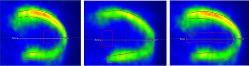
A standard MBF value was determined from 25 patients' rest/stress 13N-NH3 PET/CT images reconstructed with ordered subset expectation maximization (OSEM), 5 s time frame for the first frames without ToF, subsequently analyzed using a basal VOI and the deGrado compartment model. MBFs calculated using 2 or 10 s for the first frames, ToF, block-sequential regularized expectation maximization (BSREM), apical or large VOI, Hutchins or Krivokapich compartment models were compared to MBFstandard in Bland–Altman plots (bias ± SD).
Results
Good agreement in global rest/stress MBF (mL/min/g) was found when changing the time frame scheme or reconstruction algorithm (MBFstandard vs. MBF2s: −0.02 ± 0.06; MBF10s: 0.01 ± 0.07; MBFBSREM: 0.01 ± 0.07), while a lower level of agreement was found when altering the other factors (MBFstandard vs. MBFToF: −0.07 ± 0.10; MBFapical VOI: −0.27 ± 0.25; MBFlarge VOI: −0.11 ± 0.10; MBFHutchins: −0.08 ± 0.10; MBFKrivokapich: −0.47 ± 0.50).
Conclusions
Quantification of MBF from 13N-NH3 PET images is more affected by choice of compartment models, ToF and blood pool VOIs than by different time frame schemes and reconstruction algorithms.
期刊介绍:
Clinical Physiology and Functional Imaging publishes reports on clinical and experimental research pertinent to human physiology in health and disease. The scope of the Journal is very broad, covering all aspects of the regulatory system in the cardiovascular, renal and pulmonary systems with special emphasis on methodological aspects. The focus for the journal is, however, work that has potential clinical relevance. The Journal also features review articles on recent front-line research within these fields of interest.
Covered by the major abstracting services including Current Contents and Science Citation Index, Clinical Physiology and Functional Imaging plays an important role in providing effective and productive communication among clinical physiologists world-wide.

 求助内容:
求助内容: 应助结果提醒方式:


