{"title":"一种新的多组分纳米颗粒递送系统“SENT-seq”:支持mRNA疗法的发展","authors":"Xiaoshuang Song, Fang Nan, Dunfang Zhang","doi":"10.1002/mba2.50","DOIUrl":null,"url":null,"abstract":"<p>A publication in <i>Nature Nanotechnology</i> by James E. Dahlman et al. reported a novel screening technique for lipid nanoparticles (LNPs) delivery vectors called single-cell nanoparticle targeting-sequencing (SENT-seq).<span><sup>1</sup></span> This technology may be a significant leap forward in the realization of high-throughput screening of LNPs formulations, LNPs delivery mechanism research, and optimization of mRNA therapy.</p><p>mRNA is a transient carrier of genetic information. A wide range of diseases can be treated in clinical applications by delivering mRNA that can express infectious diseases or cancer antigens, gene-editing components, and disease-associated therapeutic proteins in the cells.<span><sup>2</sup></span> Effective mRNA therapy requires adequate cytoplasmic mRNA translation. Therefore, a series of delivery formulations have been developed to help mRNA cross multiple biological barriers and successfully enter the cytoplasm to fulfill its biological function. Among them, LNPs are the most extensively studied and clinically advanced mRNA vectors.<span><sup>3</sup></span> The formulation of LNPs include ionizable lipids (or cationic lipids), neutral auxiliary lipids, cholesterol, pegylated lipids, and nucleic acid molecules. How can the optimal delivery efficiency of nucleic acid molecules be achieved with LNPs? For example, research has screened LNP compositions with the best delivery efficiency in vitro by changing the formulation of LNP.<span><sup>4</sup></span> However, the results in vitro cannot summarize the results in vivo. In addition, the influence of different cell subsets on LNPs uptake during in vivo delivery has yet to be fully studied. Therefore, Dahlman et al. proposed a solution suitable for screening and examining the biological distribution of LNPs delivery in vivo, defining cells according to transcriptional states rather than cell surface markers, and analyzing the effects of cell subsets with different transcription states (heterogeneity) on LNPs uptake.<span><sup>1</sup></span></p><p>Dahlman et al. designed a multiomics NP delivery system, SENT-seq, to examine the effect of cell heterogeneity on LNPs delivery.<span><sup>1</sup></span> Using this technique, they were able to quantify the biodistribution (the number of LNPs entering cells), functional delivery (mRNA translated into functional proteins), and transcriptome level of cells. They used DNA barcoding technology to quantify LNPs entering the cell. They inserted different DNA sequences into different LNPs such that each LNP had a DNA barcode. The number of LNPs that entered a single cell was characterized by barcode readouts. However, one of the significant barriers in the intracellular delivery of nucleic acid molecules is that the nucleic acid molecules degrade in the endosomes, hence, the mRNA delivered into the cell does not necessarily express a functional protein.<span><sup>2</sup></span> Here, the expression of the mRNA functional aVHH protein was also detected. The expression of aVHH on the cell surface was quantified by single-cell sequencing using the cell-hashing technique. Interestingly, SENT-seq designed by the authors can simultaneously obtain barcode readouts, aVHH protein expression, and transcriptome levels in a single cell. Beads are packaged with carboxylic acids to attach nucleotide sequences through an amide reaction. Two capture sites were designed: one for capturing the LNP-carried DNA barcode and the other for capturing the cell hash oligonucleotide antibody and all the endogenous mRNAs (Figure 1).</p><p>After injecting 24 LNPs containing specific sequences of DNA barcode and the same mRNA into mice at the same time, the liver was isolated, single-cell suspensions were obtained and then mixed with modified beads. After single-cell sequencing, the delivery efficiency results of the LNPs barcode in single cells were obtained, and the expression of functional aVHH protein in each cell subset was obtained based on the localization of cell subsets using transcriptome analysis (Figure 1). The results showed that the 17 cell subsets in the mouse liver had different levels of barcode readouts, suggesting that cell heterogeneity affects the delivery efficiency of LNPs in vivo.</p><p>To explore the genes associated with LNPs different delivery efficiency, endothelial cells EC1, EC2, and EC3 with the largest significant differences in LNPs functional delivery were selected. To identify the genes related to delivery efficiency, the different genes between the cells that expressed aVHH protein were identified based on removing the background genes. Finally, 19 different expression genes were identified. At the same time, from the uptake of 24 LNPs in 17 cell subsets, LNP-12, which has the best mRNA delivery ability, was identified (Figure 1).</p><p>In conclusion, the single-cell nanoparticle targeted sequencing technology reported by James E. Dahlman et al. is based on cell transcriptomic characteristics to distinguish cell subsets rather than cell surface markers so that cells can be classified and analyzed more comprehensively, including rare cell subsets that cell surface markers cannot mark. In addition, this technique can efficiently quantify the delivery efficiency of LNPs, expression of functional proteins, and transcriptome level of single cells simultaneously by single-cell sequencing. Additionally, studying the mechanism of different uptake levels of LNPs by different cells based on the transcription level is helpful in guiding further and better-targeted application of LNPs. In addition, this technology can simultaneously test multiple LNPs, realize high-throughput LNPs delivery analysis in vivo, and contribute to the screening of new LNPs.</p><p>Currently, the target tissue of LNPs is mainly the liver. After intravenous injection, 30-99% of the LNPs eventually enter the liver and are absorbed by liver cells.<span><sup>5</sup></span> How to target nonliver tissue is a challenge. It was proven in this study that changing the formula of LNPs can affect the targeting of LNPs. Although this work only studied the effect of cell heterogeneity in the mouse liver on the uptake of different formulations of LNPs, this technique can also be used to study targeted LNPs in other tissues. By studying LNPs targeted in nonliver organs, we can determine the underlying mechanism and then design new LNPs according to the mechanism to further expand the application of LNPs and mRNA therapy. However, in this study, healthy mice were used to explore the effect of cell heterogeneity on genes related to LNPs delivery efficiency without considering the disease model. When this technology is further applied in LNPs screening studies, it should be considered to establish and explore disease models for further exploration. In conclusion, although there are some limitations to this study, studying the biological behavior of mRNA delivery with single-cell sequencing can be an effective way to accelerate the clinical transformation of mRNA therapeutic products.</p><p>Xiaoshuang Song drafted the manuscript. Xiaoshuang Song and Fang Nan painted the schematic diagram. Dunfang Zhang revised the manuscript. All authors have read and approved the final manuscript.</p><p>The authors declare no conflicts of interest.</p><p>Not applicable.</p>","PeriodicalId":100901,"journal":{"name":"MedComm – Biomaterials and Applications","volume":"2 2","pages":""},"PeriodicalIF":0.0000,"publicationDate":"2023-06-09","publicationTypes":"Journal Article","fieldsOfStudy":null,"isOpenAccess":false,"openAccessPdf":"https://onlinelibrary.wiley.com/doi/epdf/10.1002/mba2.50","citationCount":"0","resultStr":"{\"title\":\"A new multiomics nanoparticle delivery system “SENT-seq”: To support the development of mRNA therapies\",\"authors\":\"Xiaoshuang Song, Fang Nan, Dunfang Zhang\",\"doi\":\"10.1002/mba2.50\",\"DOIUrl\":null,\"url\":null,\"abstract\":\"<p>A publication in <i>Nature Nanotechnology</i> by James E. Dahlman et al. reported a novel screening technique for lipid nanoparticles (LNPs) delivery vectors called single-cell nanoparticle targeting-sequencing (SENT-seq).<span><sup>1</sup></span> This technology may be a significant leap forward in the realization of high-throughput screening of LNPs formulations, LNPs delivery mechanism research, and optimization of mRNA therapy.</p><p>mRNA is a transient carrier of genetic information. A wide range of diseases can be treated in clinical applications by delivering mRNA that can express infectious diseases or cancer antigens, gene-editing components, and disease-associated therapeutic proteins in the cells.<span><sup>2</sup></span> Effective mRNA therapy requires adequate cytoplasmic mRNA translation. Therefore, a series of delivery formulations have been developed to help mRNA cross multiple biological barriers and successfully enter the cytoplasm to fulfill its biological function. Among them, LNPs are the most extensively studied and clinically advanced mRNA vectors.<span><sup>3</sup></span> The formulation of LNPs include ionizable lipids (or cationic lipids), neutral auxiliary lipids, cholesterol, pegylated lipids, and nucleic acid molecules. How can the optimal delivery efficiency of nucleic acid molecules be achieved with LNPs? For example, research has screened LNP compositions with the best delivery efficiency in vitro by changing the formulation of LNP.<span><sup>4</sup></span> However, the results in vitro cannot summarize the results in vivo. In addition, the influence of different cell subsets on LNPs uptake during in vivo delivery has yet to be fully studied. Therefore, Dahlman et al. proposed a solution suitable for screening and examining the biological distribution of LNPs delivery in vivo, defining cells according to transcriptional states rather than cell surface markers, and analyzing the effects of cell subsets with different transcription states (heterogeneity) on LNPs uptake.<span><sup>1</sup></span></p><p>Dahlman et al. designed a multiomics NP delivery system, SENT-seq, to examine the effect of cell heterogeneity on LNPs delivery.<span><sup>1</sup></span> Using this technique, they were able to quantify the biodistribution (the number of LNPs entering cells), functional delivery (mRNA translated into functional proteins), and transcriptome level of cells. They used DNA barcoding technology to quantify LNPs entering the cell. They inserted different DNA sequences into different LNPs such that each LNP had a DNA barcode. The number of LNPs that entered a single cell was characterized by barcode readouts. However, one of the significant barriers in the intracellular delivery of nucleic acid molecules is that the nucleic acid molecules degrade in the endosomes, hence, the mRNA delivered into the cell does not necessarily express a functional protein.<span><sup>2</sup></span> Here, the expression of the mRNA functional aVHH protein was also detected. The expression of aVHH on the cell surface was quantified by single-cell sequencing using the cell-hashing technique. Interestingly, SENT-seq designed by the authors can simultaneously obtain barcode readouts, aVHH protein expression, and transcriptome levels in a single cell. Beads are packaged with carboxylic acids to attach nucleotide sequences through an amide reaction. Two capture sites were designed: one for capturing the LNP-carried DNA barcode and the other for capturing the cell hash oligonucleotide antibody and all the endogenous mRNAs (Figure 1).</p><p>After injecting 24 LNPs containing specific sequences of DNA barcode and the same mRNA into mice at the same time, the liver was isolated, single-cell suspensions were obtained and then mixed with modified beads. After single-cell sequencing, the delivery efficiency results of the LNPs barcode in single cells were obtained, and the expression of functional aVHH protein in each cell subset was obtained based on the localization of cell subsets using transcriptome analysis (Figure 1). The results showed that the 17 cell subsets in the mouse liver had different levels of barcode readouts, suggesting that cell heterogeneity affects the delivery efficiency of LNPs in vivo.</p><p>To explore the genes associated with LNPs different delivery efficiency, endothelial cells EC1, EC2, and EC3 with the largest significant differences in LNPs functional delivery were selected. To identify the genes related to delivery efficiency, the different genes between the cells that expressed aVHH protein were identified based on removing the background genes. Finally, 19 different expression genes were identified. At the same time, from the uptake of 24 LNPs in 17 cell subsets, LNP-12, which has the best mRNA delivery ability, was identified (Figure 1).</p><p>In conclusion, the single-cell nanoparticle targeted sequencing technology reported by James E. Dahlman et al. is based on cell transcriptomic characteristics to distinguish cell subsets rather than cell surface markers so that cells can be classified and analyzed more comprehensively, including rare cell subsets that cell surface markers cannot mark. In addition, this technique can efficiently quantify the delivery efficiency of LNPs, expression of functional proteins, and transcriptome level of single cells simultaneously by single-cell sequencing. Additionally, studying the mechanism of different uptake levels of LNPs by different cells based on the transcription level is helpful in guiding further and better-targeted application of LNPs. In addition, this technology can simultaneously test multiple LNPs, realize high-throughput LNPs delivery analysis in vivo, and contribute to the screening of new LNPs.</p><p>Currently, the target tissue of LNPs is mainly the liver. After intravenous injection, 30-99% of the LNPs eventually enter the liver and are absorbed by liver cells.<span><sup>5</sup></span> How to target nonliver tissue is a challenge. It was proven in this study that changing the formula of LNPs can affect the targeting of LNPs. Although this work only studied the effect of cell heterogeneity in the mouse liver on the uptake of different formulations of LNPs, this technique can also be used to study targeted LNPs in other tissues. By studying LNPs targeted in nonliver organs, we can determine the underlying mechanism and then design new LNPs according to the mechanism to further expand the application of LNPs and mRNA therapy. However, in this study, healthy mice were used to explore the effect of cell heterogeneity on genes related to LNPs delivery efficiency without considering the disease model. When this technology is further applied in LNPs screening studies, it should be considered to establish and explore disease models for further exploration. In conclusion, although there are some limitations to this study, studying the biological behavior of mRNA delivery with single-cell sequencing can be an effective way to accelerate the clinical transformation of mRNA therapeutic products.</p><p>Xiaoshuang Song drafted the manuscript. Xiaoshuang Song and Fang Nan painted the schematic diagram. Dunfang Zhang revised the manuscript. All authors have read and approved the final manuscript.</p><p>The authors declare no conflicts of interest.</p><p>Not applicable.</p>\",\"PeriodicalId\":100901,\"journal\":{\"name\":\"MedComm – Biomaterials and Applications\",\"volume\":\"2 2\",\"pages\":\"\"},\"PeriodicalIF\":0.0000,\"publicationDate\":\"2023-06-09\",\"publicationTypes\":\"Journal Article\",\"fieldsOfStudy\":null,\"isOpenAccess\":false,\"openAccessPdf\":\"https://onlinelibrary.wiley.com/doi/epdf/10.1002/mba2.50\",\"citationCount\":\"0\",\"resultStr\":null,\"platform\":\"Semanticscholar\",\"paperid\":null,\"PeriodicalName\":\"MedComm – Biomaterials and Applications\",\"FirstCategoryId\":\"1085\",\"ListUrlMain\":\"https://onlinelibrary.wiley.com/doi/10.1002/mba2.50\",\"RegionNum\":0,\"RegionCategory\":null,\"ArticlePicture\":[],\"TitleCN\":null,\"AbstractTextCN\":null,\"PMCID\":null,\"EPubDate\":\"\",\"PubModel\":\"\",\"JCR\":\"\",\"JCRName\":\"\",\"Score\":null,\"Total\":0}","platform":"Semanticscholar","paperid":null,"PeriodicalName":"MedComm – Biomaterials and Applications","FirstCategoryId":"1085","ListUrlMain":"https://onlinelibrary.wiley.com/doi/10.1002/mba2.50","RegionNum":0,"RegionCategory":null,"ArticlePicture":[],"TitleCN":null,"AbstractTextCN":null,"PMCID":null,"EPubDate":"","PubModel":"","JCR":"","JCRName":"","Score":null,"Total":0}
A new multiomics nanoparticle delivery system “SENT-seq”: To support the development of mRNA therapies
A publication in Nature Nanotechnology by James E. Dahlman et al. reported a novel screening technique for lipid nanoparticles (LNPs) delivery vectors called single-cell nanoparticle targeting-sequencing (SENT-seq).1 This technology may be a significant leap forward in the realization of high-throughput screening of LNPs formulations, LNPs delivery mechanism research, and optimization of mRNA therapy.
mRNA is a transient carrier of genetic information. A wide range of diseases can be treated in clinical applications by delivering mRNA that can express infectious diseases or cancer antigens, gene-editing components, and disease-associated therapeutic proteins in the cells.2 Effective mRNA therapy requires adequate cytoplasmic mRNA translation. Therefore, a series of delivery formulations have been developed to help mRNA cross multiple biological barriers and successfully enter the cytoplasm to fulfill its biological function. Among them, LNPs are the most extensively studied and clinically advanced mRNA vectors.3 The formulation of LNPs include ionizable lipids (or cationic lipids), neutral auxiliary lipids, cholesterol, pegylated lipids, and nucleic acid molecules. How can the optimal delivery efficiency of nucleic acid molecules be achieved with LNPs? For example, research has screened LNP compositions with the best delivery efficiency in vitro by changing the formulation of LNP.4 However, the results in vitro cannot summarize the results in vivo. In addition, the influence of different cell subsets on LNPs uptake during in vivo delivery has yet to be fully studied. Therefore, Dahlman et al. proposed a solution suitable for screening and examining the biological distribution of LNPs delivery in vivo, defining cells according to transcriptional states rather than cell surface markers, and analyzing the effects of cell subsets with different transcription states (heterogeneity) on LNPs uptake.1
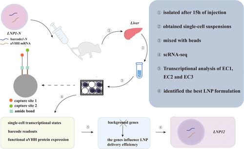
Dahlman et al. designed a multiomics NP delivery system, SENT-seq, to examine the effect of cell heterogeneity on LNPs delivery.1 Using this technique, they were able to quantify the biodistribution (the number of LNPs entering cells), functional delivery (mRNA translated into functional proteins), and transcriptome level of cells. They used DNA barcoding technology to quantify LNPs entering the cell. They inserted different DNA sequences into different LNPs such that each LNP had a DNA barcode. The number of LNPs that entered a single cell was characterized by barcode readouts. However, one of the significant barriers in the intracellular delivery of nucleic acid molecules is that the nucleic acid molecules degrade in the endosomes, hence, the mRNA delivered into the cell does not necessarily express a functional protein.2 Here, the expression of the mRNA functional aVHH protein was also detected. The expression of aVHH on the cell surface was quantified by single-cell sequencing using the cell-hashing technique. Interestingly, SENT-seq designed by the authors can simultaneously obtain barcode readouts, aVHH protein expression, and transcriptome levels in a single cell. Beads are packaged with carboxylic acids to attach nucleotide sequences through an amide reaction. Two capture sites were designed: one for capturing the LNP-carried DNA barcode and the other for capturing the cell hash oligonucleotide antibody and all the endogenous mRNAs (Figure 1).
After injecting 24 LNPs containing specific sequences of DNA barcode and the same mRNA into mice at the same time, the liver was isolated, single-cell suspensions were obtained and then mixed with modified beads. After single-cell sequencing, the delivery efficiency results of the LNPs barcode in single cells were obtained, and the expression of functional aVHH protein in each cell subset was obtained based on the localization of cell subsets using transcriptome analysis (Figure 1). The results showed that the 17 cell subsets in the mouse liver had different levels of barcode readouts, suggesting that cell heterogeneity affects the delivery efficiency of LNPs in vivo.
To explore the genes associated with LNPs different delivery efficiency, endothelial cells EC1, EC2, and EC3 with the largest significant differences in LNPs functional delivery were selected. To identify the genes related to delivery efficiency, the different genes between the cells that expressed aVHH protein were identified based on removing the background genes. Finally, 19 different expression genes were identified. At the same time, from the uptake of 24 LNPs in 17 cell subsets, LNP-12, which has the best mRNA delivery ability, was identified (Figure 1).
In conclusion, the single-cell nanoparticle targeted sequencing technology reported by James E. Dahlman et al. is based on cell transcriptomic characteristics to distinguish cell subsets rather than cell surface markers so that cells can be classified and analyzed more comprehensively, including rare cell subsets that cell surface markers cannot mark. In addition, this technique can efficiently quantify the delivery efficiency of LNPs, expression of functional proteins, and transcriptome level of single cells simultaneously by single-cell sequencing. Additionally, studying the mechanism of different uptake levels of LNPs by different cells based on the transcription level is helpful in guiding further and better-targeted application of LNPs. In addition, this technology can simultaneously test multiple LNPs, realize high-throughput LNPs delivery analysis in vivo, and contribute to the screening of new LNPs.
Currently, the target tissue of LNPs is mainly the liver. After intravenous injection, 30-99% of the LNPs eventually enter the liver and are absorbed by liver cells.5 How to target nonliver tissue is a challenge. It was proven in this study that changing the formula of LNPs can affect the targeting of LNPs. Although this work only studied the effect of cell heterogeneity in the mouse liver on the uptake of different formulations of LNPs, this technique can also be used to study targeted LNPs in other tissues. By studying LNPs targeted in nonliver organs, we can determine the underlying mechanism and then design new LNPs according to the mechanism to further expand the application of LNPs and mRNA therapy. However, in this study, healthy mice were used to explore the effect of cell heterogeneity on genes related to LNPs delivery efficiency without considering the disease model. When this technology is further applied in LNPs screening studies, it should be considered to establish and explore disease models for further exploration. In conclusion, although there are some limitations to this study, studying the biological behavior of mRNA delivery with single-cell sequencing can be an effective way to accelerate the clinical transformation of mRNA therapeutic products.
Xiaoshuang Song drafted the manuscript. Xiaoshuang Song and Fang Nan painted the schematic diagram. Dunfang Zhang revised the manuscript. All authors have read and approved the final manuscript.

 求助内容:
求助内容: 应助结果提醒方式:


