{"title":"3D打印可降解羟基磷灰石生物活性陶瓷用于颅骨再生","authors":"Xingyu Gui, Boqing Zhang, Zixuan Su, Zhigang Zhou, Zhihong Dong, Pin Feng, Chen Fan, Ming Liu, Qingquan Kong, Changchun Zhou, Yujiang Fan, Xingdong Zhang","doi":"10.1002/mba2.41","DOIUrl":null,"url":null,"abstract":"<p>Hydroxyapatite (HA) bioceramics have been extensively employed as bone tissue scaffolds owing to their biodegradability and osteoinductivity. In our work, HA, a significant component of natural bone tissue used as the raw material to produce porous scaffolds employing three-dimensional (3D)-printing technology. Physical and chemical properties, porosity, and compression resistance of the scaffolds were investigated in vitro. The scaffold was confirmed to have a large number of interconnected pore structures on the surface and inside HA scaffolds showed good cell compatibility and cell adhesion in cell text. To analyze the effect of the scaffold on bone repair and regeneration in vivo, the large-size defect of beagle skull was repaired with a 3D printing group and an autologous bone group (ABG) for 8 months. Images and histological analysis of the 3D printing group indicated better integration with adjacent tissues. However, there were obvious gaps in the ABG, which indicates weak bone regeneration ability of this group due to unmatched implant dimension. Immunohistochemistry and immunofluorescence results showed that 3D-printed scaffolds had a highly vascularized structure. This study indicates that 3D-printed bioceramics scaffolds that are osteoinductivity and biodegradable have great potential in maxillofacial bone regeneration.</p>","PeriodicalId":100901,"journal":{"name":"MedComm – Biomaterials and Applications","volume":"2 2","pages":""},"PeriodicalIF":0.0000,"publicationDate":"2023-06-04","publicationTypes":"Journal Article","fieldsOfStudy":null,"isOpenAccess":false,"openAccessPdf":"https://onlinelibrary.wiley.com/doi/epdf/10.1002/mba2.41","citationCount":"3","resultStr":"{\"title\":\"3D-printed degradable hydroxyapatite bioactive ceramics for skull regeneration\",\"authors\":\"Xingyu Gui, Boqing Zhang, Zixuan Su, Zhigang Zhou, Zhihong Dong, Pin Feng, Chen Fan, Ming Liu, Qingquan Kong, Changchun Zhou, Yujiang Fan, Xingdong Zhang\",\"doi\":\"10.1002/mba2.41\",\"DOIUrl\":null,\"url\":null,\"abstract\":\"<p>Hydroxyapatite (HA) bioceramics have been extensively employed as bone tissue scaffolds owing to their biodegradability and osteoinductivity. In our work, HA, a significant component of natural bone tissue used as the raw material to produce porous scaffolds employing three-dimensional (3D)-printing technology. Physical and chemical properties, porosity, and compression resistance of the scaffolds were investigated in vitro. The scaffold was confirmed to have a large number of interconnected pore structures on the surface and inside HA scaffolds showed good cell compatibility and cell adhesion in cell text. To analyze the effect of the scaffold on bone repair and regeneration in vivo, the large-size defect of beagle skull was repaired with a 3D printing group and an autologous bone group (ABG) for 8 months. Images and histological analysis of the 3D printing group indicated better integration with adjacent tissues. However, there were obvious gaps in the ABG, which indicates weak bone regeneration ability of this group due to unmatched implant dimension. Immunohistochemistry and immunofluorescence results showed that 3D-printed scaffolds had a highly vascularized structure. This study indicates that 3D-printed bioceramics scaffolds that are osteoinductivity and biodegradable have great potential in maxillofacial bone regeneration.</p>\",\"PeriodicalId\":100901,\"journal\":{\"name\":\"MedComm – Biomaterials and Applications\",\"volume\":\"2 2\",\"pages\":\"\"},\"PeriodicalIF\":0.0000,\"publicationDate\":\"2023-06-04\",\"publicationTypes\":\"Journal Article\",\"fieldsOfStudy\":null,\"isOpenAccess\":false,\"openAccessPdf\":\"https://onlinelibrary.wiley.com/doi/epdf/10.1002/mba2.41\",\"citationCount\":\"3\",\"resultStr\":null,\"platform\":\"Semanticscholar\",\"paperid\":null,\"PeriodicalName\":\"MedComm – Biomaterials and Applications\",\"FirstCategoryId\":\"1085\",\"ListUrlMain\":\"https://onlinelibrary.wiley.com/doi/10.1002/mba2.41\",\"RegionNum\":0,\"RegionCategory\":null,\"ArticlePicture\":[],\"TitleCN\":null,\"AbstractTextCN\":null,\"PMCID\":null,\"EPubDate\":\"\",\"PubModel\":\"\",\"JCR\":\"\",\"JCRName\":\"\",\"Score\":null,\"Total\":0}","platform":"Semanticscholar","paperid":null,"PeriodicalName":"MedComm – Biomaterials and Applications","FirstCategoryId":"1085","ListUrlMain":"https://onlinelibrary.wiley.com/doi/10.1002/mba2.41","RegionNum":0,"RegionCategory":null,"ArticlePicture":[],"TitleCN":null,"AbstractTextCN":null,"PMCID":null,"EPubDate":"","PubModel":"","JCR":"","JCRName":"","Score":null,"Total":0}
3D-printed degradable hydroxyapatite bioactive ceramics for skull regeneration
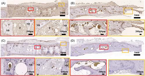
Hydroxyapatite (HA) bioceramics have been extensively employed as bone tissue scaffolds owing to their biodegradability and osteoinductivity. In our work, HA, a significant component of natural bone tissue used as the raw material to produce porous scaffolds employing three-dimensional (3D)-printing technology. Physical and chemical properties, porosity, and compression resistance of the scaffolds were investigated in vitro. The scaffold was confirmed to have a large number of interconnected pore structures on the surface and inside HA scaffolds showed good cell compatibility and cell adhesion in cell text. To analyze the effect of the scaffold on bone repair and regeneration in vivo, the large-size defect of beagle skull was repaired with a 3D printing group and an autologous bone group (ABG) for 8 months. Images and histological analysis of the 3D printing group indicated better integration with adjacent tissues. However, there were obvious gaps in the ABG, which indicates weak bone regeneration ability of this group due to unmatched implant dimension. Immunohistochemistry and immunofluorescence results showed that 3D-printed scaffolds had a highly vascularized structure. This study indicates that 3D-printed bioceramics scaffolds that are osteoinductivity and biodegradable have great potential in maxillofacial bone regeneration.

 求助内容:
求助内容: 应助结果提醒方式:


