{"title":"基于无标记策略的LC-MS/MS†分析诱导多能干细胞生成的大骨节病软骨细胞n -糖基化蛋白","authors":"Sen Wang, Peilin Meng, Linlin Yuan and Xiong Guo","doi":"10.1039/D3MO00018D","DOIUrl":null,"url":null,"abstract":"We aimed to compare N-glycosylation proteins in Kashin-Beck disease (KBD) chondrocytes and normal chondrocytes derived from induced pluripotent stem cells (iPSCs). KBD and normal iPSCs were reprogrammed from human KBD and normal dermal fibroblasts, respectively. Subsequently, chondrocytes were differentiated from KBD and normal iPSCs separately. Immunofluorescence was utilized to assay the protein markers of iPSCs and chondrocytes. Differential N-glycosylation proteins were screened using label-free strategies with LC-MS/MS. Bioinformatics analyses were utilized to interpret the functions of differential N-glycosylation proteins. Immunofluorescence staining revealed that both KBD-iPSCs and normal-iPSCs strongly expressed pluripotency markers OCT4 and NANOG. Meanwhile, chondrocyte markers collagen II and SOX9 are presented in KBD-iPSC-chondrocytes and normal-iPSC-chondrocytes. We obtained 87 differential N-glycosylation sites which corresponded to 68 differential proteins, which were constructed into 1 cluster. We obtained collagen type I trimer and 9 other biological processes; polysaccharide binding and 9 other molecular functions; regulation of transcription by RNA polymerase II and 9 other cellular components from GO; the Pl3K-Akt signaling pathway and 9 other KEGG pathways; peroxisome and 7 other subcellular locations; and integrin alpha chain, C-terminal cytoplasmic region, conserved site and 9 other classifications of domain annotations, and 2 networks. FGFR3 and LRP1 are expressed at higher levels in KBD-iPSC-chondrocytes, while the expressions of COL2A1, TIMP1, UNC5B, NOG, LEPR, and ITGA1 were down-regulated in KBD-iPSC-chondrocytes. The differential expressions of these N-glycosylation proteins may lead to the abnormal function of KBD chondrocytes.","PeriodicalId":19065,"journal":{"name":"Molecular omics","volume":" 6","pages":" 454-463"},"PeriodicalIF":3.0000,"publicationDate":"2023-04-26","publicationTypes":"Journal Article","fieldsOfStudy":null,"isOpenAccess":false,"openAccessPdf":"","citationCount":"0","resultStr":"{\"title\":\"Analysis of N-glycosylation protein of Kashin–Beck disease chondrocytes derived from induced pluripotent stem cells based on label-free strategies with LC-MS/MS†\",\"authors\":\"Sen Wang, Peilin Meng, Linlin Yuan and Xiong Guo\",\"doi\":\"10.1039/D3MO00018D\",\"DOIUrl\":null,\"url\":null,\"abstract\":\"We aimed to compare N-glycosylation proteins in Kashin-Beck disease (KBD) chondrocytes and normal chondrocytes derived from induced pluripotent stem cells (iPSCs). KBD and normal iPSCs were reprogrammed from human KBD and normal dermal fibroblasts, respectively. Subsequently, chondrocytes were differentiated from KBD and normal iPSCs separately. Immunofluorescence was utilized to assay the protein markers of iPSCs and chondrocytes. Differential N-glycosylation proteins were screened using label-free strategies with LC-MS/MS. Bioinformatics analyses were utilized to interpret the functions of differential N-glycosylation proteins. Immunofluorescence staining revealed that both KBD-iPSCs and normal-iPSCs strongly expressed pluripotency markers OCT4 and NANOG. Meanwhile, chondrocyte markers collagen II and SOX9 are presented in KBD-iPSC-chondrocytes and normal-iPSC-chondrocytes. We obtained 87 differential N-glycosylation sites which corresponded to 68 differential proteins, which were constructed into 1 cluster. We obtained collagen type I trimer and 9 other biological processes; polysaccharide binding and 9 other molecular functions; regulation of transcription by RNA polymerase II and 9 other cellular components from GO; the Pl3K-Akt signaling pathway and 9 other KEGG pathways; peroxisome and 7 other subcellular locations; and integrin alpha chain, C-terminal cytoplasmic region, conserved site and 9 other classifications of domain annotations, and 2 networks. FGFR3 and LRP1 are expressed at higher levels in KBD-iPSC-chondrocytes, while the expressions of COL2A1, TIMP1, UNC5B, NOG, LEPR, and ITGA1 were down-regulated in KBD-iPSC-chondrocytes. The differential expressions of these N-glycosylation proteins may lead to the abnormal function of KBD chondrocytes.\",\"PeriodicalId\":19065,\"journal\":{\"name\":\"Molecular omics\",\"volume\":\" 6\",\"pages\":\" 454-463\"},\"PeriodicalIF\":3.0000,\"publicationDate\":\"2023-04-26\",\"publicationTypes\":\"Journal Article\",\"fieldsOfStudy\":null,\"isOpenAccess\":false,\"openAccessPdf\":\"\",\"citationCount\":\"0\",\"resultStr\":null,\"platform\":\"Semanticscholar\",\"paperid\":null,\"PeriodicalName\":\"Molecular omics\",\"FirstCategoryId\":\"99\",\"ListUrlMain\":\"https://pubs.rsc.org/en/content/articlelanding/2023/mo/d3mo00018d\",\"RegionNum\":4,\"RegionCategory\":\"生物学\",\"ArticlePicture\":[],\"TitleCN\":null,\"AbstractTextCN\":null,\"PMCID\":null,\"EPubDate\":\"\",\"PubModel\":\"\",\"JCR\":\"Q3\",\"JCRName\":\"BIOCHEMISTRY & MOLECULAR BIOLOGY\",\"Score\":null,\"Total\":0}","platform":"Semanticscholar","paperid":null,"PeriodicalName":"Molecular omics","FirstCategoryId":"99","ListUrlMain":"https://pubs.rsc.org/en/content/articlelanding/2023/mo/d3mo00018d","RegionNum":4,"RegionCategory":"生物学","ArticlePicture":[],"TitleCN":null,"AbstractTextCN":null,"PMCID":null,"EPubDate":"","PubModel":"","JCR":"Q3","JCRName":"BIOCHEMISTRY & MOLECULAR BIOLOGY","Score":null,"Total":0}
Analysis of N-glycosylation protein of Kashin–Beck disease chondrocytes derived from induced pluripotent stem cells based on label-free strategies with LC-MS/MS†
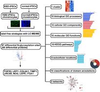
We aimed to compare N-glycosylation proteins in Kashin-Beck disease (KBD) chondrocytes and normal chondrocytes derived from induced pluripotent stem cells (iPSCs). KBD and normal iPSCs were reprogrammed from human KBD and normal dermal fibroblasts, respectively. Subsequently, chondrocytes were differentiated from KBD and normal iPSCs separately. Immunofluorescence was utilized to assay the protein markers of iPSCs and chondrocytes. Differential N-glycosylation proteins were screened using label-free strategies with LC-MS/MS. Bioinformatics analyses were utilized to interpret the functions of differential N-glycosylation proteins. Immunofluorescence staining revealed that both KBD-iPSCs and normal-iPSCs strongly expressed pluripotency markers OCT4 and NANOG. Meanwhile, chondrocyte markers collagen II and SOX9 are presented in KBD-iPSC-chondrocytes and normal-iPSC-chondrocytes. We obtained 87 differential N-glycosylation sites which corresponded to 68 differential proteins, which were constructed into 1 cluster. We obtained collagen type I trimer and 9 other biological processes; polysaccharide binding and 9 other molecular functions; regulation of transcription by RNA polymerase II and 9 other cellular components from GO; the Pl3K-Akt signaling pathway and 9 other KEGG pathways; peroxisome and 7 other subcellular locations; and integrin alpha chain, C-terminal cytoplasmic region, conserved site and 9 other classifications of domain annotations, and 2 networks. FGFR3 and LRP1 are expressed at higher levels in KBD-iPSC-chondrocytes, while the expressions of COL2A1, TIMP1, UNC5B, NOG, LEPR, and ITGA1 were down-regulated in KBD-iPSC-chondrocytes. The differential expressions of these N-glycosylation proteins may lead to the abnormal function of KBD chondrocytes.
Molecular omicsBiochemistry, Genetics and Molecular Biology-Biochemistry
CiteScore
5.40
自引率
3.40%
发文量
91
期刊介绍:
Molecular Omics publishes high-quality research from across the -omics sciences.
Topics include, but are not limited to:
-omics studies to gain mechanistic insight into biological processes – for example, determining the mode of action of a drug or the basis of a particular phenotype, such as drought tolerance
-omics studies for clinical applications with validation, such as finding biomarkers for diagnostics or potential new drug targets
-omics studies looking at the sub-cellular make-up of cells – for example, the subcellular localisation of certain proteins or post-translational modifications or new imaging techniques
-studies presenting new methods and tools to support omics studies, including new spectroscopic/chromatographic techniques, chip-based/array technologies and new classification/data analysis techniques. New methods should be proven and demonstrate an advance in the field.
Molecular Omics only accepts articles of high importance and interest that provide significant new insight into important chemical or biological problems. This could be fundamental research that significantly increases understanding or research that demonstrates clear functional benefits.
Papers reporting new results that could be routinely predicted, do not show a significant improvement over known research, or are of interest only to the specialist in the area are not suitable for publication in Molecular Omics.