Clémence Simon, Oriane Morel, Godfrey Neutelings, Fabien Baldacci-Cresp, Marie Baucher, Corentin Spriet, Christophe Biot, Simon Hawkins and Cédric Lion*,
{"title":"利用airscan超分辨显微镜和生物正交化学研究植物细胞壁木质素化的复杂性","authors":"Clémence Simon, Oriane Morel, Godfrey Neutelings, Fabien Baldacci-Cresp, Marie Baucher, Corentin Spriet, Christophe Biot, Simon Hawkins and Cédric Lion*, ","doi":"10.1021/cbmi.3c00052","DOIUrl":null,"url":null,"abstract":"<p >In this paper, we present the use of multiplex click/bioorthogonal chemistry combined with super-resolution Airyscan microscopy to track biomolecules in living systems with a focus on studying lignin formation in plant cell walls. While laser scanning confocal microscopy (LSCM) provided insights into the tissue-scale dynamics of lignin formation and distribution in our previous reports, its limited resolution precluded an in-depth analysis of lignin composition at the unique cell wall or substructure level. To overcome this limitation, we explored the use of Airyscan microscopy, which, among the super-resolution techniques available, offers an optimal balance between performance, cost, accessibility, and ease of implementation. Our study demonstrates that a triple labeling strategy using copper-catalyzed azide–alkyne cycloaddition (CuAAC), strain-promoted azide–alkyne cycloaddition (SPAAC), and inverse electronic-demand Diels–Alder cycloaddition (IEDDA) to label modified lignin metabolic precursors can be combined with Airyscan microscopy to reveal the zones of active lignification at the single cell level with improved sensitivity and resolution. This approach enables insights into the lignin composition in wall substructures, such as pits or in wall layers that are otherwise not distinguishable by classical LSCM. Our work emphasizes the importance of studying lignin formation in plant cell walls and demonstrates the potential of combining bioorthogonal chemistry and super-resolution microscopy techniques for studying biomolecules in living systems.</p>","PeriodicalId":53181,"journal":{"name":"Chemical & Biomedical Imaging","volume":null,"pages":null},"PeriodicalIF":0.0000,"publicationDate":"2023-07-06","publicationTypes":"Journal Article","fieldsOfStudy":null,"isOpenAccess":false,"openAccessPdf":"https://pubs.acs.org/doi/epdf/10.1021/cbmi.3c00052","citationCount":"1","resultStr":"{\"title\":\"Exploring Lignification Complexity in Plant Cell Walls with Airyscan Super-resolution Microscopy and Bioorthogonal Chemistry\",\"authors\":\"Clémence Simon, Oriane Morel, Godfrey Neutelings, Fabien Baldacci-Cresp, Marie Baucher, Corentin Spriet, Christophe Biot, Simon Hawkins and Cédric Lion*, \",\"doi\":\"10.1021/cbmi.3c00052\",\"DOIUrl\":null,\"url\":null,\"abstract\":\"<p >In this paper, we present the use of multiplex click/bioorthogonal chemistry combined with super-resolution Airyscan microscopy to track biomolecules in living systems with a focus on studying lignin formation in plant cell walls. While laser scanning confocal microscopy (LSCM) provided insights into the tissue-scale dynamics of lignin formation and distribution in our previous reports, its limited resolution precluded an in-depth analysis of lignin composition at the unique cell wall or substructure level. To overcome this limitation, we explored the use of Airyscan microscopy, which, among the super-resolution techniques available, offers an optimal balance between performance, cost, accessibility, and ease of implementation. Our study demonstrates that a triple labeling strategy using copper-catalyzed azide–alkyne cycloaddition (CuAAC), strain-promoted azide–alkyne cycloaddition (SPAAC), and inverse electronic-demand Diels–Alder cycloaddition (IEDDA) to label modified lignin metabolic precursors can be combined with Airyscan microscopy to reveal the zones of active lignification at the single cell level with improved sensitivity and resolution. This approach enables insights into the lignin composition in wall substructures, such as pits or in wall layers that are otherwise not distinguishable by classical LSCM. Our work emphasizes the importance of studying lignin formation in plant cell walls and demonstrates the potential of combining bioorthogonal chemistry and super-resolution microscopy techniques for studying biomolecules in living systems.</p>\",\"PeriodicalId\":53181,\"journal\":{\"name\":\"Chemical & Biomedical Imaging\",\"volume\":null,\"pages\":null},\"PeriodicalIF\":0.0000,\"publicationDate\":\"2023-07-06\",\"publicationTypes\":\"Journal Article\",\"fieldsOfStudy\":null,\"isOpenAccess\":false,\"openAccessPdf\":\"https://pubs.acs.org/doi/epdf/10.1021/cbmi.3c00052\",\"citationCount\":\"1\",\"resultStr\":null,\"platform\":\"Semanticscholar\",\"paperid\":null,\"PeriodicalName\":\"Chemical & Biomedical Imaging\",\"FirstCategoryId\":\"1085\",\"ListUrlMain\":\"https://pubs.acs.org/doi/10.1021/cbmi.3c00052\",\"RegionNum\":0,\"RegionCategory\":null,\"ArticlePicture\":[],\"TitleCN\":null,\"AbstractTextCN\":null,\"PMCID\":null,\"EPubDate\":\"\",\"PubModel\":\"\",\"JCR\":\"\",\"JCRName\":\"\",\"Score\":null,\"Total\":0}","platform":"Semanticscholar","paperid":null,"PeriodicalName":"Chemical & Biomedical Imaging","FirstCategoryId":"1085","ListUrlMain":"https://pubs.acs.org/doi/10.1021/cbmi.3c00052","RegionNum":0,"RegionCategory":null,"ArticlePicture":[],"TitleCN":null,"AbstractTextCN":null,"PMCID":null,"EPubDate":"","PubModel":"","JCR":"","JCRName":"","Score":null,"Total":0}
Exploring Lignification Complexity in Plant Cell Walls with Airyscan Super-resolution Microscopy and Bioorthogonal Chemistry
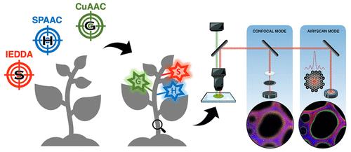
In this paper, we present the use of multiplex click/bioorthogonal chemistry combined with super-resolution Airyscan microscopy to track biomolecules in living systems with a focus on studying lignin formation in plant cell walls. While laser scanning confocal microscopy (LSCM) provided insights into the tissue-scale dynamics of lignin formation and distribution in our previous reports, its limited resolution precluded an in-depth analysis of lignin composition at the unique cell wall or substructure level. To overcome this limitation, we explored the use of Airyscan microscopy, which, among the super-resolution techniques available, offers an optimal balance between performance, cost, accessibility, and ease of implementation. Our study demonstrates that a triple labeling strategy using copper-catalyzed azide–alkyne cycloaddition (CuAAC), strain-promoted azide–alkyne cycloaddition (SPAAC), and inverse electronic-demand Diels–Alder cycloaddition (IEDDA) to label modified lignin metabolic precursors can be combined with Airyscan microscopy to reveal the zones of active lignification at the single cell level with improved sensitivity and resolution. This approach enables insights into the lignin composition in wall substructures, such as pits or in wall layers that are otherwise not distinguishable by classical LSCM. Our work emphasizes the importance of studying lignin formation in plant cell walls and demonstrates the potential of combining bioorthogonal chemistry and super-resolution microscopy techniques for studying biomolecules in living systems.
期刊介绍:
Chemical & Biomedical Imaging is a peer-reviewed open access journal devoted to the publication of cutting-edge research papers on all aspects of chemical and biomedical imaging. This interdisciplinary field sits at the intersection of chemistry physics biology materials engineering and medicine. The journal aims to bring together researchers from across these disciplines to address cutting-edge challenges of fundamental research and applications.Topics of particular interest include but are not limited to:Imaging of processes and reactionsImaging of nanoscale microscale and mesoscale materialsImaging of biological interactions and interfacesSingle-molecule and cellular imagingWhole-organ and whole-body imagingMolecular imaging probes and contrast agentsBioluminescence chemiluminescence and electrochemiluminescence imagingNanophotonics and imagingChemical tools for new imaging modalitiesChemical and imaging techniques in diagnosis and therapyImaging-guided drug deliveryAI and machine learning assisted imaging
 求助内容:
求助内容: 应助结果提醒方式:


