Jereme C. Wingert, Jonathan N. Anguiano, Jonathan D. Ramos, Jordan M. Blacktop, Angela E. Gonzalez, Lynn Churchill, Barbara A. Sorg
{"title":"大鼠延长可卡因自给药后内侧前额叶皮层细小白蛋白和会阴神经网的表达增强。","authors":"Jereme C. Wingert, Jonathan N. Anguiano, Jonathan D. Ramos, Jordan M. Blacktop, Angela E. Gonzalez, Lynn Churchill, Barbara A. Sorg","doi":"10.1111/adb.13334","DOIUrl":null,"url":null,"abstract":"<p>The medial prefrontal cortex (mPFC) drives cocaine-seeking behaviour in rodent models of cocaine use disorder. Parvalbumin (PV)-containing GABAergic interneurons powerfully control the output of the mPFC, yet few studies have focused on how these neurons modulate cocaine-seeking behaviour. Most PV neurons are surrounded by perineuronal nets (PNNs), which regulate the firing of PV neurons. We examined staining intensity and number of PV and PNNs after long-access (6 h/day) cocaine self-administration in rats followed by either 8–10 days extinction ± cue-induced reinstatement or short-term (1–2 days) or long-term (30–31 days) abstinence ± cue-induced reinstatement. The intensity of PNNs was increased in the prelimbic and infralimbic PFC after long-term abstinence in the absence of cue reinstatement and after cue reinstatement following both daily extinction sessions and after a 30-day abstinence period. PV intensity was increased after 30 days of abstinence in the prelimbic but not infralimbic PFC. Enzymatic removal of PNNs with chondroitinase ABC (ABC) in the prelimbic PFC did not prevent incubation of cue-induced reinstatement but decreased cocaine-seeking behaviour at both 2 and 31 days of abstinence, and this decrease at 31 days was accompanied by reduced c-Fos levels in the prelimbic PFC. Increases in PNN intensity have generally been associated with the loss of plasticity, suggesting that the persistent and chronic nature of cocaine use disorder may in part be attributed to long-lasting increases in PNN intensity that reduce the ability of stimuli to alter synaptic input to underlying PV neurons.</p>","PeriodicalId":7289,"journal":{"name":"Addiction Biology","volume":"28 11","pages":""},"PeriodicalIF":3.1000,"publicationDate":"2023-10-02","publicationTypes":"Journal Article","fieldsOfStudy":null,"isOpenAccess":false,"openAccessPdf":"https://onlinelibrary.wiley.com/doi/epdf/10.1111/adb.13334","citationCount":"0","resultStr":"{\"title\":\"Enhanced expression of parvalbumin and perineuronal nets in the medial prefrontal cortex after extended-access cocaine self-administration in rats\",\"authors\":\"Jereme C. Wingert, Jonathan N. Anguiano, Jonathan D. Ramos, Jordan M. Blacktop, Angela E. Gonzalez, Lynn Churchill, Barbara A. Sorg\",\"doi\":\"10.1111/adb.13334\",\"DOIUrl\":null,\"url\":null,\"abstract\":\"<p>The medial prefrontal cortex (mPFC) drives cocaine-seeking behaviour in rodent models of cocaine use disorder. Parvalbumin (PV)-containing GABAergic interneurons powerfully control the output of the mPFC, yet few studies have focused on how these neurons modulate cocaine-seeking behaviour. Most PV neurons are surrounded by perineuronal nets (PNNs), which regulate the firing of PV neurons. We examined staining intensity and number of PV and PNNs after long-access (6 h/day) cocaine self-administration in rats followed by either 8–10 days extinction ± cue-induced reinstatement or short-term (1–2 days) or long-term (30–31 days) abstinence ± cue-induced reinstatement. The intensity of PNNs was increased in the prelimbic and infralimbic PFC after long-term abstinence in the absence of cue reinstatement and after cue reinstatement following both daily extinction sessions and after a 30-day abstinence period. PV intensity was increased after 30 days of abstinence in the prelimbic but not infralimbic PFC. Enzymatic removal of PNNs with chondroitinase ABC (ABC) in the prelimbic PFC did not prevent incubation of cue-induced reinstatement but decreased cocaine-seeking behaviour at both 2 and 31 days of abstinence, and this decrease at 31 days was accompanied by reduced c-Fos levels in the prelimbic PFC. Increases in PNN intensity have generally been associated with the loss of plasticity, suggesting that the persistent and chronic nature of cocaine use disorder may in part be attributed to long-lasting increases in PNN intensity that reduce the ability of stimuli to alter synaptic input to underlying PV neurons.</p>\",\"PeriodicalId\":7289,\"journal\":{\"name\":\"Addiction Biology\",\"volume\":\"28 11\",\"pages\":\"\"},\"PeriodicalIF\":3.1000,\"publicationDate\":\"2023-10-02\",\"publicationTypes\":\"Journal Article\",\"fieldsOfStudy\":null,\"isOpenAccess\":false,\"openAccessPdf\":\"https://onlinelibrary.wiley.com/doi/epdf/10.1111/adb.13334\",\"citationCount\":\"0\",\"resultStr\":null,\"platform\":\"Semanticscholar\",\"paperid\":null,\"PeriodicalName\":\"Addiction Biology\",\"FirstCategoryId\":\"3\",\"ListUrlMain\":\"https://onlinelibrary.wiley.com/doi/10.1111/adb.13334\",\"RegionNum\":3,\"RegionCategory\":\"医学\",\"ArticlePicture\":[],\"TitleCN\":null,\"AbstractTextCN\":null,\"PMCID\":null,\"EPubDate\":\"\",\"PubModel\":\"\",\"JCR\":\"Q3\",\"JCRName\":\"BIOCHEMISTRY & MOLECULAR BIOLOGY\",\"Score\":null,\"Total\":0}","platform":"Semanticscholar","paperid":null,"PeriodicalName":"Addiction Biology","FirstCategoryId":"3","ListUrlMain":"https://onlinelibrary.wiley.com/doi/10.1111/adb.13334","RegionNum":3,"RegionCategory":"医学","ArticlePicture":[],"TitleCN":null,"AbstractTextCN":null,"PMCID":null,"EPubDate":"","PubModel":"","JCR":"Q3","JCRName":"BIOCHEMISTRY & MOLECULAR BIOLOGY","Score":null,"Total":0}
Enhanced expression of parvalbumin and perineuronal nets in the medial prefrontal cortex after extended-access cocaine self-administration in rats
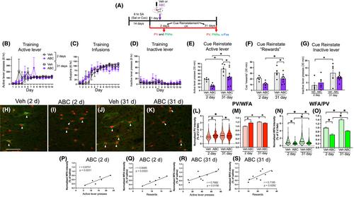
The medial prefrontal cortex (mPFC) drives cocaine-seeking behaviour in rodent models of cocaine use disorder. Parvalbumin (PV)-containing GABAergic interneurons powerfully control the output of the mPFC, yet few studies have focused on how these neurons modulate cocaine-seeking behaviour. Most PV neurons are surrounded by perineuronal nets (PNNs), which regulate the firing of PV neurons. We examined staining intensity and number of PV and PNNs after long-access (6 h/day) cocaine self-administration in rats followed by either 8–10 days extinction ± cue-induced reinstatement or short-term (1–2 days) or long-term (30–31 days) abstinence ± cue-induced reinstatement. The intensity of PNNs was increased in the prelimbic and infralimbic PFC after long-term abstinence in the absence of cue reinstatement and after cue reinstatement following both daily extinction sessions and after a 30-day abstinence period. PV intensity was increased after 30 days of abstinence in the prelimbic but not infralimbic PFC. Enzymatic removal of PNNs with chondroitinase ABC (ABC) in the prelimbic PFC did not prevent incubation of cue-induced reinstatement but decreased cocaine-seeking behaviour at both 2 and 31 days of abstinence, and this decrease at 31 days was accompanied by reduced c-Fos levels in the prelimbic PFC. Increases in PNN intensity have generally been associated with the loss of plasticity, suggesting that the persistent and chronic nature of cocaine use disorder may in part be attributed to long-lasting increases in PNN intensity that reduce the ability of stimuli to alter synaptic input to underlying PV neurons.
期刊介绍:
Addiction Biology is focused on neuroscience contributions and it aims to advance our understanding of the action of drugs of abuse and addictive processes. Papers are accepted in both animal experimentation or clinical research. The content is geared towards behavioral, molecular, genetic, biochemical, neuro-biological and pharmacology aspects of these fields.
Addiction Biology includes peer-reviewed original research reports and reviews.
Addiction Biology is published on behalf of the Society for the Study of Addiction to Alcohol and other Drugs (SSA). Members of the Society for the Study of Addiction receive the Journal as part of their annual membership subscription.
 求助内容:
求助内容: 应助结果提醒方式:


