Abstracts of the 13th International Conference on Cachexia, Sarcopenia and Muscle Wasting, 11-13 December 2020
1-01
Rehabilitating cachexia: development and functional characterization of a novel longitudinal and translational model of cancer-associated cachexia
Ishan Roy1,2, Ben Binder-Markey1,2, Danielle Sychowski1, Donna McAllister3, Dominic D'Andrea1, Colin Franz1,2, Michael B. Dwinell3 and Richard L. Lieber1,2
1Shirley Ryan AbilityLab (formerly known as Rehabilitation Institute of Chicago), Chicago, IL, USA;2Department of Physical Medicine and Rehabilitation, Northwestern University, Chicago, IL, USA;3Department of Microbiology & Immunology, Medical College of Wisconsin, Milwaukee, WI, USA
Introduction: The physiologic mechanisms that drive functional changes due to cachexia are poorly understood. Existing cachexia models limit our ability to address these mechanisms because they either express a rare tumour type or cause rapid death. Such models are not amenable to multi-domain functional analysis of rehabilitation protocols, which typically require 6–8 weeks. The goal of this study was to develop and functionally characterize a longitudinal model of cancer-associated cachexia.
Methods: The “KPC orthotopic injection” pancreatic cancer mouse model was selected for optimization. We tested multiple cell clones, cell doses, and vehicle types in order to maximize survival. Ex vivo analysis included skeletal and cardiac muscle mass. Functional characterization included: hind-limb grip strength for muscle function; open-field arena for ambulation and anxiety; Y-maze for spatial memory; and Morris Water Maze and Rotarod for endurance.
Results: Serial dilution of multiple KPC clones yielded optimal conditions for extending median survival, from 3 weeks up to 8.5 weeks post-injection using the KPC orthotopic model (p < 0.0001). In weekly ex vivo analysis, the optimized model resulted in progressive skeletal and cardiac muscle mass loss at 5 weeks post-injection and continued through 9 weeks (p < 0.01). Starting 5 weeks post-injection, animals had 8% decline in grip strength (p < 0.01) and a sustained decrease in distance ambulated and gait speed of 30–50% (p < 0.05). Animal subjects retained spatial memory similar to controls, indicating that functional deficits were not confounded by behavioural change. There was also a trend towards decreased motor endurance (p = 0.07) at 5 weeks and increased open-field anxiety (p = 0.06) at 8 weeks.
Conclusion: This optimized model of cancer-associated cachexia demonstrates progressive loss of muscle and function in multiple domains while accounting for potential confounding factors of cognition and behaviour. The model is tailored for longitudinal studies and sets the stage for mechanistic and translational studies of cachexia rehabilitation.
1-02
Alterations in mitochondrial turnover during the development of cancer cachexia in tumour-bearing female mice
Seongkyun Lim1, Megan E. Rosa-Caldwell1, Wesley S. Haynie1, Lisa T. Jansen1, Kirsten R. Dunlap1, Francielly Morena da Silva1, J. William Deaver1, Michael Wiggs2, Tyrone A. Washington1 and Nicholas P. Greene1
1Health, Human Performance, and Recreation, University of Arkansas, Fayetteville, AR, USA;2Health, Human Performance, and Recreation, Baylor University, Waco, TX, USA
Introduction: Cachexia is prevalent in ~80% of cancer patients and responsible for ~20% of cancer-related deaths. While the exact mechanisms leading to the onset of cancer cachexia (CC) are unknown, it has been reported previously that mitochondrial degeneration could be a culprit, as its appearance precedes skeletal muscle atrophy. We have recently reported that mitochondrial degeneration occurs during the early stages of CC in tumour-bearing male mice. However, alterations to mitochondrial health during CC in female animals are not fully elucidated. Therefore, the purpose of this study is to characterize alterations in mitochondrial health during the development of CC in female mice.
Methods: Fifty female C57BL6/J mice were injected with Lewis Lung Carcinoma cells at 8-weeks age. The tumours were allowed to develop for 1, 2, 3, and 4 weeks, and PBS-injected control group (n = 10) were age-matched with the 4 weeks. The 3 and 4 weeks were combined and reclassified by low tumour (LT) weight (≤1.2 g) and high tumour (HT) weight (≥2 g). Gastrocnemius tissue was collected for analysis of mitochondrial turnover via immunoblotting, RT-qPCR, and histological assessment of MitoTimer (Redox sensitive reporter gene).
Results: Both body and muscle weights were reduced in HT compared to other groups (p < 0.05). Both mRNA and protein content of Bnip3 (mitophagy) were ~53% (p < 0.05) and ~147% (p < 0.001) higher in HT compared to other groups, respectively. MitoTimer pure red puncta (completely degenerated mitochondria) was ~107% higher in HT compared to PBS and 1 week (p < 0.01). MitoTimer red:green ratio (oxidative stress) was ~40% higher in HT compared to 1 week and 2 week (p < 0.01). mRNA abundance of PGC-1α (mitochondrial biogenesis) was ~45% lower in HT compared to PBS and 1 week (p < 0.001) although protein content of COX-IV (mitochondrial content) remained unchanged.
Conclusions: Our data suggest alterations in mitochondrial turnover occurred in HT suggesting muscle mitochondrial degeneration is associated with tumour burden in female mice.
Acknowledgements: This study was funded by the National Institutes of Health, Award number: R15 AR069913/AR/NIAMS.
1-03
Alterations in extracellular matrix remodelling during early stages of cancer cachexia in tumour-bearing female mice
Francielly Morena da Silva1, Seongkyun Lim1, Megan E. Rosa-Caldwell1, Wesley S. Haynie1, Lisa T. Jansen1, Kristen R. Dunlap1, J. William Deaver1, Michael Wiggs2, Tyrone A. Washington1 and Nicholas P. Greene1
1Health, Human Performance, and Recreation, University of Arkansas, Fayetteville, AR, USA;2Health, Human Performance, and Recreation, Baylor University, Waco, TX, USA
Introduction: Cancer cachexia (CC) is responsible for up to 30–40% of cancer-associated deaths. Defined by muscle atrophy, CC if further characterized by reduced muscular strength, increased collagen deposition and ECM dysregulation. The ECM is critical for proper force transmission and is also a reservoir for growth factors and other signalling molecules. Matrix metalloproteinases (MMPs) are a family of enzymes capable of degrading ECM components such as non-contractile proteins, and are elevated during CC development, along with enhanced collagen deposition in cachectic muscle. Further investigation of ECM remodelling during CC progression is needed.
Purpose: To investigate how ECM dynamics change during CC development.
Methods: Fifty 8-week-old female C57BL6/J mice were injected with Lewis Lung Carcinoma cells into their hind-flank. The tumour was allowed to grow for 1, 2, 3 and 4 weeks, with a 4-week age-matched PBS control group (n = 10/group). The 3 and 4 weeks were combined and reclassified by low (tumour weight-TW ≤ 1.2 g; LT) and high (TW ≥ 2 g; HT) tumour mass. Gastrocnemius tissue was collected and prepared for mRNA abundance analysis using RT-qPCR.
Results: Collagen-1 and collagen-3 mRNA abundances were ~2-fold (p < 0.0001) and ~1-fold (p < 0.0001) higher in 1 week compared to the other groups, respectively. The ratio of collagen 3:1 mRNA abundance was decreased ~37% from 1 week compared to control, LT, and HT (p < 0.008), but not different compared to 2 weeks. MMP-2 mRNA abundance was ~50% higher at 1 week compared to LT and HT (p < 0.01) but was not different from control and 2 weeks. However, MMP-9 mRNA abundance was ~3-fold greater in HT compared to all the other conditions (p < 0.0001).
Conclusions: Altered collagen-3:1 ratio and increased MMP-2 in early stages of tumour growth precede CC and are followed by elevated MMP-9 in CC suggests changes in ECM remodelling during early stages of CC. This could hinder muscle contractile function in cancer patients.
Acknowledgements: This study was funded by the National Institutes of Health, Award number: R15 AR069913/AR/NIAMS.
1-04
Quercetin administration attenuates cancer-related cachexia and increases cancer-related survival and in C57BL/6 mice bearing syngeneic cutaneous melanoma model
Magda Mendes Vieira1, Amanda Mota Lacerda1, Valéria Couto Quintão1, Amanda Rodrigues Santos1, Andréia de Souza Brito1, Otavio Cardoso Filho1,2, Vinícius Rodrigues Dias1,3, Ludmila Regina de Souza1 and Alfredo Maurício Batista de Paula1,4
1Nucleus of Epidemiologic and Molecular Research Catrumano (NUPEMOC), Health Research Laboratory, Post-graduate Programme in Health Sciences, Universidade Estadual de Montes Claros (UNIMONTES), Montes Claros, Brazil;2Department of Chemical Engineering, Instituto Federal do Norte de Minas Gerais (IFNMG), Montes Claros, Brazil;3Department of Physical Education, Universidade Estadual de Montes Claros (UNIMONTES), Montes Claros, Brazil;4Department of Dentistry, Universidade Estadual de Montes Claros (UNIMONTES), Montes Claros, Brazil
Introduction: During late stages of the tumour progression, it might occur cancer-related cachexia (CRC), a paraneoplastic syndrome characterized as a progressive, systemic, physical consumption state in an individual with cancer. Quercetin (Querc) is a flavanol that exhibits a wide range of modulatory effects on disturbed molecular pathways related to cancer progression, including those related to CRC.
Aim: We investigated the effects of Querc on tumour volume, CRC diagnosis time, skeletal muscle volume and strength, count of skeletal muscle fibres and white adipocytes, and cancer-related survival (CRS) of the C57BL/6 mice bearing a syngeneic, cutaneous melanoma model (SCMM).
Methods: B16F10 cells were injected into flank of the 69 female C57BL/6 mice to establish the SCMM. After the clinical appearance of the tumour, the animals were divided into four groups: (i) mice no-tumour induced, oral gavage with placebo (PBS, control-non-tumour); (ii) tumour-bearing mice and administration of placebo (control-tumour); (iii) tumor-bearing mice treated with Querc 20 mg/kg body weight (Querc20); and (iv) tumour-bearing mice treated with Querc 50 mg/kg (Querc50). Animals received placebo or Querc 20 and 50 mg/kg body weight using oral gavage for 12 days. For CRS analysis, animals were divided in three groups: (i) tumour-bearing mice and administration of placebo; (ii) tumour-bearing mice treated with Querc20; and (iii) tumor-bearing mice treated with Querc50. Groups were monitored for 18 days. Diagnosis of CRC was established when body weight loss ≥5%. Serum levels of albumin and C-reactive protein (CRP) were measured by enzyme immunoassays. Forelimb skeletal muscle volume and strength were assessed using a high-frequency power Doppler ultrasound and dynamometer devices, respectively. Skeletal muscle tissue (SMT) samples were collected and subjected to gene expression analysis using qPCR. The present study was analysed and approved by an ethics committee on research and animal welfare (CEEBEA/UNIMONTES: protocol number: 102/2016).
Results: Control mice with tumour exhibited a significant increase of tumour volume compared to mice treated with Querc20 and Querc50. Mice treated with Querc50 significantly exhibited inhibition of body weight loss, higher time for CRC occurrence, and showed a better overall CRS. Querc20 administration significantly reduced CRP serum expression in mice bearing cutaneous with TMSMCM. SMT from mice treated with Querc50 exhibited a significant increase of myokine gene expression of TRIM55 but significant downregulation of TRIM63.
Conclusions: Querc exhibited anti-tumour and anti-cachectic effects and improved the overall CRS of mice bearing SCMM. Further animal and human studies are needed to validate the use of Querc as a phytonutrient as a neoadjuvant agent during cancer treatment.
1-05
Molecular and physiologic characterization of a novel murine model of metastatic head and neck cancer cachexia
Brennan Olson1,2, Mason A. Norgard1, Peter Levasseur1 and Xinxia Zhu and Daniel L. Marks1,3,4
1Papé Family Pediatric Research Institute, Oregon Health & Science University, Portland, OR, USA;2Medical Scientist Training Program, Oregon Health & Science University, Portland, OR, USA;3Brenden-Colson Center for Pancreatic Care, Oregon Health and & Science University Portland, OR, USA;4Knight Cancer Institute, Oregon Health & Science University, Portland, OR, USA
Introduction: Effective therapies for cachexia are lacking, potentially owing to the mismatch in clinically relevant models of cachexia. Cachexia observed in a clinical setting is commonly associated with advanced or late-stage cancers that are metastatic. With nearly half of all patients with head and neck cancer (HNC) experiencing cachexia at diagnosis, and with an even larger proportion of patients developing cachexia after metastatic progression, establishing a robust model of metastatic HNC cachexia may provide a valuable translational tool to the field. To this end, we sought to establish and characterize a metastatic model of human papilloma virus (HPV) positive head and neck squamous cell carcinoma that recapitulates the cardinal clinical and molecular features of cachexia.
Methods: Male and female C57BL/6 mice were implanted subcutaneously with an oropharyngeal squamous cell carcinoma cells stably transformed with HPV16 E6 and E7 together with hRas and luciferase (mEERL) that metastasizes to the lungs (MLM).1 We then robustly characterize the physiologic, behavioural, and molecular signatures during cachexia development.
Results: MLM-implanted mice rapidly developed primary tumours and eventual metastatic lesions to the lungs. Behaviorally, mice progressively lost lean and fat mass, displayed decreased locomotor activities, and mild appetite suppression during tumour development. Canonical muscle catabolism programs associated with cachexia, including E3 ubiquitin ligase and autophagy pathways, along with browning of adipose tissue, are observed and correlate closely with metastatic development. Finally, we observe both neuroendocrine and autonomic aberrations during MLM-cachexia, including activation of the hypothalamic–pituitary adrenal axis and sympathetic nervous system.
Conclusions: This syngeneic MLM allograft model of cancer cachexia is reliable, consistent, and readily recapitulates the key clinical and molecular features of cachexia. Since few metastatic or HNC models of cachexia exist, we believe this model is well suited for future mechanistic studies and preclinical therapy development of metastatic disease-associated cachexia.
Reference
1
Vermeer, DW, Coppock, JD, Zeng, E, et al. Metastatic model of HPV+ oropharyngeal squamous cell carcinoma demonstrates heterogeneity in tumor metastasis. Oncotarget2016; 7(17): 24194–24207.
1-06
Characterizing biological mechanisms of muscle wasting in a clinically relevant model of colorectal cancer and sequential chemotherapy treatment
Gauhar Ali1, Vera Mazurak2, Bhumi Bhatt3, Sambasivarao Damaraju3 and Vickie E. Baracos1
1Department of Oncology, Faculty of Medicine and Dentistry, University of Alberta, Edmonton, Canada;2Division of Human Nutrition, University of Alberta, Edmonton, Canada;3Department of Laboratory Medicine and Pathology, University of Alberta, Edmonton, Canada
Introduction: Cancer patients experience muscle wasting that may be attributable to their tumour or chemotherapy, the effect of which occur simultaneously in the clinical setting. These are often studied separately, so their possible synergistic effects in the development and progression of muscle wasting and effect on dynamic changes in gene expression in muscle over time have not been investigated. We address this question in a pre-clinical model with sequential chemotherapy treatments, as experienced clinically.
Methods: Chemotherapy (irinotecan plus 5-fluorouracil) was initiated 2 weeks after Ward colorectal carcinoma implantation in female Fischer 344 rats. mRNA profiling and expression were determined in gastrocnemius muscle by RNA sequencing. Differential gene expression (DE) analysis was performed (fold-change cut-off ≥1.5 and p-value <0.05), and Ingenuity Pathway Analysis (IPA) was used for functional annotation of the identified DE mRNAs.
Results: Of the 230 total significant (p < 0.05) canonical pathways identified in IPA analysis, 16 were attributed to tumour alone; the most significant being Transcriptional Regulatory Network in Embryonic Stem Cells (p = 2.45E-05), DNA Methylation and Transcriptional Repression Signalling (p = 7.59E-05) and NER Pathway (p = 4.17E04). Ninety additional pathways were prominent by one cycle of chemotherapy; Type II Diabetes Mellitus Signalling (p = 4.79E-06), Small Cell Lung Cancer Signalling (p = 9.33E-05) and FAT10 Cancer Signalling Pathway (p = 1.78E-04) were most significant. A 2nd second cycle of chemotherapy identified 124 additional pathways, exclusive of those earlier induced by the tumour and 1st cycle of chemotherapy; Protein Ubiquitination Pathway (p = 1.62E-07), Oestrogen Receptor Signalling (p = 4.57E-06) and Integrin Signalling (p = 5.37E-06) were among the significant pathways identified.
Conclusion: The tumour and sequential cycles of chemotherapy treatments resulted in a succession of changes in skeletal muscle transcriptome. This study contributes to an understanding of muscle biology at the level of gene expression changes along the treatment trajectory and supports the model of dynamic gene expression networks contributing to possible outcomes and negates the assumptions of a steady-state model.
1-07
The role of adipose tissue breakdown in the acute phase of sepsis: a comparison of interorgan fluxes of amino acids and glycerol in a Pseudomonas aeruginosa induced septic pig model
Ryan Morse1, Gabriella A.M. ten Have1, John J. Thaden1, Marielle P. Engelen1, Martin Hagve2 and Nicolaas E.P. Deutz1
1Center for Translational Research in Aging & Longevity, Department of Health & Kinesiology, Texas A&M University, College Station, TX, USA;2Department of Gastrointestinal Surgery, University Hospital North-Norway, Tromso, Norway
Rationale: Sepsis leads to an acute breakdown of muscle to support increased caloric and amino acid requirements. Little is known about the role of adipose tissue breakdown in glucose substrate supply during the acute phase of sepsis. Therefore, in a translational porcine model of sepsis, we explored uptake and release of total amino acids (AA), glycerol, and glucose across gut, liver and muscle.
Methods: Acute sepsis was induced in 13 pigs (±25 kg) by iv infusion of Pseudomonas aeruginosa for 18 h and eight controls (C). Plasma samples were obtained across Hindquarter (HQ), Portal Drained Viscera (PDV), and liver. Concentrations of glucose, glycerol, and AA were measured by mass spectrometry. Net organ balances were calculated using (Arterial–Venous difference) * plasma flow. Data are expressed as mean [95% CI]. Statistics: Release and uptake: Wilcoxon; Comparison between groups: ANOVA. Significance: p < 0.05.
Results: Arterial concentrations: In sepsis (S) plasma glucose decreased (C: 4.4 [4.0, 4.8] vs. S: 3.1 [2.7, 3.5] mM, p < 0.0001), and there was a trend towards increased AA (p = 0.0563), with no difference in glycerol. HQ: We observed AA release (p < 0.001), with unchanged release of glycerol or uptake of glucose. PDV: We found a decrease in glycerol (p = 0.0072) and glucose (p = 0.0027) uptake. Liver: Increased AA uptake (p = 0.0068) and a trend towards decreased glucose release (p = 0.0803), but unchanged glycerol uptake.
Conclusions: In the acute phase of sepsis, the expected increase in muscle glycerol production through lipolysis to provide substrate for gluconeogenesis is not observed. We hypothesize that the increased demand for glucogenic substrate by the liver is not supplied by lipolysis, but instead by the breakdown of muscle tissue.
Sponsor: NIH R01GM084447, S10RR027047.
1-08
Chronic cachexia is dependent on sustained IL-1R signalling during parasite infection
Stephanie J. Melchor1, Daniel Abebayehu1, Sheryl Coutermarsh-Ott2, Thomas Barker1 and Sarah E. Ewald1
1University of Virginia, Charlottesville, USA;2Virginia Polytechnic Institute, USA
Cachexia is an immune-metabolic disease of progressive muscle wasting that impairs patient survival and quality of life across a range of chronic diseases. T. gondii is a protozoan parasite that causes lifelong infection in many warm-blooded organisms, including humans and mice. Here, we show that mice infected with T. gondii develop robust, sustained cachexia that lasts for at least 18 weeks. Consistent with an emerging role for the IL-1 axis in disease tolerance, we show that mice deficient in the Type 1 IL-1 receptor (IL-1R) have more severe acute muscle wasting, adipocyte and hepatocyte necrosis, independent of parasite burden. Unexpectedly, IL-1R−/− mice rapidly recover from acute disease, despite sustained parasite infection, and are protected from chronic cachexia as well as perivascular liver and muscle fibrosis. These data are consistent with a model where IL-1R signalling benefits cell survival and tissue integrity over short periods of inflammation, but sustained reliance on IL-1 mediated tolerance programs come at the cost of fibrosis and cachexia.
1-09
GDF15 neutralization does not impact anorexia or survival in the lipopolysaccharide (LPS) acute inflammation mouse model
Zhidan Wu1, Olivier Bezy1, Srinath Jagarlapudi1, Anita Patel2, Chang Zou1, Donald Bennett1, Laura Lin1, Randy J. Seeley2, Bei B. Zhang1 and Danna M. Breen1
1Pfizer Inc, Cambridge, USA;2University of Michigan, Ann Arbor, USA
Growth differentiation factor 15 (GDF-15) causes anorexia and weight loss in animal models. Higher circulating levels are associated with cachexia and increased mortality in cancer and other chronic diseases such as sepsis. In preclinical tumour models, GDF-15 inhibition reverses cachexia and improves survival; however, the role of infection- and sepsis-induced GDF-15 in mouse models is controversial based on published reports. In order to investigate the role of sepsis-induced GDF-15, we examined whether GDF-15 neutralization via a validated, highly potent monoclonal antibody (mAB2) modulates lipopolysaccharide (LPS)-induced anorexia, weight loss, and mortality in rodents. mAB2 efficacy was confirmed by reversing AAV-GDF-15-induced weight loss in wildtype mice. LPS (sub-lethal dose, 5 mg/kg) injection transiently increased circulating GDF-15 in wildtype mice, reaching concentrations like those reported in septic patients within 90 min and remaining elevated after 48 h (~1 ng/mL). LPS decreased food intake and body weight and increased illness behaviour and mortality. GDF-15 neutralization with mAB2 did not prevent or exacerbate any of the effects of LPS. Similarly, in GDF-15 knockout mice, the LPS effect on food intake and survival was comparable to that observed in wildtype controls. Therefore, effective inhibition of circulating active GDF-15 via antibody or gene knockout demonstrated that survival was independent of GDF-15 in the LPS acute inflammation model.
1-10
Metabolomic biomarker candidates for skeletal muscle loss in the collagen-induced arthritis (CIA) model
Marianne S. Oliveira1,2, Jordana M.S. Silva1, Rafaela C.E. Santo1,2, Paulo V.G. Alabarse3 and Ricardo M. Xavier1,2
1Laboratório de Doenças Autoimunes, Serviço de Reumatologia, Hospital de Clínicas de Porto Alegre, Porto Alegre, Brazil;2Faculdade de Medicina, Universidade Federal do Rio Grande do Sul, Porto Alegre, Brazil;3VA San Diego Healthcare System, San Diego, CA, USA
Background: Rheumatoid arthritis (RA) is an autoimmune disease characterized by synovial joint degradation and extra-articular manifestations, such as loss of muscle mass. Currently, there is no consensus for the diagnosis or treatment of the debilitating RA muscle loss. Metabolomics analysis may provide a systematic overview of metabolic changes during the development of the disease. The aim of this study was to investigate metabolites in urine of mice submitted to collagen-induced arthritis model as possible biomarkers of disease-related muscle loss.
Methods: DBA1/J male mice were divided in two groups: collagen-induced arthritis (CIA; n = 13) and healthy controls (CO; n = 11). During the experimental period, urine samples were collected at days 0, 18, 35, 45, 55, and 65 days after disease induction. Subsequently, urine samples were subjected to metabolomics analysis using nuclear magnetic resonance spectroscopy (NMR). Metabolites were identified using the Chenomx and the Birmingham Metabolite libraries. Statistical model was performed using principal component analysis (PCA), partial least-squares discriminant analysis (PLSDA) and partial least-squares regression analysis (PLSR), followed by metabolic pathway analysis. The final list of metabolites was filtered to identify muscle metabolism related pathways.
Results: Almost 400 metabolites were identified in the whole set of urine samples, and there were statistically significant differences between CIA vs. CO, and time-dependent differences as the disease developed. Twenty-eight metabolites were associated with pathways related to muscle tissues, including muscle catabolic and anabolic processes. The following list of metabolites is suggested as potential biomarkers of muscle loss in arthritis: 3-methylhistidine, 4-aminobutyric acid, acetylcholine, arginine, aspartate, carnosine, creatine, creatinine, glutamine, histamine, histidine, isoleucine, leucine, l-methionine, lysine, myo-inositol, N,N-dimethylglycine, N-acetyl alanine, N-acetylmethionine, pantothenate, phenylalanine, phosphocholine, phosphocreatine, pyridoxine, sarcosine, succinyl acetone, thiamine and urocanate.
Conclusions: Several metabolites related to muscle metabolism were found in urine of CIA mice, with significant differences in comparison with CO mice. Also, altered metabolites were identified relatively to the stage of disease development. These data suggest that most metabolic alterations occurring in muscle tissues may be detected in the urine of arthritic mice, enabling further validation in the urine of RA patients, targeting prognosis, diagnosis, and monitoring of muscle loss mediated by the disease.
1-11
Expression and role of microRNAs associated with inflammation in cancer cachexia
Joana M.O. Santos1,2, Sara Peixoto da Silva1,2, Margarida M.S.M. Bastos3, Paula A. Oliveira4, Rui M. Gil da Costa1,3,4,5 and Rui Medeiros1,2,6,7,8
1Molecular Oncology and Viral Pathology Group, IPO Porto Research Center (CI-IPOP), Portuguese Oncology Institute of Porto (IPO Porto), Porto, Portugal;2Faculty of Medicine of the University of Porto (FMUP), Porto, Portugal;3Laboratory for Process Engineering, Environment, Biotechnology and Energy (LEPABE), Faculty of Engineering of the University of Porto (FEUP), Porto, Portugal;4Center for the Research and Technology of Agro-Environmental and Biological Sciences (CITAB), University of Trás-os-Montes and Alto Douro (UTAD), Vila Real, Portugal;5Postgraduate Programme in Adult Health (PPGSAD), Tumour and DNA Biobank (BTMA), Federal University of Maranhão (UFMA), São Luís, Brazil;6Research Department of the Portuguese League Against Cancer – Regional Nucleus of the North (Liga Portuguesa Contra o Cancro – Núcleo Regional do Norte), Porto, Portugal;7Virology Service, Portuguese Oncology Institute of Porto (IPO Porto), Porto, Portugal;8Biomedical Research Center (CEBIMED), Faculty of Health Sciences of the Fernando Pessoa University, Porto, Portugal
Introduction: Systemic inflammation is one of the main drivers of cachexia which is a wasting syndrome present in some cancer patients. This syndrome has a high impact on the quality of life and survival of cancer patients. MicroRNAs have been emerging as important players on cancer cachexia, and it is well known that there is reciprocal regulation of microRNAs and pro-inflammatory signalling pathways. K14-HPV16 mice have been recently reported by our group as a model of cancer cachexia, presenting lesions ranging from hyperplasia to invasive carcinoma in several anatomic locations, systemic inflammation, and muscle wasting.
Methods: Thus, the aim of this work is to study the expression of inflammation-related microRNAs in gastrocnemius of K14-HPV16 mice and to explore their role on muscle wasting using bioinformatic tools.
Results: Our results showed that miR-223-3p (p = 0.004), let-7b-5p (p = 0.034), miR-21a-5p (p = 0.034), miR-150-5p (p = 0.027), and miR-155-5p (p = 0.011) were overexpressed in gastrocnemius samples from cachectic K14-HPV16 mice compared to the control group. Our bioinformatic analysis showed that these microRNAs' targets participate in several process related to muscle wasting, such as muscle structure development, response to growth factor, regulation of cell differentiation, muscle cell proliferation, and regulation of MAPK signalling. In fact, references to MAPK signalling were constantly present in all of our analysis. When analysing protein–protein interaction-network, two major functional modules were obtained. Among the top 10 hub proteins there are Kras, Igf1r, and Nras that participate in MAPK pathway.
Conclusions: Thus, we can conclude that inflammation-related microRNAs miR-223-3p, let-7b-5p, miR-21a-5p, miR-150-5p, and miR-155-5p play a role on muscle wasting in K14-HPV16 transgenic mice. To the best of our knowledge, this study is the first to report an association between let-7b-5p, miR-150 and miR-155 with muscle wasting caused by cancer. Bioinformatic data indicate that these microRNAs converge in regulating the MAPK signalling pathway, an important player in muscle biology.
Funding sources: This study was funded by the Portuguese League Against Cancer—Regional Nucleus of the North (Liga Portuguesa Contra o Cancro—Núcleo Regional do Norte), by the Research Center of the Portuguese Oncology Institute of Porto (project no. PI86-CI-IPOP-66-2017 and PI127-CI-IPOP-118-2019), by Base Funding—UIDB/00511/2020 of the Laboratory for Process Engineering, Environment, Biotechnology and Energy—LEPABE—funded by national funds through the FCT/MCTES (PIDDAC), by European Investment Funds from FEDER/COMPETE/POCI—Operational Competitiveness and Internationalization Program, and by National Funds from FCT—Portuguese Foundation for Science and Technology, under the project UID/AGR/04033/2019. Joana M.O. Santos is supported by a PhD fellowship (SFRH/BD/135871/2018) from FCT—Fundação para a Ciência e a Tecnologia.
1-12
Distinct tissue-specific gene regulation and potential inter-organ communication in a mouse model of cancer cachexia
Ji-Won Heo and Sung-Eun Kim
Department of Food and Nutrition, Sookmyung Women's University, Seoul, Korea
Introduction: Cancer cachexia is a multifactorial syndrome characterized by body weight loss, which negatively affects chemotherapy efficiency and quality of life of cancer patients. Accumulating evidence reveals that cancer cachexia is a systemic disorder influencing and/or affected by various organs including skeletal muscle, adipose tissue, and liver. In the present study, we performed bioinformatics analysis of gene expression profiles in skeletal muscle, white adipose tissue, and liver of cachetic mice in order to understand inter-organ crosstalk in cancer cachexia.
Methods: Genome-wide expression changes in skeletal muscle, adipose tissue, and liver of CT26-tumour bearing mice were analysed by SurePrint G3 Mouse Gene Expression 8x60K v2 (Agilent, Inc.) Bioinformatics analysis was performed using Gene Set Enrichment Analysis, Database for Annotation, Visualization and Integrated Discovery, and Ingenuity Pathway Analysis (QIAGEN) to explore communications between different organs. Gene expression results of selected genes were validated by qRT-PCR.
Results: We identified 299, 508, and 1311 genes differentially regulated in skeletal muscle, adipose tissue, and liver, respectively. Seventy-eight genes, including 77 up-regulated and 1 down-regulated, involved in response to stimulus and immune system process were differentially regulated in all organs. The top networks matched by the genes commonly differentially regulated in all organs were (1) cellular function and maintenance, haematological system development and function, immunological disease, (2) cardiovascular disease, cell death and survival, cell-to-cell signalling and interaction, and (3) antimicrobial response, infectious diseases, inflammatory response. These top networks included Bcl3, Csf2rb, Fcgr2a, Lilrb3, Cebpd, Cxcl14, Osmr, Serpina3, and Socs3, which interacted strongly with surrounding genes associated with inflammation and energy metabolism induced by cancer cachexia.
Conclusions: This study provided evidence that several genes up-regulated in skeletal muscle, adipose tissue, and liver might be candidate early biomarkers for early detection and prevention of cancer cachexia.
(This research was supported by the Basic Science Research Program through the National Research Foundation of Korea (NRF) funded by the Ministry of Education (NRF-2016R1A6A3A11934151 to S-EK).)
1-13
Multi-compartment metabolomics and metagenomics reveal new metabolic targets in cancer cachexia
Sarah A. Pötgens1, Morgane M. Thibaut1, Nicolas Joudiou2, Martina Sboarina1, Audrey M. Neyrinck1, Patrice D. Cani1,3, Sandrine P. Claus4, Nathalie M. Delzenne1 and Laure B. Bindels1
1Metabolism and Nutrition Research Group, Louvain Drug Research Institute, UCLouvain, Université Catholique de Louvain, Brussels, Belgium;2Nuclear and Electron Spin Technologies Platform (NEST), Louvain Drug Research Institute, UCLouvain, Université Catholique de Louvain, Brussels, Belgium;3Walloon Excellence in Life Sciences and BIOtechnology (WELBIO), Louvain Drug Research Institute, Université Catholique de Louvain, Brussels, Belgium;4School of Chemistry, Food and Pharmacy, Department of Nutritional Sciences, University of Reading, Reading, UK
Introduction: Cancer cachexia is a multifactorial syndrome characterized by multiple metabolic dysfunctions. Besides the muscle, other organs such as the liver and the gut microbiota may also contribute to this syndrome and deserve further investigation.
Methods: We combined proton Nuclear Magnetic Resonance (1H-NMR) metabolomics in multiple compartments with 16S rDNA sequencing. These analyses were complemented by molecular and biochemical analyses, as well as hepatic transcriptomics.
Results:1H-NMR revealed major changes between control and cachectic (C26) mice in the four analysed compartments (i.e. cecal content, portal vein, liver, and vena cava). More specifically, glucose metabolism pathways in the C26 model were altered with a reduction in glycolysis and gluconeogenesis and an activation of the hexosamine pathway, arguing against the existence of a Cori cycle in this model. In parallel, amino acid uptake by the liver, with an up to fourfold accumulation of nine amino acids, was mainly used for acute phase response protein synthesis rather than to fuel the tricarboxylic acid cycle and gluconeogenesis. We also identified a 35% reduction in hepatic carnitine levels and a lower activation of the phosphatidylcholine pathway as potential contributors to the hepatic steatosis present in this model. Our work also reveals a reduction of different beneficial intestinal bacterial activities in cancer cachexia with decreased levels of acetate, butyrate, and aromatic amino acid metabolites, which may contribute to the altered intestinal homeostasis in these mice. Finally, we report a twofold intestinal transit acceleration as a key factor shaping the gut microbiota composition and activity in cancer cachexia, which together lead to a faecal loss of proteins and amino acids.
Conclusions: Our work highlight new metabolic pathways potentially involved in cancer cachexia and further supports the interest of exploring the gut microbiota composition and activity, as well as intestinal transit, in cancer patients with and without cachexia.
1-14
Characterization of a novel, mouse orthotopic lung cancer model to study lung cancer cachexia
Wouter R.P.H. van de Worp1, Jan Theys2, Alba Sanz González1, Brent van der Heyden3, Frank Verhaegen3, Annemie M.W.J. Schols1, Ardy van Helvoort1,4 and Ramon C.J. Langen1
1Department of Respiratory Medicine, NUTRIM–School of Nutrition and Translational Research in Metabolism, Maastricht University Medical Center+, Maastricht, The Netherlands;2Department of Precision Medicine, GROW–School for Oncology and Developmental Biology, Maastricht University Medical Center+, Maastricht, The Netherlands;3Department of Radiation Oncology (MAASTRO), GROW–School for Oncology and Developmental Biology, Maastricht University Medical Centre+, Maastricht, The Netherlands;4Danone Nutricia Research, Utrecht, The Netherlands
Introduction: Although various lung tumour-bearing animal models have been used to explore underlying mechanisms of cancer cachexia, these models do not recapitulate anatomical and immunological features key to lung cancer and associated muscle wasting. As these shortcomings may hamper translating experimental findings into the clinic, we characterized a syngeneic, orthotopic lung cancer mouse model to study the aetiology of lung cancer-induced muscle wasting.
Methods: Immune competent, male 129S2/Sv mice, 11 weeks old, were randomly allocated to either (1) sham control group or (2) tumour-bearing group. Syngeneic lung epithelium-derived adenocarcinoma cells (K-rasG12D; p53R172HΔG) were inoculated intrapulmonary into the left lung lobe of the mice. Body weight and food intake were measured daily. At baseline and weekly after surgery, grip strength was measured and tumour growth and muscle volume were assessed using micro cone beam CT imaging. At the end of the study, animals were euthanized and skeletal muscles of the lower hind limbs were collected.
Results: Approximately 60% of the tumour-bearing mice developed cachexia. The cachectic mice showed reduced final body weight (13.7 ± 5.7%) and CT-based muscle mass (13.8 ± 8.1%) compared to sham controls and had a median survival time of 33.5 days post-surgery until humane endpoint. In cachectic mice, markers for proteolysis, both ubiquitin proteasome system (Atrogin-1 and MuRF-1) and autophagy-lysosomal pathway (GABARAPL and Bnip3), were significantly upregulated, whereas markers for protein synthesis (e.g. p-4E-BP1) were significantly decreased. Furthermore, cachectic mice showed increased glucocorticoid signalling indicated by increased expression of direct targets of the glucocorticoid receptor (KLF15 and Glul) compared to sham controls.
Conclusions: We developed an orthotopic model of lung cancer cachexia in immune competent mice. This model will contribute to understanding the underlying mechanisms of lung cancer or treatment-induced cachexia and can be deployed to test the efficacy of intervention strategies.
1-15
Nutraceutical role of leucine in the protein imbalance of the cachectic heart
Gabriela de Matuoka e Chiocchetti, Luana Carolina Souza Lima and Maria Cristina Cintra Gomes-Marcondes
Laboratory of Nutrition and Cancer, Department of Structural and Functional Biology, Biology Institute, University of Campinas (UNICAMP), Brazil
Background: The relationship between cancer cachexia and heart disease implies in a decreased size of the heart, managed by the relative rates of protein synthesis and degradation. However, there is a lack of studies directly measuring these rates in the heart of experimental models of cancer cachexia. On the other hand, recent studies have elucidated that leucine diet has beneficial effects on skeletal muscle in tumour-bearing animals, especially by reversing the protein imbalance caused by cancer cachexia. Nevertheless, there is no sufficient knowledge about the nutraceutical effect of leucine in the cachectic heart. Therefore, we evaluated the rates of protein synthesis and degradation in the heart of cachectic rats supplemented with leucine, in other to correlate them with heart size and cachexia index.
Materials and methods: Wistar adult rats were distributed into 4 groups: control (C) and Walker 256 tumour-bearing (W), both groups fed a control diet (18% protein), and leucine (L) and leucine tumour-bearing (LW) groups fed a leucine-rich diet (18% protein + 3% leucine). After 21 days or pre-agonistic period, the rats were euthanised and morphometric parameters were assessed. The was dissected and incubated with cycloheximide for protein degradation assay. Tyrosine released in incubation buffer were assayed. The statistical analysis was performed by one-way ANOVA, followed by post-hoc Bonferroni's test. The correlation of Pearson was also calculated.
Results and conclusion: The heart weight was significant lower in W group in comparison to C group (P = 0.0092) and negatively correlated with cachexia index (r = −0.6222), indicating that cancer cachexia was affecting the heart size in our experimental model. Regarding the protein degradation, the tyrosine release was curiously lower in the W group in comparison with C group (P = 0.0162), and positively correlated with the heart size (r = 0.4688), suggesting a protection of the heart tissue, in comparison with the skeletal muscle. No effect of leucine supplementation was observed. Pathways analysis are needed to better understand if the leucine has an positive effect minimizing the protein imbalance in the cachectic heart.
Funding: This research was funded by FAPESP (#2017/02739-4; #2019/14803-4).
1-16
Relationship between leucine and cancer in the process of sarcopenia and cachexia in ageing Walker 256 tumour-bearing rats
Leisa Lopes-Aguiar, Gabriela de Matuoka e Chiocchetti, Rogério Willians dos Santos and Maria Cristina Cintra Gomes-Marcondes
Laboratory of Nutrition and Cancer, Department of Structural and Functional Biology, Biology Institute, University of Campinas, São Paulo, Brazil
Introduction: The elucidation of sarcopenia and cachexia mechanisms is important to prevent the process of skeletal muscle atrophy. Current evidence has shown that nutritional supplementation with leucine can preserve the muscle mass in cancer cachexia. However, the role of leucine in the process of sarcopenia and cancer cachexia is not yet totally known. Therefore, we evaluated the relationship between leucine and cancer in ageing Walker 256 tumour-bearing rats.
Methods: Twenty-two ageing Wistar rats were distributed into the following different experimental groups: control (C) and Walker 256 tumour-bearing (W) both subjected to a normoproteic diet (18% protein); leucine (L) and Walker 256 tumour-bearing (WL) both subjected to leucine-rich diet (18% protein plus 3% L-leucine). The tumour implant was performed by subcutaneous inoculation of viable neoplastic cells from Walker 256 tumour. After 21 days or pre-agonistic period, all animals were euthanised and the morphometric and metabolic parameters were assessed. The statistical analysis were performed by one-way ANOVA, followed by post-hoc Tukey's test.
Results: The body weight was significantly decreased in both tumour-bearing groups, with deeply reduction in W (110%, P = 0.006), but little lighter in WL (71%, P = 0.024) in comparison to C and L groups, respectively. The normalized gastrocnemius and tibialis anterior muscles showed the same pattern of muscle mass reduction in W (gastrocnemius: 29%, P = 0.003; tibialis: 24%, P = 0.004), since it was a lighter reduction in WL (gastrocnemius: 14%, P = 0.010; tibialis: 15%, P = 0.036) when compared to C and L groups, respectively. In addition, the metabolic rate was also lower in W (151%, P = 0.024) and WL (185%, P = 0.007) groups in comparison to L group.
Conclusions: Our preliminary results suggest that the process of sarcopenia and cancer cachexia led to reduced morphometric and metabolic parameters. However, additional studies are needed to assess the relationship between leucine and cancer in ageing Walker 256 tumour-bearing rats.
Funding: This research was funded by FAPESP (#2017/02739-4; #2019/20558-2).
1-17
A nutritional supplementation with leucine improved walking, behaviour, and strength tests of cachectic Walker 256 tumour-bearing Wistar rats
Laís Rosa Viana1, Gabriela de Matuoka, Chiocchetti1, Willians Fernando Vieira2, Carla de Moraes Salgado1, Alexandre Leite Rodrigues de Oliveira2 and André Schwambach Vieira3 and Maria Cristina Cintra Gomes-Marcondes1
1Laboratory of Nutrition and Cancer, Department of Structural and Functional Biology, Biology Institute, University of Campinas (UNICAMP), Sao Paulo, Brazil;2Laboratory of Nerve Regeneration, Department of Structural and Functional Biology, Institute of Biology, State University of Campinas (UNICAMP), Sao Paulo, Brazil;3Laboratory of Electrophysiology, Neurobiology and Behaviour–LENC, Department of Structural and Functional Biology, Biology Institute, University of Campinas (UNICAMP), Sao Paulo, Brazil
Background: Muscle loss associated with cancer cachexia jeopardizes the quality of life, reducing muscle function. Branched-chain amino acid leucine has a high interest mainly for maintaining lean body mass in experimental model of cancer cachexia. We aimed to evaluate the effects of a leucine-rich diet on muscular functional activity, through CatWalk (walking test), EthoVisionXT12 (behaviour test) and Grip strength (strength test) in tumour-bearing cachectic rats.
Methods: Wistar adult rats were distributed into 4 groups: control (C) and Walker 256 tumour-bearing (W), both groups fed a control diet (18% protein), and leucine (L) and leucine tumour-bearing (LW) groups fed a leucine-rich diet (18% protein + 3% leucine). Their functional activity tests were assessed a week before tumour inoculation and at the endpoint moment.
Results: The Catwalk test revealed that the walking pattern was significant altered at the endpoint moment in both W and LW groups. Despite not having completely preventing the functional decline, leucine-rich diet minimized this decay in some of the analysed parameters. The maximum contact area and the print area of forelimb paws were just decreased in W group. Despite having a decreased on the maximum intensity mean of the footprint in both W and LW groups, the decline was lighter in LW (4.38% (P < 0.01) and 3.26% (P < 0.05), fore and hindlimb paws, respectively) in comparison to W group (5.79% (P < 0.001) and 6.79% (P < 0.001), respectively). In a similar way, the behaviour analysis has shown that Walker 256 tumour evolution led to a negative impact on the mobility of tumour-bearing rats. In W group, the distance moved, velocity and time moving were significantly reduced, as well as the muscle strength. On the other hand, the distance moved and the velocity remained unchanged in LW group. Although the time moving was also diminished, in LW was lighter (35.71% (P < 0.01)) than in W group (50.04% (P < 0.001)). Additionally, the LW group not only maintained the muscle strength but was also similar to both control groups, C and L.
Conclusion: Walker 256 tumour-bearing rats fed a leucine-rich diet had better functional activity (faster, more mobile, and resilient), tended to be protected against impaired gait pattern and were protected against muscle strength loss (Financial support: #2015/21890-0; #2017/02739-4).
1-18
Changes in browning of white adipose tissue in cancer cachexia
Alessio Molfino1, Giovanni Imbimbo1, Raffaella Carletti1, Roberta Belli1, Maria Ida Amabile1, Cesarina Ramaccini1, Giuseppe Nigri2 and Maurizio Muscaritoli1
1Department of Translational and Precision Medicine, Sapienza University of Rome, Rome, Italy;2Department of Medical and Surgical Sciences and Translational Medicine, St Andrea University Hospital, Sapienza University of Rome, Rome, Italy
Introduction: Browning of white adipose tissue (WAT) contributes to the progression of cachexia involving several pathways including uncoupling protein 1 (UCP1). Our study aimed at assessing molecular phenotype of browning in cancer patients with or without cachexia compared to healthy controls.
Methods: We enrolled gastrointestinal cancer patients and controls undergoing surgery for gastrointestinal tumours and for non-malignant diseases. We diagnosed anorexia by FAACT score and pre-cachexia and cachexia according to international criteria. During surgery, we collected subcutaneous adipose tissue samples. By RT-PCR, we analysed markers of browning including CIDEA, UCP1, TMEM26, and PGC-1α mRNA levels, and we conducted histomorphological analysis of adipose tissue cells (cross sectional area-CSA).
Results: We studied 25 gastrointestinal cancer patients and 15 controls. In cancer patients, cachexia and anorexia accounted for 56% and 76%, respectively. UCP1 levels resulted lower in cancer patients compared to controls (P = 0.003), in patients with cachexia + pre-cachexia group vs. controls (P = 0.009) and in non-cachectic vs. controls (P = 0.01). Moreover, CIDEA mRNA levels were increased in cancer patients compared to controls, in particular in pre-cachexia + cachexia group vs. controls (P = 0.003), whereas no differences were observed between non-cachectic and controls. TMEM26 levels were increased in cachectic patients vs. non-cachectic (p = 0.027) and vs. controls (P = 0.046), and PGC-1α levels were slightly up-regulated in cancer cachexia compared to controls (P = 0.048). The CSA of adipose cells was decreased in cancer patients with respect to controls (P < 0.01), but we did not observe differences among cancer patients with and without cachexia.
Conclusion: Our study showed perturbation in browning process in WAT of cancer patients with and without cachexia compared to controls. Interestingly, we showed a downregulation of UCP1 mRNA levels in WAT of cancer patients, indicating changes in thermogenic pathways during the disease.
1-20
Resolvin E1 attenuates endotoxin induced muscle atrophy in human derived muscle cells
Luke Baker1, Neil Martin2, Emma Watson1, Mark Lewis2 and Martin Lindley2
1University of Leicester, Leicester, UK;2Loughborough University, Loughborough, UK
Loss in skeletal muscle size and function is a common debilitating co-morbidity in an array of chronic disease states as well as during the ageing process. This can lead to a loss of physical activity and ability to perform everyday tasks, leading those affected into a downward spiral of muscle loss and inactivity which has been strongly linked to increased rates of morbidity and mortality. Many factors have been linked to induce such processes, one of which is inflammation, with therapeutic research looking for ways to resolve chronic inflammation to subsequently alleviate related muscle atrophy. Resolvin E1 (RvE1) is a specialized pro-resolving lipid mediator, derived from the metabolism of the omega-3 fatty acid EPA, which has shown to have beneficial pro-resolving properties in an array of cell types, including our previous work in immortalized skeletal muscle cell lines. Our work is the first to show beneficial pro-resolving properties of RvE1 in human skeletal muscle cells. RvE1 was seen to attenuate lipopolysaccharide (LPS) induced inflammatory related gene expression of both IL-6 (LPS 7.82 ± 0.52 vs. RvE1 3.93 ± 0.32, p = 0.015) and MCP-1 (LPS 21.45 ± 0.92 vs. RvE1 17.31 ± 0.52, p = 0.023) leading to an alleviation in downstream endotoxin induced myotube atrophy (μm) (LPS 20.29 ± 1.36 vs. RvE1 28.76 ± 1.13, p = 0.003). Further to this preliminary evidence suggests that RvE1 may induce its effects through the inhibition of classical canonical inflammatory signalling. Our novel findings provide initial rational for further investigation of RvE1 as a naturally occurring nutritional therapeutic in chronic conditions characterized with a degree of inflammatory induced skeletal muscle atrophy.
1-21
C2C12 incubated with cancer conditioned medium as a model for functional mitochondrial measurements
Miranda van der Ende1,2, Mieke Poland1, Klaske van Norren1 and Sander Grefte2
1Division Human Nutrition, Wageningen University & Research, Wageningen, The Netherlands;2Human and Animal Physiology, Wageningen University & Research, Wageningen, The Netherlands
Cancer cachexia is a complex metabolic syndrome characterized by clinically relevant loss of muscle mass with or without loss of fat mass. From extensive literature research, we know that gene and protein expression of muscle mitochondrial dynamics are altered during cancer cachexia in animal models.1 Additionally, we know that the regeneration of muscle is hampered.2 To study the changes in mitochondrial dynamics in relation to muscle regeneration a cell model is being developed. Here, we show the first results of this model. C2C12 myoblast were incubated with conditioned medium of genetically similar pancreatic tumour cell lines that differ in cachexia-inducing capacity in vivo (kind gift of Elizabeth Jaffee and Daniel Marks). Additionally, a new, and probably more realistic, control was added. This control is 33% of used (old) differentiation medium to correct for the usage of nutrients by the cells while making conditioned medium. The C2C12 cells were exposed to 33% conditioned tumour medium during a 7-day differentiation period after which creatine kinase (CK) activity was measured. CK transports ATP from the mitochondrial membrane to the parts of the cell that need the energy.3,4 It has been shown that myotubes have a bigger energy expenditure than myoblasts and thus need more CK activity.5 Therefore, CK activity can be used as an indirect measure for differentiated C2C12 myotubes and mitochondrial activity. Results are shown in Figure 1 (n = 3 experiments in triplo) and suggests that compared to control (both fresh and old medium) the CK activity is significantly decreased (P < 0.05 on day 5 and 7) after incubation with 4662 and KPC conditioned medium. It could be suggested that mitochondrial activity is influenced by the treatment. Next, we want to do RNAseq and further study the mitochondrial dynamics by functional assays, such as sea horse.
References
1
Ende, M, Grefte, S, Plas, R, Meijerink, J, Witkamp, RF, Keijer, J, Norren, K. Mitochondrial dynamics in cancer-induced cachexia. Biochim Biophys Acta2018; 1870: 137–150.
2
Burckart, K, Beca, S, Urban, RJ, Sheffield-Moore, M. Pathogenesis of muscle wasting in cancer cachexia: targeted anabolic and anticatabolic therapies. Curr Opin Clin Nutr Metab Care2010; 13: 410–416.
3
Bessman, SP, Geiger, PJ. Transport of energy in muscle: the phosphorylcreatine shuttle. Science1981; 211: 448–452.
4
Delaporte, C, Dautreaux, B, Fardeau, M. Human myotube differentiation in vitro in different culture conditions. Biol Cell1984; 57: 17–22.
5
Veliça, P, Bunce, CM. A quick, simple and unbiased method to quantify C2C12 myogenic differentiation. Muscle Nerve2011; 44: 366–370.
1-22
Phosphorylation of dystrophin S3059 protects against C2C12 myotube atrophy
Kristy Swiderski, Christopher J. Brock, Jennifer Trieu, Annabel Chee, Savant S. Thakur, Dale M. Baum, Paul Gregorevic, Kate T. Murphy and Gordon S. Lynch
Centre for Muscle Research, Department of Physiology, The University of Melbourne, VIC, Australia
Introduction: The dystrophin-glycoprotein complex (DGC) is a multi-protein structure required to maintain muscle fibre membrane integrity, transmit force, and maintain muscle proteostasis. Dystrophin membrane localisation is perturbed in muscles wasting as a consequence of cancer cachexia, tenotomy, and advanced ageing, which are all associated with inflammation. Through proteomics and mutagenesis studies, we identified novel phosphorylated residues within endogenous dystrophin and showed that phosphorylation at serine 3059 (S3059) enhanced interaction between dystrophin and β-dystroglycan. We hypothesised that dystrophin S3059 phosphorylation is fundamental to the aetiology of muscle wasting and investigated the role of S3059 phosphorylation on DGC protein interactions and muscle cell size.
Methods: Phospho-null (mutation to alanine) and phosphomimetic (mutation to glutamine) mutations were made in dystrophin constructs which were transfected into C2C12 muscle cells or AAV-293 cells in the presence or absence of various kinase inhibitors to assess effects on myotube diameter and protein function.
Results: Over-expression of a dystrophin construct unable to be phosphorylated at S3059 (SA) reduced myotube size in C2C12 cells. Furthermore, over-expression of a dystrophin construct with a phosphomimetic mutation at S3059 (SE) attenuated myotube atrophy in the presence of C-26 cells. Addition of inhibitors of extracellular regulated kinase 2 (ERK2) and cyclin-dependent kinase 1 (Cdk1) or the ERK activator phorbol myristate acetate (PMA), indicated that ERK2 and/or Cdk1 may phosphorylate the dystrophin protein to increase the association between dystrophin and β-dystroglycan.
Conclusions: These findings demonstrate a link between loss of dystrophin S3059 phosphorylation and destabilization of the DGC which may be mediated by ERK2 and/or Cdk1. Determining the mechanisms underlying post-translational modification of S3059 will identify novel targets to restore DGC interactions to preserve and protect muscles and improve clinical outcomes for patients whose muscles are wasting and seemingly unresponsive to other treatments.
Supported by the National Health & Medical Research Council (GNT1144772).
1-23
The role of 11β-HSD1 in glucocorticoid signalling and muscle atrophy in a model of acute exacerbation of COPD
Justine Michelle Webster2,4,5, Wouter van de Worp5, Sara Lambrichts5, Gareth Lavery2, Annemie M.W.J. Schols5, Rowan S. Hardy1,2,3,6 and Ramon Langen5
1Institute of Inflammation and Ageing, University of Birmingham, Birmingham, UK;2Institute of Metabolism and Systems Research, University of Birmingham, Birmingham, UK;3MRC Arthritis Research UK Centre for Musculoskeletal Ageing Research, University of Birmingham, Birmingham, UK;4Centre for Endocrinology, Diabetes and MetabolismBirmingham Health Partners, Birmingham, UK;5Department of Respiratory Medicine, NUTRIM School of Nutrition and Translational Research in Metabolism, Faculty of Health, Medicine and Life Sciences, Maastricht University, Maastricht, Netherlands;6Institute of Clinical Sciences, University of Birmingham, Birmingham, UK
Background and objective: Chronic obstructive pulmonary disease (COPD) is associated with skeletal muscle atrophy. Disease progression is accelerated by episodes of acute exacerbation (AE), with increased inflammation levels of glucocorticoids (GCs) as potential triggers for muscle atrophy. Eleven beta-hydroxysteroid dehydrogenase 1 (11β-HSD1) activates GCs within muscle and is induced by inflammation. Using animals with a global knock out (KO) of 11β-HSD1, we aimed to delineate the contribution of GC activation by 11β-HSD1 to skeletal muscle atrophy in a model of COPD-AE and assess its potential as a therapeutic target.
Methods: Emphysema was induced by two intra-tracheal (IT) instillations of elastase in WT and global 11β-HSD1 KO animals, followed with a single bolus of IT-LPS or vehicle control (AE). CT scans were obtained to confirm elastase-induced emphysema, and to determine muscle mass changes following LPS. After 48 h, animals were sacrificed and muscles, serum and lungs collected. Muscle (gastrocnemius) and lung tissue were assessed for anabolic, catabolic (muscle only), inflammatory, and GC-sensitive mRNA and protein expression using RT-qPCR and western blot. ELISA was used to assess IL-6 levels in serum.
Results & conclusion: Muscle wasting was evident in both WT and 11β-HSD1/KO relative to vehicle treated controls in response to LPS. This was characterized by increased expression of atrophy markers FoxO1, Atrogin-1 and Mstn and upregulation of the inflammatory markers CXCL1 and IL-1β in muscle of both groups. Serum IL-6 was significantly increased in LPS treated mice, though no changes were seen in IL-6 expression in muscle.
Severity of muscle wasting was exacerbated in 11β-HSD1/KO animals in response to AE, where muscle weights and CT-derived muscle volumes were significantly lower relative to WT counterparts. This was accompanied by increased expression of MuRF1 in 11β-HSD1/KO relative to WTs in response to LPS.
These findings suggest muscle atrophy in response to pulmonary inflammation is increased in emphysematous 11β-HSD1/KO mice compared to WT controls, highlighting the important role of 11β-HSD1 and GC signalling in skeletal muscle wasting during COPD-AE.
3-01
Low cholinesterase levels at diagnosis of pancreatic cancer are associated with cachexia and pancreatic cancer prognosis
Seiko Miura
Department of General and Digestive Surgery, School of Medicine, Kanazawa Medical University, Uchinada, Japan
Introduction: The relationship between cachexia and prognosis of pancreatic cancer patients has been unclear. In this study, we aimed to discover new prognostic factors and the effect of cachexia on the survival rates of pancreatic cancer patients. We look into cholinesterase has a half-life of about 10 days, which is the enzyme produced in hepatocytes and rapidly secreted into the blood without being stored and is measured as an index of hepatic protein synthesis.
Methods: A total of 106 pancreatic cancer patients were enrolled. Patients who did not show symptoms of cachexia (77 non-cachexia) at the time of cancer diagnosis had overall longer survival than those who were cachexic (29 cachexia). We selected 30 possible prognostic variables including history, nutritional status, treatment modalities, and clinicopathologic characteristics.
Results: Twelve variables showed statistically significant association with patient survival (univariate analysis) and seemed to be the important prognostic factors. Potentially confounding factors of cachexia were selected by checking the relationship to cachexia, mutual relationships among variables. Ten variables were finally studied by Cox model (multivariate analysis); i.e. albumin, cholinesterase (ChE), C-reactive protein (CRP), total lymphocyte count (TLC), Resection, BSA, clinical stage, gender, age, and cachexia. Four prognostically significant variables were found: clinical stage (1.031), TLC (1.787) and ChE (2.764) and Gender (1.833).
Conclusions: Although cachexia was no longer significant after adjusting confounding factors, cachexia alone effects the prognosis of pancreatic cancer patients. TLC and ChE were found to be robust markers for prognostic factors of pancreatic cancer patients. These findings suggest that at the time of diagnosis of pancreatic cancer patients, the presence of cachexia is considered to be a useful prognostic measure. Since ChE is hardly affected by biliary atresia that can occur in pancreatic cancer, it is considered to be useful as an index of nutritional status.
Katherine R. Pelz1, Heike Mendez1, Brennan Olsen2, Xinxia Zhu2, Daniel L. Mark1,2 and Aaron J. Grossberg1,3,4
1Brenden Colson Center for Pancreatic Care, Oregon Health & Science University, Portland, OR, USA;2Department of Paediatrics, Oregon Health & Science University, Portland, OR, USA;3Department of Radiation Medicine, Oregon Health & Science University, Portland, OR, USA;4Cancer Early Detection Advanced Research Center, Oregon Health & Science University, Portland, OR, USA
Introduction: Cachexia, defined by muscle and adipose tissue wasting, affects between 50–80% of patients with pancreatic adenocarcinoma (PDAC) and corresponds with decreased quality of life, survival, and ability to tolerate therapeutic interventions. The loss of adipose tissue has been attributed to altered pancreatic exocrine function, suggesting that malabsorption drives cachexia. However, nutritional interventions cannot fully reverse cachexia and enzyme replacement paradoxically leads to worsened survival in preclinical models. By modifying nutritional challenges at different stages of cachexia development, we sought to understand the relative contributions of undernutrition and metabolic reprogramming to adipose and skeletal muscle wasting.
Methods: Adult C57BL/6J mice received orthotopic PDAC tumour or sham injections. We nutritionally challenged mice using 50% food restriction (FR) or overnight fasting using 2 × 2 factorial designs. Gonadal adipose and gastrocnemius muscle mass were quantified. Blood glucose and ketones were measured using a point-of-care glucometer and ketometer, respectively. Ketogenic and gluconeogenic potential were evaluated by fasting mice overnight, followed by octanoate (0.2 g/kg) or alanine challenge (2 g/kg), respectively. Liver metabolic gene expression was measured using qPCR. Exocrine pancreatic function was estimated by quantifying faecal protein, fat, and protease activity. Statistical comparisons were performed using t-tests and one-way, two-way, and repeated measures ANOVAs, as appropriate.
Results: Food restricted PDAC mice exhibited no difference in fat mass compared to their sham counterparts, but lost significantly more muscle mass. Faecal lipid, protease, and protein analyses at this timepoint show no evidence of malabsorption. Both our pre-cachectic and cachectic mice show decreased fasting blood glucose and ketones compared to sham, as well as impaired gluconeogenic and ketogenic potential. Expression of genes involved in ketogenesis and gluconeogenesis was decreased in livers of PDAC mice compared to sham.
Conclusions: PDAC increases susceptibility to undernutrition by impairing gluconeogenic and ketogenic capacity, supporting a paraneoplastic aetiology for PDAC cachexia.
3-03
The human pancreatic tumour organoid secretome suppresses macrophage mitochondrial respiration without affecting macrophage function
Min Deng, Rianne Vaes, Steven Olde Damink and Sander Rensen
Department of Surgery and NUTRIM School of Nutrition and Translational Research in Metabolism, Maastricht University, Maastricht, The Netherlands
Background: Systemic inflammation induced by tumour-derived factors is considered to play a key role in the pathogenesis of cancer cachexia. Pro-inflammatory immune responses are generally characterized by a metabolic shift from oxidative phosphorylation to aerobic glycolysis, but nothing is known about the potential impact of tumour factors on immunometabolism in the context of cancer cachexia. We hypothesized that tumour factors from cachectic pancreatic cancer patients reduce oxidative phosphorylation in macrophages.
Methods: Pancreatic tumour organoids from cachectic (n = 3) and non-cachectic patients (n = 3) were incubated with RPMI for 24 h to generate conditioned medium (CM). Human monocytes were isolated from peripheral blood mononuclear cells by positive selection using magnetic CD14 microbeads and differentiated to macrophages. The macrophages were incubated with RPMI or CM (50% v/v) for 24 h. We then analysed mitochondrial respiration using the Agilent Seahorse XF96 extracellular flux analyser (Seahorse Bioscience). Furthermore, we conducted functional assays, focusing on macrophage apoptosis, lipid uptake, and phagocytosis.
Results: Macrophages exposed to CM from pancreatic tumour organoids showed a reduced commitment to oxidative phosphorylation, as evident from their strongly suppressed basal oxygen consumption rate (91.6 ± 26.9 pmol/min vs. 120.2 ± 21.6 pmol/min for macrophages incubated with control medium, p = 0.017). This reduction was comparable to that seen after LPS stimulation (85.8 ± 32.0 pmol/min). In line with this, we observed significantly decreased levels of ATP production in macrophages exposed to organoid CM. Interestingly, the basal oxygen consumption rate of macrophages exposed to CM from organoids established from cachectic vs. non-cachectic patients also differed significantly (81.7 ± 29.0 vs. 100.8 ± 21.4 pmol/min). Similar observations were made for the spare respiratory capacity of the macrophages (control: 309.6 ± 92.1 vs. non-cachectic: 142.0 ± 53.7 vs. cachectic: 111.0 ± 51.5 pmol/min) as well as the maximal respiratory capacity. However, initial analyses of the functionality of macrophages that were exposed to CM from cachectic vs. non-cachectic organoids revealed no significant differences.
Conclusion: Factors released by pancreatic tumour organoids from cachectic patients suppress macrophage respiration, but this does not appear to majorly affect key macrophage functions.
3-04
Prevalence and severity of cancer cachexia by BMI-weight loss (WL) grades in advanced stage gastrointestinal and lung cancers: a population-based study
Lisa Martin, Sean Kazemi, Hailey Fedoruk, Quincy Chu, Michael Sawyer and Vickie Baracos
University of Alberta, Edmonton, Canada
Background: Prevalence and severity of cachexia in contemporary cancer patient populations are not known. Our aim was to report real-world population-based BMI and WL data from patients receiving cancer care in Alberta, Canada (population ~4 million).
Method: Population-based data were acquired from provincial electronic medical records and the Alberta Cancer Registry (ACR), certified by the North American Association for Central Cancer Registries. ACR Search criteria included individuals starting 1st-line palliative chemotherapy for cancers of pancreas, biliary tract, or lung (non-small cell (NSCLC) or small cell (SCLC)) of advanced stage, between 1/1/2013 and 12/31/2017. Data collected age, sex, diagnosis, stage, height, weight, and BMI (kg/m2). Weight change was calculated from start of systemic therapy. Mortality-based BMI-WL Grades (0–4; Martin et al. J Clin Oncol 2015; 33:90) were applied.
Results: The search identified n = 208, n = 313, n = 534, n = 236 consecutive patients with biliary tract, pancreas, NSCLC and SCLC, respectively. A total of 18 067 records (~14/patient) of weight/BMI were evaluated. Overall, BMI at start of systemic therapy was variable (13–50 kg/m2) with a prominent right skew: 3.8% were underweight (BMI < 18.5), while 15.1%, 5.9% and 3.8% were obese Class I, II and III, respectively, with little variation by tumour site (P > 0.1). Body weight over time showed intra-individual variability. Some patients remined within BMI-WL Grade 0: biliary tract (13.9%ab), pancreas (9.6%a), NSCLC (8.8%a) and SCLC (16.7%b; P = 0.003), respectively. By contrast, the proportion (P = 0.0004) and median days (P < 0.01) to attain BMI-WL Grade 4 differed by tumour site: biliary tract (25.5%b, 182 days [95% CI 96–267]), pancreas (41.2%c; 104 [62–145]), NSCLC (27.5%b; 244 [180–307]) and SCLC (17.2%a, 161 [67–255]), respectively.
Conclusions: Population-based data provide evidence of cachexia prevalence and severity, and this is tumour site specific, with severe and rapidly evolving cachexia in pancreatic cancer, followed by NSCLC, biliary tract cancer and SCLC.
3-05
Inflammation-induced cholestasis in cancer cachexia
Morgane M. Thibaut1, Martina Sboarina1, Martin Roumain2, Sarah A. Pötgens1, Audrey M. Neyrinck1, Florence Destrée1, Justine Gillard1,3, Isabelle A. Leclercq3, Guillaume Dachy4, Jean-Baptiste Demoulin4, Anne Tailleux5, Sophie Lestavel5, Marialetizia Rastelli1,6, Amandine Everard1,6, Patrice D. Cani1,6, Paolo Porporato7, Audrey Loumaye8, Jean-Paul Thissen8, Giulio G. Muccioli2, Nathalie M. Delzenne1 and Laure B. Bindels1
1Metabolism and Nutrition Research Group, Louvain Drug Research Institute, UCLouvain, Université Catholique de Louvain, Brussels, Belgium;2Bioanalysis and Pharmacology of Bioactive Lipids Research Group, Louvain Drug Research Institute, UCLouvain, Université Catholique de Louvain, Brussels, Belgium;3Laboratory of Hepato-Gastroenterology, Institut de Recherche Expérimentale et Clinique, UCLouvain, Université Catholique de Louvain, Brussels, Belgium;4Experimental Medicine Unit, de Duve Institute, UCLouvain, Université Catholique de Louvain, Brussels, Belgium;5European Genomic Institute for Diabetes (EGID), Univ. Lille, Inserm, CHU Lille, Institut Pasteur de Lille, Lille, France;6Walloon Excellence in Life Sciences and BIOtechnology (WELBIO), Louvain Drug Research Institute, UCLouvain, Université Catholique de Louvain, Brussels, Belgium;7Department of Molecular Biotechnology and Health Science, Molecular Biotechnology Center, University of Torino, Torino, Italy;8Endocrinology, Diabetology and Nutrition Department, Institut de Recherches Expérimentales et Cliniques, UCLouvain, Université Catholique de Louvain, Cliniques Universitaires Saint-Luc, Brussels, Belgium
Introduction: Cancer cachexia is a debilitating metabolic syndrome contributing to cancer death. Organs other than the muscle may contribute to the pathogenesis of cancer cachexia. This work explores new mechanisms underlying hepatic alterations in cancer cachexia.
Methods: We used transcriptomics to reveal the hepatic gene expression profile in the C26 cachectic mouse model. We performed bile acid, tissue mRNA, histological, biochemical and western blot analyses. Two interventional studies were performed using a neutralizing IL-6 antibody and a bile acid sequestrant, cholestyramine. Our findings were evaluated in a cohort of 94 colorectal cancer patients with or without cachexia (43/51).
Results: In C26 cachectic mice, we discovered alterations in five inflammatory pathways as well as in other pathways, including bile acid, fatty acid and xenobiotic metabolism. The hepatobiliary transport system was deeply impaired in cachectic mice, leading to increased systemic and hepatic bile acid levels, increased hepatic inflammatory cytokines and neutrophil recruitment to the liver of cachectic mice. Adaptive mechanisms were set up to counteract this bile acid accumulation by repressing bile acid synthesis and by enhancing alternative routes of basolateral bile acid efflux. Targeting bile acids using cholestyramine reduced hepatic inflammation, without affecting the hepatobiliary transporters. Reducing IL-6 levels counteracted the change in expression of genes involved in the hepatobiliary transport, bile acid synthesis and inflammation. Serum bile acid levels were increased in cachectic versus non-cachectic cancer patients and were strongly correlated to systemic inflammation.
Conclusion: We show alterations in bile acid metabolism and hepatobiliary secretion in cancer cachexia. In this context, we demonstrate the contribution of systemic inflammation to the impairment of the hepatobiliary transport system, and the role played by bile acids in the hepatic inflammation. This work paves the way to a better understanding of the role of the liver in cancer cachexia.
3-06
Grey and white matter morphology in cachectic colorectal cancer patients: a voxel-based morphometry MRI study
Estefanía Simoes1, Joanna Correia-Lima1, Amanda S. Santos1, Larissa Santana1, Naomi Antunes6, Fang Bin2, José Pinhata1,3,4, Paulo Sergio Martins de Alcantara3, Ricardo Uchida2, Alessandro Laviano7, Fabio Duran6, Geraldo Busatto5,6 and Marília Seelaender1,4,5
1Cancer Metabolism Research Group, University of São Paulo, São Paulo, Brazil;2Santa Casa de Misericórdia de São Paulo, São Paulo, Brazil;3Department of Clinical Surgery, University Hospital, University of São Paulo, São Paulo, Brazil;4LIM 26, Hospital das Clínicas of the University of São Paulo, São Paulo, Brazil;5Faculdade de Medicina, University of São Paulo, São Paulo, Brazil;6Neuroimaging Laboratory (LIM-21) - Institute Psychiatry University of São Paulo, São Paulo, Brazil;7Department of Clinical Medicine, Sapienza University of Rome, Rome, Italy
Background: Cancer cachexia is a clinically challenging multifactorial and multiorgan syndrome, characterized by loss of appetite and weight loss, ultimately leading to poor outcome of the disease. Systemic inflammation which characterizes the syndrome leads to central nervous system (CNS) dysregulation and neuroinflammation, with impact on neural circuits controlling feeding behaviour and body composition. However, how precisely cachexia affects the morphology and function of the human central nervous system is unknown.
Aim: To evaluate possible alterations of grey and white matter in the CNS (central nervous system) of cachectic patient's comparing with weight-stable counterparts. Methods– Patients with colorectal cancer were divided into Weight-Stable Cancer (WSC, n = 12), and Cachectic Cancer (CC, n = 10) groups. Structural magnetic resonance imaging (Philips Achieva Scanner 3 Tesla) was performed after signature of the informed consent form. Voxel-based morphometry analyses were carried out with Statistical Parametric Mapping Package (SPM12).
Results: Grey matter (GM) of CC presented significant structural differences when compared with WSC (pFWE: p-value corrected using small volume correction). Cachectic patients showed increased GM in areas related to food intake, satiety and behaviour: caudate nucleus (right pFWE = 0.048; left pFWE = 0.0402), putamen (left pFWE = 0.011) and orbitofrontal cortex (right pFWE = 0.001; left pFWE = 0.005). Also, decreased GM in CC was found in Insula (right pFWE = 0.025) and in the temporal gyrus (left pFWE = 0.001). Considering white matter volumes, no significant differences were found between CC and WSC.
Conclusions: The results indicate that cachexia compromises CNS morphology mostly causing changes in GM of cachectic patients, not affecting WM. Cachexia leads to alterations in regional volume patterns, that reflect neuroinflammation and neuronal damage. The described changes in the neuroanatomy may contribute to loss of homeostatic functions and deficient information processing, as well as metabolic and behavioural derangements in human cachexia.
3-07
The impact of circulating tumour cells on cancer cachexia in small-cell lung cancer
Tateaki Naito, Haruyasu Murakami, Hirotsugu Kenmotsu and Toshiaki Takahashi
Division of Thoracic Oncology, Shizuoka, Japan
Introduction: The severity of cancer cachexia may associate with the aggressiveness of cancer itself. Enumeration of circulating tumour cells (CTCs) reflects the tumour burden and metastatic potential of small-cell lung cancer (SCLC). This study aimed to explore the relationship between cancer cachexia and CTCs in SCLC.
Methods: In total, 51 patients with newly diagnosed SCLC, starting chemotherapy or chemoradiotherapy, were prospectively enrolled (Trial No. UMIN000003333). CTCs were isolated from the baseline blood samples using the CellSearch System (Veridex LLC). In this post-hoc analysis, we evaluated the presence of cancer cachexia (Fearon K, 2011) and the weight-loss grading system (Martin L, 2013).
Results: Among 29 evaluable patients, 19 (65.5%) had cancer cachexia with a median of cancer cachexia grade 3 (range, 0 to 4). Median CTCs was 4 (range, 0 to 1683) per 7.5 mL of blood. The extensive disease showed more CTCs than the limited disease (9 vs. 2, p < 0.05). There is no difference in CTCs between cachectic and non-cachectic patients (5 vs. 3, p = 0.75). CTCs did not correlate to weight-loss grade (Spearman rho = 0.04, p = 0.86).
Conclusions: CTCs has no impacts on either presence or severity of cancer cachexia. The metastatic potential of SCLC may not be the determinant of cancer cachexia.
3-08
SARC-F questionnaire score is associated with mortality of cancer patients receiving palliative care
1Department of Palliative and Supportive Medicine, Graduate School of Medicine, Aichi Medical University;2Department of Nutrition, Aichi Medical University Hospital;3Department of Geriatric Medicine, National Center for Geriatrics and Gerontology;4Department of Pharmacy, Aichi Medical University Hospital;5Department of Nutrition, Hamamatsu City Rehabilitation Hospital;6Department of Oral and Maxillofacial Surgery, Graduate School of Medicine, Aichi Medical University;7Department of Clinical Nutrition and Food Service, NTT Medical Center Tokyo
Background: Predicting life expectancy is essential for cancer patients receiving palliative care. Sarcopenia is associated with their prognosis, but the relationship between mortality and the Simple Questionnaire to Rapidly Diagnose Sarcopenia (SARC-F) score is unclear. We investigated the prognostic efficacy of SARC-F in cancer patients receiving palliative care.
Methods: The subjects comprised adult solid cancer patients receiving palliative care from May 2019 to April 2020. We retrospectively analysed biochemical laboratory findings (serum albumin [Alb]) and C-reactive protein [CRP]), edema status, hand grip strength (HGS), leg circumference (CC), and SARC-F score, routinely recorded at admission. Cox's proportional hazards model was used to estimate the median mortality hazard ratio (HR) of each finding.
Results: Of the 304 patients (28.6% male, median age 68 years), median survival was 96 (range 72–127, 95% CI) days. Univariate analysis found mortality correlated with Alb (HR 0.517, range 0.423–0.634, 95% CI, p < 0.001), CRP (1.069 1.047–1.092, 95% CI, p < 0.001), edema (1.365 1.218–1.530, 95% CI, p < 0.001), HGS (0.969 0.953–0.985, 95% CI, p < 0.001), CC (0.964 0.933–0.997, 95% CI, p = 0.034) and SARC-F score (1.158 1.108–1.210, 95% CI, p < 0.001). Multivariate analysis adjusted for age and sex found SARC-F score (HR 1.120, range 1.053–1.191, 95% CI, p < 0.001), CRP (1.042 1.010–1.074, 95% CI, p = 0.010) and edema (1.427, 1.199–1.697, 95% CI, p < 0.001), independently predicted survival duration.
Conclusions: Along with prognostic factors CRP and edema, SARC-F score was associated with mortality in patients with advanced cancer. As a minimally invasive clinical measure, SARC-F is considered a useful predictor of prognosis for cancer patients in palliative care.
3-09
Growth differentiation factor 11 (GDF11) is not a key regulator of cancer cachexia
Brianna LaCarubba, Susie Collins, Aaron D. Antona, Joe Palandra, Greg Weber, Zhidan Wu, Bei Zhang, Ja Young Kim-Muller and Danna Breen
Pfizer, Amesbury, USA
Introduction: GDF11 is a member of the transforming growth factor β superfamily of cytokines and induces signalling through the activin type 2 (ActRII) receptor to regulate muscle mass. Supraphysiological levels of GDF11 have been reported to decrease food intake in preclinical models by elevating growth differentiation factor 15 (GDF15) a cytokine associated with cachexia in cancer patients. It is unknown however if GDF11 plays a role in cancer cachexia. Therefore, we examined whether circulating GDF11 and GDF15 levels were associated with weight loss in non-small-cell lung carcinoma (NSCLC) patients and in preclinical cachexia models.
Methods: GDF11 concentrations from plasma of NSCLC patients and preclinical cachexia models were measured using a novel immunoaffinity LC–MS/MS assay. GDF15 levels were measured using ELISA.
Results: In NSCLC patients, the circulating GDF11 concentration was similar between patients that lost body weight (WL) (> 5% WL (n = 125); 0–5% WL (n = 116)) and those that were weight stable and/or gained weight (WS) (n = 86) (~0.5 ng/mL in all groups) during chemotherapy treatment. GDF11 was not associated with weight loss (p = 0.45, >5% WL vs. WS). In contrast, circulating GDF15 was higher in patients that experienced weight loss (> 5% WL: 3.2 ng/mL; 0–5% WL: 2.3 ng/mL; WS: 2.1 ng/mL) and was associated with weight loss (p = 0.005, >5% WL vs. WS). In cachectic mouse tumour models reported to be GDF15-dependent (human fibrosarcoma (HT-1080); mouse renal cell carcinoma (RENCA)), we confirmed a weight loss phenotype and a robust elevation of plasma GDF15 (HT-1080: ~ 3 ng/mL; RENCA: ~ 5 ng/mL). GDF11 was unchanged in both models (non-tumour-bearing: ~0.5 ng/mL; tumour-bearing: ~0.4 ng/mL).
Conclusions: These data suggest that GDF11 is not a key regulator of cancer cachexia and add to the growing body of evidence supporting an important role of GDF15.
3-10
Oxytocin, the neurohypophyseal hormone has an anticachectic potential
Alexandra Benoni1,2, Medhi Hassani1,2, Viviana Moresi1, Zhenlin Li2, Onnik Agbulut2, Dario Coletti1,2, Zhigang Xue2 and Sergio Adamo1
1DAHFMO Unit of Histology and Medical Embryology, and Interuniversity Institute of Myology, Sapienza University of Rome, Italy;2Dept. Of Biological Adaptation and Ageing B2A (CNRS UMR 8256 - INSERM ERL U1164 - UPMC P6), Sorbonne University, France
Introduction: Oxytocin (OT) affects the CNS, the uterus and the mammary gland; only lately, OT was also shown to promote myogenic differentiation, thus affecting skeletal muscle cells. OT levels decrease with ageing and its exogenous administration counteracts sarcopenia in aged mice. Several pharmacological and hormonal treatments are currently proposed against cancer cachexia, with, for instance, Anamorelin currently in phase III-clinical trial. However, to date, the cachexia syndrome remains incurable.
Methods: We exploited an in vitro model, based on L6 myoblast cell cultures exposed to C26 tumour-conditioned medium (C26-CM). In vivo, a fragment of C26 tumour was grafted to 8 weeks of age male BALB/C mice. A regeneration assay was performed by freeze injury on the Tibialis anterioris in mice, followed by treatments with either OT, TNF or both by intramuscular injection. Gastrocnemius muscle and myogenic cells were analysed by morphometric/morphologic and molecular analyses.
Results: We observed an inhibition of differentiation by the C26-CM, reversed by the addition of OT. We observed in vivo, in C26-tumour-bearing mice, that daily OT injections counteract the C26-dependent skeletal muscle wasting in skeletal muscle. We observed a rescue of skeletal mass, muscle fibre cross-sectional area and markers of protein catabolism, resulting in a recovery of the body weight of the animals. OT accelerated muscle regeneration following focal injury, a process that was inhibited by the pro-inflammatory cytokine TNF, used to mimic the pro-cachectic environment.
Conclusions: Since hampered muscle regeneration and satellite cell function are important phenomena contributing to muscle wasting in cachexia, our data indicate that OT treatment positively affects myogenic cultures as well as muscle homeostasis in tumour bearing mice, fully reverting muscle atrophy. Since OT is authorized for clinical use, these results could readily be translated into effective clinical practice to prevent and/or treat cachexia in cancer patients.
3-11
Role of miR-223-3p in cancer cachexia
Lorena Garcia-Castillo1, Marc Beltrà1, Fabrizio Pin1, Giovanni Birolo2,3, Barbara Pardini2,3, Giuseppe Matullo2,3, Fabio Penna1 and Paola Costelli1
1Department of Clinical and Biological Sciences, University of Turin, Italy;2Italian Institute for Genomic Medicine, IIGM (formerly Human Genetics Foundation, HuGeF), Turin, Italy;3Department of Medical Sciences, University of Turin, Turin, Italy
Cancer cachexia is a multifactorial syndrome characterized by anorexia and body weight loss, mainly due to muscle and fat wasting. MicroRNAs (miRNAs) are a class of non-coding post-transcriptional regulators that play, among other functions, a central role in myogenic progenitor commitment, proliferation and differentiation by inhibiting the translation of specific genes. There is strong evidence that miRNAs would function as an effective system that regulates muscle protein degradation system but their role in cachexia remains unclear.
The aim of the present study was to perform a miRNA profiling in the skeletal muscle of mice bearing the C26 tumour, characterized by progressive skeletal muscle wasting.
sRNA-Seq data revealed that miRNA expression is dysregulated in the skeletal muscle of C26-bearing animals. Some miRNAs were statistically upregulated in the tumour-bearing mice (miR-21a-5p, miR-29a-3p, miR-29c-3p, miR-223-3p). Gene ontology analysis highlighted miR-223-3p as a central regulator of muscle homeostasis since it targets genes involved in both muscle development and muscle wasting, including Mef2c and Foxo3. MiR223-3p is upregulated in early stages of muscle regeneration after injury but its role in cancer cachexia is still unknown. To clarify this point, miR-223-3p was overexpressed in the skeletal muscle of healthy and C26-bearing animals. Briefly, a plasmid coding for miR-223-3p was locally transfected by means of in vivo electroporation in the tibialis anterior (TA) muscle. The contralateral TA was transfected with a plasmid encoding for a scrambled sequence and used as control. Plasmid transfection was successful as GFP + myofibers were observed throughout the muscle. The overexpression of miR-223-3p plasmid did not rescue tibialis anterior weight loss in tumour-bearing mice.
Further analysis must be performed to better understand the local effect of miR-223-3p overexpression, such as GFP + myofiber cross-sectional area (CSA) assessment and gene expression of some pathways related to proteolytic systems and muscle regeneration.
3-12
RNA bio-profiling studies from human skeletal muscle biopsies in cancer cachexia research: an update
Bhumi Bhatt1, Sunita Ghosh2, Vera Mazurak3, Vickie E. Baracos2 and Sambasivarao Damaraju1
1Department of Laboratory Medicine and Pathology, University of Alberta, Edmonton, Canada;2Department of Oncology, University of Alberta, Edmonton, Canada;3Division of Human Nutrition, University of Alberta, Edmonton, Canada
Background: Cancer-associated muscle wasting is associated with poor clinical outcomes. Bio-profiling studies using human skeletal muscle (HSM) biopsies are limited and a comprehensive review of literature in this area may potentially identify the gaps and help design studies towards an understanding of the molecular mechanisms of cancer cachexia (CC).
Methods: A Scopus web search was conducted from 1976–2020 using cachexia, sarcopenia (defined by low Skeletal Muscle Index), myosteatosis (low Skeletal Muscle Radiodensity), and related search terms; 5582 research articles were retrieved and excluded for non-cancer related, and non-HSM biopsy studies leaving 74 original articles of interest.
Results and conclusions: 35 of 74 studies were on bio-profiling (transcriptomic, proteomics, metabolomics) and morphology of skeletal muscle in cachexic cancer patients compared with either non-cachexic cancer patients or healthy controls. Variations in CC classifications e.g., weight loss, sarcopenia, myosteatosis, and/or international consensus diagnostic criteria were identified. Low sample size, heterogeneous cancer types, stratifications on weight loss alone, or comparisons with reference to healthy controls were evident. 10 of 35 were whole transcriptome or proteomic profiles (hypothesis generating studies) and the remaining (n = 25) utilized candidate gene or protein biomarker approaches (hypothesis testing studies) to validate the biomarkers identified in rodent models. However, these approaches may not capture the complex interplay of biomolecules in CC. Only two studies were on miRNAs, and none addressed other small non-coding or long non-coding (lnc) RNAs. Expression analysis was based on aggregated samples and sex-related differences were not accounted.
Future directions: Integrative analysis of mRNA/splice variants, miRNAs, snoRNAs, tRNAs, piRNAs, and lncRNAs (RNAome) may identify complex regulatory networks, pathways and post-transcriptional mechanisms underlying the pathophysiology of CC. Establishing a common pipeline for analysis of sequencing datasets and analysis accounting for clinical characteristics, and body composition may offer comprehensive insights into the molecular mechanisms of CC.
3-13
C-reactive protein and its relationship with pain in advanced cancer cachexia
Koji Amano
Department of Palliative Medicine, National Cancer Center Hospital, Tokyo, Japan
Introduction: To investigate relationships between serum levels of C-reactive protein (CRP) and subtypes of pain in patients with advanced cancer cachexia.
Methods: This study involved a secondary analysis of a multicenter prospective cohort study conducted in 23 palliative care units across Japan. Patients rated the severity of pain on the Numerical Rating Scale (NRS) and physicians evaluated pain on the Integrated Palliative care Outcome Scale (IPOS). Physicians assessed neuropathic pain and breakthrough pain based on their presence or absence. Patients were divided into four groups according to CRP levels: low (CRP < 1 mg/dL), moderate (1 ≤ CRP < 5 mg/dL), high (5 ≤ CRP < 10 mg/dL), and very high (10 mg/dL ≤ CRP). Comparisons were performed using the Kruskal-Wallis test or chi-squared test. To evaluate the relationship between CRP, pain NRS, pain IPOS, neuropathic pain, and breakthrough pain, adjusted odds ratios (ORs) and 95% confidence intervals (CIs) in the logistic models were calculated.
Results: A total of 1513 patients were divided into the four groups: low (n = 234), moderate (n = 513), high (n = 352), and very high (n = 414). Spearman's correlation coefficient between pain NRS and pain IPOS was 0.66 (p < 0.001). Spearman's correlation coefficients between CRP, pain NRS, and pain IPOS were 0.15 (p < 0.001) and 0.16 (p < 0.001), respectively. In the models of pain NRS and pain IPOS, significantly higher adjusted ORs than in the low CRP group were observed in the very high CRP group (1.81 [95% CI 1.14–2.88], P = 0.01; 1.74 [95% CI 1.18–2.57], P = 0.005, respectively). There were no relationships between CRP, neuropathic pain, and breakthrough pain.
Conclusion: This study indicated the relationships between CRP, pain NRS, and pain IPOS. However, the results did not demonstrate a causal relationship among them.
3-14
Assessment of nutritional status on hospital admission, a Portuguese oncology center study
Carolina Trabulo1,2, Joanna Lopes1, David Dias2,3, Joao Gramaça1, Idilia Pina1 and Paula Ravasco2,4
1Hospital Center Barreiro-Montijo, Lisbon, Portugal;2Center for Interdisciplinar Research in Health, Universidade Católica Portuguesa;3Hospital Universitário Algarve, Portugal;4Hospital Universitário de Santa Maria, CHULN & Universidade de Lisboa, Portugal
Background: Malnutrition is a substantial predictor of reduced survival, as well as quality of life, increased frequency of hospital readmission and length of stay. The nutritional status of the oncology patients has a determining role in the evolution of the disease, allowing to signal those in need of nutritional intervention. The aim of the present study was to stratify the nutritional risk in an oncology unit through the application of the Patient Generated Subjective Global Assessment score (PG-SGA), designed and validated for oncology patients.
Methods: Observational study of 601 cancer patients, assessed during admission to an Oncology Unit (OU) in the period from November 2016 to February 2020. All patients were considered eligible for inclusion, except non-collaborating and/or comatose. The nutritional status was assessed using PG-SGA.
Results: A total of 561 patients admitted to the OU where assessed, with an average age of 65 ± 13 years, 54% of whom were female. 83% had a score> 9, with a critical need for nutritional support. More than half of the sample had some degree of malnutrition: 75,8% moderate and 12,7% severe.
Conclusion: The scored PG-SGA is an easy tool used determine nutrition assessment that allows quick identification and prioritization of malnutrition in hospitalized patients with cancer.
3-15
Tumour-derived PTHrP in patients with cachexia
Tanja Krauss1, Simone Heisz1, Claudine Seeliger1, Olga Prokopchuk2, Klaus-Peter Janssen2, Marc E. Martignoni2, Melina Claussnitzer3 and Hans Hauner1
1Else Kroener-Fresenius-Center of Nutritional Medicine, School of Life Sciences, Technical University of Munich, Freising, Germany;2Department of Surgery, Klinikum rechts der Isar, Technical University of Munich, Munich, Germany;3Broad Institute of MIT and Harvard, Cambrige, MA, USA
Background: Cancer cachexia is characterized by an ongoing loss of skeletal muscle mass with or without loss of fat mass in patients with cancer. The metabolic alterations and inflammation contribute to adipose tissue wasting. There is evidence that white adipose tissue may undergo a browning process, resulting in lipid mobilization and energy expenditure by increased thermogenesis. Tumour-derived PTHrP seems to be a key molecule playing multiple roles in cachexia, from fat “browning” to a possible therapeutic target.
Methods: To identify potential therapeutic targets, we built a biobank (blood, liver tissue, muscle tissue and fat tissue) in cooperation with the Surgical Clinic and Polyclinic of the “Klinikum rechts der Isar”. In this cancer cachexia study, plasma PTHrP, clinical parameters and routine blood values were measured by ELISA in patients with benign and malign diseases of the gastrointestinal tract.
Results: Ninety-two patients with cancerous diseases and 18 control patients were included in the study. Plasma PTHrP levels were associated with significantly higher levels of leucocytes and thrombocytes (p < 0.05). Additionally, patients with higher PTHrP levels exhibited a decrease in values of the liver enzymes gamma-glutamyl transferase (GGT), glutamic pyruvate transaminase (GPT) and glutamic oxaloacetic transaminase (GOT) (p < 0.05). There were no significant differences between patients with and without cancer or cancer cachexia.
Conclusion: This data show a negative relationship between plasma concentrations of PTHrP and liver enzymes. Plasma PTHrP levels did not predict cancer-associated weight-loss in this heterogeneous, diverse cohort. These findings suggest further investigation about the role of PTHrP in circulatory regulation and its potential therapeutic applications.
3-16
Circulating lipids are defining features of murine and human cancer cachexia
J. Zuber1,2,3, P. Morigny1,2,3, M. Haid4, M. Seelaender5, M. Berriel Diaz1,2,3 and M. Rohm1,2,3
1Institute for Diabetes and Cancer, Helmholtz Center Munich, Neuherberg, Germany;2Joint Heidelberg-IDC Translational Diabetes Program, Inner Medicine 1, Heidelberg University Hospital, Heidelberg, Germany;3German Center for Diabetes Research (DZD), Neuherberg, Germany;4Research Unit Molecular Endocrinology and Metabolism, Helmholtz Center Munich, German Research Center for Environmental Health, Neuherberg, Germany;5Cancer Metabolism Research Group, LIM 26 HC, Medical School, University of São Paulo, São Paulo, Brazil
Introduction: Cancer cachexia (CCx) is a multifactorial, energy-wasting syndrome reducing the efficiency of anti-cancer therapies and survival of cancer patients. To find novel therapies, previous studies focused on the identification of tumour and host-derived proteins involved in tissue wasting. However, there is still a lack of studies addressing the changes in bioactive lipids in CCx. The aim of this study was to identify specific lipid species as hallmark of CCx by performing a broad range lipid analysis of plasma from well-established CCx mouse models, and cachectic and weight stable cancer patients.
Methods: Plasma from non-cachectic, pre-cachectic and cachectic mice, and weight stable and cachectic cancer patients, was analysed using the Lipidyzer™ platform. We quantified 13 lipid classes and over 1100 individual lipid species.
Results: We found a decrease in several lysophosphatidylcholine (LPC) species and an increase in numerous sphingolipids as mutual features of CCx in mice and cancer patients. Notably, sphingolipid levels gradually increased during cachexia development, suggesting they might be potential biomarkers. Additionally, correlations between specific lipid species and readouts of CCx were performed. LPC(16:1), LPC(20:3), SM(16:0), SM(24:1), CER(16:0), CER(24:1), HCER(16:0), and HCER(24:1) were the most consistently affected lipid species between mice and humans, and correlated negatively (LPCs) or positively (SMs, CERs and HCERs) with the severity of body weight loss. Moreover, the study describes a mechanistic insight into ceramide metabolism in cachexia, as we identified liver ceramide synthesis pathways as the likely origin of elevated circulating ceramides in wasting.
Conclusion: High levels of sphingolipids are a defining feature of murine and human CCx and may contribute to tissue wasting through the inhibition of anabolic signals. The progressive increase in sphingolipids during cachexia development supports their potential as early biomarkers. Thus, this study may pave the way for future research on lipids as biomarkers and mediators of CCx [1].
4-02
The validity of SARC-F on screening sarcopenia defined by AWGS 2019 in hospitalized older adults
1Department of Geriatric Medicine, National Center for Geriatrics and Gerontology;2Department of Palliative and Supportive Medicine, Graduate School of Medicine, Aichi Medical University;3Department of Nutrition, Aichi Medical University Hospital;4Department of Pharmacy, Aichi Medical University Hospital;5Department of Nutrition, Hamamatsu City Rehabilitation Hospital;6Department of Oral and Maxillofacial Surgery, Graduate School of Medicine, Aichi Medical University;7Department of Clinical Nutrition and Food Service, NTT Medical Center Tokyo;8Biostatistics Center, Kurume University
Introduction: The importance of early detection of sarcopenia in older adults has been proposed. However, the validity of the SARC-F screening tool for sarcopenia detection in older adult in-patients has not been investigated. In this study, we aimed to examine the accuracy of the SARC-F ≥ 4 for detecting sarcopenia when administered at hospital admission.
Methods: This cross-sectional, retrospective study enrolled hospitalized older adults (age ≥65 years) who underwent a nutritional assessment by the nutrition support team during their hospitalization. The SARC-F was recorded at the time of admission. The criteria proposed by the Asia Working Group for Sarcopenia in 2019 were applied to diagnose sarcopenia and possible sarcopenia. Appendicular muscle mass was estimated using validated equations and three different models for sarcopenia diagnosis. Sensitivity, specificity, and positive/negative likelihood ratios were calculated to determine the accuracy of the SARC-F ≥ 4 for sarcopenia detection. Receiver-operating characteristic analyses were performed to calculate the area under the curve (AUC).
Results: We enrolled 1689 patients (mean age: 77.2 ± 7.3 years; male: 54.4%) in this study, of which, 636 (37.7%) exhibited SARC-F ≥ 4. Patients with SARC-F ≥ 4 had a significantly higher prevalence of sarcopenia than those with SARC-F < 4 (65.4–78.9% vs. 40.9–45.2%, p < 0.001). Sensitivity, specificity, and positive/negative likelihood ratios of SARC-F ≥ 4 for sarcopenia were 49.1–51.3%, 73.9–81.2%, and 1.88–2.72/0.60–0.69 and those for possible sarcopenia were 48.0%, 84.5%, and 3.11/0.62, respectively. The AUC for sarcopenia and possible sarcopenia was 0.644–0.695 and 0.708, respectively. The AUC of SARC-F for possible sarcopenia was equivalent to or larger than that for sarcopenia (DeLong test p = 0.438, 0.088, and <0.001 vs. the three models).
Conclusions: SARC-F ≥ 4 is a suitable screening tool for sarcopenia in hospitalized older adults. SARC-F assessment could facilitate the detection and exclusion of sarcopenia at hospitalization and may lead to early adoption of a therapeutic and preventive approach.
4-03
Comparison of diagnostic performance of SARC-F and its two modified versions (SARC-CalF and SARC-F + EBM) in community-dwelling older adults from Poland using two sets of diagnostic criteria of sarcopenia developed by the European working group on sarcopenia in older people (EWGSOP1 and EWGSOP2)
Roma Krzymińska-Siemaszko, Ewa Deskur-Śmielecka, Aleksandra Kaluźniak-Szymanowska, Marta Lewandowicz and Katarzyna Wieczorowska-Tobis
Laboratory of Geriatric Medicine, Department of Palliative Medicine, Poznan University of Medical Sciences
Introduction: The most popular screening tool for sarcopenia diagnosis is the SARC-F questionnaire. As its sensitivity is unsatisfactory, two modified versions of the questionnaire have been published: SARC-CalF (including calf circumference as an additional item) and SARC-F + EBM (assessing additionally age and Body Mass Index). The aim of the analysis was to compare the performance of SARC-F, SARC-CalF, and SARC-F + EBM questionnaires against two reference standards of sarcopenia diagnosis -EWGSOP1 and EWGSOP2 criteria.
Methods: We performed the sensitivity/specificity analysis and compared the overall diagnostic accuracy of SARC-F, SARC-CalF(33/34 cm)[cut-off points: 33 cm for women (W) and 34 cm for men (M)], and SARC-F + EBM in 160 community-dwelling volunteers aged ≥60 years from Poland (W:55.6%). According to the EWGSOP1 criteria, sarcopenia was defined as low muscle mass (LMM) [ALM index and Polish cut off points: ≤7.4 kg/m2 (M) and ≤5.6 kg/m2 (W)] combined with low muscle strength (LMS) [handgrip strength (HGS) < 30 kg(M) and <20 kg(W)] or low physical performance (LPP) (gait speed ≤0.8 m/s). According to the EWGSOP2 criteria, sarcopenia was defined as LMS [HGS < 27 kg(M) and <16 kg(W), and/or chair stand test (CST) > 15 s] combined with LMM [ALM index ≤7.0 kg/m2 (M) and ≤5.5 kg/m2 (W)].
Results: Depending on the version of the SARC-F questionnaire used, from 18.8% (SARC-F) to 29.4% (SARC-F + EBM) subjects were identified as having a risk of sarcopenia. Sarcopenia was identified in 20.6% by the EWGSOP1 criteria and in 11.3% by the EWGSOP2 criteria. With respect to the two reference standards used, the sensitivity of SARC-F, SARC-CalF(33/34 cm), and SARC-F + EBM ranged 33.3–50.0%, 60.6–72.2%, 57.6–72.2%, respectively. The specificity ranged 85.0–85.2%, 86.6–90.6%, 76.1–78.0%, respectively. The AUC of SARC-F, SARC-CalF(33/34 cm) and SARC-F + EBM ranged 0.652–0.711, 0.796–0.829, 0.771–0.803, respectively.
Conclusions: The modified versions of SARC-F have better diagnostic performance as compared to the original questionnaire. The SARC-CalF(33/34 cm) seems to be the best screening tool for sarcopenia in community-dwelling older adults.
4-04
Clinimetric properties of the newly developed short form sarcopenia quality of life (SF-SarQoL®) questionnaire
Anton Geerinck1, Charlotte Beaudart1, Jean-Yves Reginster1,2, Médéa Locquet1, Christian Monseur3 and Sophie Gillain and Olivier Bruyère1
1Division of Public Health, Epidemiology and Health Economics, World Health Organization Collaborating Center for Public Health aspects of musculoskeletal health and ageing, University of Liège, Belgium;2Chair for Biomarkers of Chronic Diseases, Biochemistry Department, College of Science, King Saud University, Riyadh, Saudi Arabia;3Department of Education Sciences, University of Liège, Liège, Belgium; 4Geriatrics Department, University Hospital of Liège, Liège, Belgium
Introduction: A short version of the Sarcopenia Quality of Life (SarQoL®) questionnaire has recently been developed, reducing the number of items from 55 to 14, significantly easing response burden. To support the evaluation of quality of life in sarcopenia and the use of the SF-SarQoL® in clinical studies, we investigated its clinimetric properties.
Methods: The SF-SarQoL®, EQ-5D and the original SarQoL® questionnaire were administered via a postal-based survey to a sample of older, community-dwelling people who had previously participated in the SarcoPhAge study, a Belgian cohort. Two weeks later, they completed the SF-SarQoL® a second time. Sarcopenia was diagnosed in a clinical setting according to the EWGSOP2 criteria. We investigated the discriminative power, internal consistency, criterion and construct validity, test–retest reliability and factor structure.
Results: A total of 214 people, with a median age of 76 (73–81) years old and mostly female (63.1%), participated. Excellent discriminative power was found between people with low (n = 70) and normal grip strength (n = 143) [33.33 (20.25–41.35) vs. 47.44 (29.49–69.44); p < 0.001] and between sarcopenic (n = 21) and non-sarcopenic individuals (n = 193) [34.62 (16.03–42.95) vs. 41.03 (27.78–62.82); p = 0.043]. Internal consistency was high (α = 0.915 and ω = 0.917) indicating that the SF-SarQoL® is homogeneous. A very strong correlation between the short and long versions of the SarQoL®, at ICC = 0.837 (0.791–0.873), confirmed criterion validity. We also found strong correlations with the EQ-5D index score (r = 0.664; p < 0.001) and the EQ-VAS (r = 0.699; p < 0.001), reinforcing the construct validity claim. Test–retest reliability, calculated among 133 participants, was high with an ICC of 0.910 (0.842–0.944). A confirmatory factor analysis confirmed a one-dimensional model (CFI = 1.000; TLI = 1.008; RMSEA < 0.001; SRMR = 0.050).
Conclusions: The 14-item short form of the SarQoL® questionnaire is a valid and reliable tool, and could be used to measure quality of life in sarcopenia in epidemiological studies and clinical trials.
4-05
Consequence of SARC-CalF on SARC-F's screening sensitivity and specificity among community-dwelling older adults: a systematic review
Rômulo Roosevelt da Silva Filho, Aline de Bastos Ferreira and Erika Aparecida Silveira
Medicine Faculty, Post-Graduation Program in Health Sciences, Universidade Federal de Goiás
Introduction: Recent studies suggest SARC-F + calf circumference (SARC-CalF) could enhance sarcopenia screening accuracy, compared to solely SARC-F. We aimed to analyse the scientific evidence assessing the effect of SARC-CalF on SARC-F's sensitivity and specificity in community-dwelling elders.
Methods: Systematic review of cross-sectional studies, conducted according to PRISMA guidelines. PubMed and Scielo databases were consulted. Sensitivity and specificity of SARC-F and SARC-CalF were established towards sarcopenia diagnosis according to the revised European Consensus (EWGSOP2). The search strategy applied was: SARC-F AND (SARC-CalF OR “calf circumference” OR calf) AND (diagnosis OR screening OR risk OR sensitivity OR specificity) AND (community-dwelling OR non-institutionalized OR community OR dwelling) AND (EWGSOP2 OR “revised European Consensus”). Inclusion criteria: cross-sectional studies; SARC-F and SARC-CalF screening; EWGSOP2 diagnosis of sarcopenia; adults ≥60 years. Exclusion criteria: nursing home residents; hospitalized.
Results: We found three studies. Two of them were conducted with Polish elderly of both sexes, ≥65 years. One study was conducted with Brazilian female-only subjects ≥60 years. Low calf circumference and SARC-F risk score were determined either by standardized or validated cut-offs. Analysing those three studies, the prevalence of overall sarcopenia ranged from 13.9% to 17.0%; from 2.1% to 15.2% in women and from 14.7% to 23.8% in men. Sensitivity of SARC-F ranged from 0.0 to 41.2 and specificity ranged from 85.9 to 95.4. For SARC-CalF, sensitivity ranged from 37.5 to 83.3 and specificity ranged from 79.0 to 93.9. SARC-CalF did not significantly alter sensitivity and specificity of SARC-F in two studies. In the female-only study, SARC-CalF enhanced sensitivity (0.0 to 83.3 [95% CI: 53.5–100.0]), but decreased specificity (95.4 [95% CI: 92.9–97.8] to 79.0 [95% CI: 74.3–83.8]).
Conclusion: Based on EWGSOP2 criteria, SARC-CalF does not enhance SARC-F's sensitivity or specificity. However, there is evidence suggesting SARC-CalF could enhance sensibility and decrease specificity among aged women.
4-06
Establishing a hierarchy of importance for different aspects of quality of life in sarcopenia from a patient perspective, a best-worst scaling survey
Anton Geerinck1, Médéa Locquet1, Mickael Hilligsmann2, Jean-Yves Reginster1,3, Olivier Bruyère1 and Charlotte Beaudart1
1Division of Public Health, Epidemiology and Health Economics, World Health Organization Collaborating Center for Public Health aspects of musculoskeletal health and ageing, University of Liège, Belgium;2Department of Health Services Research, University of Maastricht, The Netherlands;3Chair for Biomarkers of Chronic Diseases, Biochemistry Department, College of Science, King Saud University, Riyadh, Saudi Arabia
Introduction: Information on the quality of life (QOL) of sarcopenic individuals is slowly accumulating. However, our understanding of what patients find the most and least important with regards to QOL in sarcopenia is still in its infancy. We therefore sought to establish an importance hierarchy among 14 aspects of QOL in sarcopenia using a best-worst scaling (BWS) approach, facilitating a more nuanced, item-based, interpretation of future results.
Methods: The 14 items in the short form Sarcopenia Quality of Life (SF-SarQoL®) questionnaire were used to design 24 choice tasks of 4 items each. Participants indicated the most and least important item for a set of 12 choice tasks. The BWS survey was distributed by post to a sample of older, community-dwelling people who had previously participated in the Belgian SarcoPhAge cohort. Relative importance scores (RIS) were estimated using the Hierarchical Bayes method, and were rescaled so that the sum of all RIS was 100.
Results: In total, 163 people were included in the analysis. Participants had a median age of 75 (73–81) years old, and most were women (n = 107–65.6%). The 3 most important items were: “feeling a reduction of your physical capacity” (RIS = 11.26), “having problems with balance” (RIS = 11.09) and “feeling a reduction of the strength in your legs” (RIS = 9.03). The 3 least important items were: “feeling a reduction of the strength in your arms” (RIS = 4.35), “feeling a reduction of your muscle mass” (RIS = 3.82) and “having difficulty carrying heavy objects” (RIS = 2.89). The complete hierarchy of importance is presented in figure 1.
Conclusion: This is the first study to report a hierarchy of importance of QOL aspects in sarcopenia, and shows that some aspects are more important than others. These results might help bypass the focus on summary scores and inform a weighted approach to changes in QoL measured with the SF-SarQoL®.
4-07
Screening for the risk of sarcopenia in hospitalized individuals
Mara Rubia Areco Cristaldo1, Valdete Regina Guandalini2, Sheilla de Oliveira Faria3 and Maria Claudia Bernardes Spexoto4
1Nutrition Student, Federal University of Grande Dourados, Dourados, Brazil;2Adjunct Professor, Federal University of Espírito Santo, Vitória, Brazil;3Nutritionist, Department of Preventive Medicine, Faculty of Medicine FMUSP, University of São Paulo, São Paulo, Brazil;4Adjunct Professor, Faculty of Health Sciences, Federal University of Grande Dourados, Dourados, Brazil
Introduction: SARC-F is recommended by European Working Group on Sarcopenia in Older People (EWGSOP2) as a convenient tool for tracking the risk of sarcopenia in communities and clinical settings. The use of SARC-Calf (SARC-F + Calf Circumference-CC) seems to improve the screening performance of sarcopenia in clinical practice. Thus, we aimed to evaluate the risk of sarcopenia using the SARC-F and SARC-Calf instruments and estimate the association between the risk of sarcopenia and the variables of interest.
Methods: Cross-sectional study evaluating a convenience sample of individuals hospitalized between April 2019 and March 2020 at a University Hospital in Brazil. Risk of sarcopenia was assessed with SARC-F and SARC-Calf. Handgrip strength (HGS), muscle mass and physical performance were evaluated using dynamometry, CC and 4-meter gait speed (GS), respectively.
Results: A total of 90 patients were evaluated with a mean age of 65.4(SD = 9.7) years. The majority of the sample were men, elderly, hospitalized for surgical procedure and without current labor activity. Most patients (61.1%) had an adequate CC. The risk of sarcopenia was 41.1% using SARC-F and 31.1% using SARC-Calf. In addition, diagnosis of sarcopenia using the SARC-F was found to be associated with sex (p = .032), HGS (p < .001) and GS (p < .001), while associations were found between SARC-Calf and the variables age group (adult/elderly) (p = .029), labor activity (p = .008), HGS (p < .001) and GS (p = .007).
Conclusion: The risk of sarcopenia was observed in about a third of the patients evaluated. As in both instruments the risk was associated with HGS and GS, they may be a satisfactory instrument to assess muscle function and strength in hospitalized individuals. We recommend that SARC-F, with or without the CC measurement, should be incorporated into daily practice in order to allow early intervention and thus reduce complications and negative clinical outcomes.
4-09
The joint association of frailty and sarcopenia with incidence health outcomes: findings from the UK biobank prospective cohort study
Fanny Petermann-Rocha1,2, Stuart R. Gray2, Jill P. Pell1, Frederick K. Ho1 and Carlos Celis-Morales1,2
1Institute of Health and Wellbeing, University of Glasgow, Glasgow, UK;2British Heart Foundation Glasgow Cardiovascular Research Centre, Institute of Cardiovascular and Medical Sciences, University of Glasgow, Glasgow, UK
Introduction: Frailty and sarcopenia independently predict worse health-related outcomes. However, there is limited evidence regarding their joint association with incident health outcomes. This study aimed to investigate the joint association of frailty and sarcopenia with cardiovascular disease (CVD), respiratory disease and cancer incidence in middle-aged and older adults in the UK Biobank study. Methods: 316 980 UK Biobank participants were included in this prospective study. Sarcopenia was defined according to the EWGSOP2 2019. Frailty was defined using a modified version of the Fried criteria. Combined classifications of sarcopenia and frailty were generated with the following seven subgroups derived: i) normal, ii) no-sarcopenic/pre-frail, iii) no-sarcopenic/frail, iv) pre-sarcopenic/pre-frail, v) pre-sarcopenic/frail, vi) sarcopenic/pre-frail, and vii) sarcopenic/frail. No participants had (pre)sarcopenia but not frailty. Associations between these exposures and incident health outcomes were investigated using Cox-proportional hazard models.
Results: 51.7% of the participants were not sarcopenic nor frail (normal), 41.3% were pre-frail or frail, 6.5% pre-sarcopenia and frail (including pre-frail) and 0.5% as having both sarcopenia and frailty (including pre-frailty). All combinations of frailty and sarcopenia were associated with incidence for CVD and respiratory disease. The combination sarcopenic/frail showed the strongest association with CVD (HR: 1.68 [95% CI: 1.22 to 2.30]) and respiratory disease incidence (HR: 1.77 [95% CI: 1.40 to 2.24]). No associations were identified between the combinations of sarcopenia and frailty and cancer incidence.
Conclusions: Our findings indicate that different combinations of frailty and sarcopenia were associated with incident health outcomes, highlighting the joint association between both conditions. However, those individuals with frailty and sarcopenia showed the strongest associations with CVD and respiratory disease incidence.
4-10
GripBMI: a fast and simple sarcopenia screening tool in post acute inpatient rehabilitation
Irina Churilov1, Leonid Churilov1, Kim Brock2, David Murphy2, Richard J. MacIsaac2 and Elif I. Ekinci3
1St Vincent's Hospital Melbourne; The University of Melbourne, Fitzroy, Australia;2Austin Health, St Vincent's Hospital Melbourne;3Austin Health, The University of Melbourne
Introduction: Sarcopenia is prevalent in post acute inpatient rehabilitation. An easy to administer screening test may improve identification of sarcopenia in this population, which may promote its early detection and treatment.
Methods: This cross-sectional study with prospective data collection recruited patients admitted to rehabilitation in a metropolitan tertiary referral hospital in Australia. Participant's true sarcopenia status was ascertained using EWGSOP2 cut offs for grip strength and muscle mass. Two SARC-F questionnaires were administered, for participants' current and premorbid status. To develop GripBMI screening tool, BMI test positivity cut off was established on training sample and validated in conjunction with the established grip strength cut off on validation sample using area under the Receiver Operating Curve (ROC) analysis.
Results: True prevalence of sarcopenia in 277 participants (median age 64 years (IQR 53–72), 52% male) was 14% (95% CI 11%–19%). Screening utility for sarcopenia of SARC-F positive status at the time of admission had ROC of 0.50, and of premorbid SARC-F positive status had ROC of 0.51.
Of 42 participants positive on the GripBMI screen, 33 had sarcopenia, and out of 235 participants negative on the GripBMI screen, 7 participants had sarcopenia, resulting in GripBMI ROC area 0.89, sensitivity 83%, specificity 96%, positive predictive value 79%, negative predictive value 97%, diagnostic OR 119 (95% CI 42–338).
Conclusions: The GripBMI screening tool uses the combination of EWGSOP2 recommended low grip strength cut offs and Body Mass Index of less than 25 as a positive screening test for sarcopenia.
Clinical Nutrition 2020; in press
4-11
Development of appendicular muscle mass estimating formulas for older adults considering paralysis
1Department of Clinical Nutrition and Food Service, NTT Medical Center Tokyo;2Department of Geriatric Medicine, National Center for Geriatrics and Gerontology;3Biostatistics Center, Kurume University;4Department of Nutrition, Hamamatsu City Rehabilitation Hospital;5Department of Nursing, Nishinomiya Kyoritsu Neurosurgical Hospital;6Okinawa Chuzan Clinical Research Center, Chuzan Hospital;7Department of Nutrition, Aichi Medical University Hospital;8Department of Palliative and Supportive Medicine, Graduate School of Medicine, Aichi Medical University
Introduction: Sarcopenia is a progressive, systemic skeletal muscle disease with poor outcomes associated with public health. This study aimed to develop appendicular skeletal muscle mass (ASM) estimating formulas using anthropometric measurements while considering paralysis to accommodate older adults with disabilities.
Methods: This retrospective study included stroke patients aged ≥65 years who were admitted to Chuzan Hospital between August 2018 and December 2019. A total of 315 patients were analyzed. We used the fivefold cross-validation method to develop six different ASM estimating formulas. These formulas included age, gender, height, weight, arm circumference, triceps skinfold, calf circumference, and presence of paralysis. The correlation between the ASM calculated from the developed equation (estimated ASM) and the ASM measured by bioelectrical impedance analysis (measured ASM) and the precision of the estimating formulas in detecting muscle mass loss was verified. Pearson's correlation coefficient (r) and intraclass correlation coefficient (ICC) were used to examine the correlation between the estimated and measured ASMs. The precision of detection of muscle mass loss calculated from the ASM estimating formula was assessed on the basis of sensitivity, specificity, accuracy, F-value, and Matthew's correlation coefficient (MCC).
Results: The mean measured ASM was 13.7 ± 4.3 kg. 241 (76.5%) patients had decreased measured ASM. The mean adjusted R2 of the six formulas that were developed was 0.861–0.871. The r and ICC of the ASM estimated by all formulas and measured ASM were strongly correlated (r = 0.929–0.936 and ICC = 0.926–0.934). These formulas demonstrated excellent sensitivity (86.0%–88.2%), specificity (72.5%–81.1%), accuracy (0.838–0.870), F-value (0.899–0.918), and MCC (0.509–0.612) for measured ASM depletion.
Conclusion: In this study, we developed six formulas to estimate ASM using anthropometric measurements that can be measured in daily clinical practice with consideration on the presence of paralysis.
4-12
Body composition of long-living patients with coronary artery disease
Svetlana V. Topolyanskaya and Leonid I. Dvoretski
Department of Hospital Therapy N2, First Moscow State Medical University (Sechenov University), Moscow, Russia
Introduction: Very limited data are available on body composition of long-living patients with coronary artery disease (CAD), therefore, we evaluated body composition of long-living patients with CAD.
Methods: 200 hospitalized patients with CAD (females – 69,3%, males – 30,7%) aged 90–106 years were enrolled in this cross-sectional study. Body composition was assessed by dual-energy X-ray absorptiometry.
Results: 70.3% of patients were overweight or obese. Obesity was observed in 30.2% of patients; in 93.5% of them it was 1st degree obesity, 2nd degree was only in 6.5%, and 3rd degree was never met. The body weight deficit was found in only one patient (0.49%). Mean body mass index was 27.6 (18.2–38.8) kg/m2. Women had more fat then men: total fat – 39.8% vs. 30.0% (p < 0.0001), lower extremities fat −42.4% vs. 27.4% (p < 0.0001). The mean total T-score was −1.75SD. The greatest BMD was recorded in lumbar spine (1005.6 ± 190.6 mg/cm3), the lowest BMD – in ribs (626.2 ± 83.9 mg/cm3). As expected, female patients had lower BMD in all parts of the body (p < 0.0001). Mean total mass of lean tissue in women was 38.4 kg, and in men - 48.8 kg (p < 0.000001). The musculoskeletal index remained within the normal range in 77.2% and was below normal in 22.8%. A decrease in the musculoskeletal index was observed in 22.9% of men and 19.1% of women (p = 0.5). Significant positive correlations were found between lean tissue and BMD (p < 0.000001). Muscle strength (according to dynamometry data) positively correlated with lean tissue content (r = 0.55; p < 0.000001). The content of lean tissue was positively correlated with the distance covered in the 6-minute walk test (p = 0.007.
Conclusion: Study results demonstrated some features of body composition in patients with CAD aged 90 years or older. Significant associations between bone, fat and lean tissue were observed in the study population.
4-13
Sarcopenia detection using a handheld dynamometer in fracture neck of femur patients presenting to a district general hospital
Sanjay Suman, Faisal Jamil and William Ogburn
Medway NHS Foundation Trust, Gillingham, UK
Introduction: Frail elderly with Sarcopenia (loss of muscle strength and muscle mass) represent the highest proportion of those who fall and fracture Neck of Femur (NOF), but Sarcopenia is often not formally diagnosed. This is despite tools and a published diagnostic algorithm being available (European Working Group on Sarcopenia in Older People, EWGSOP). Sarcopenia often co-exists with frailty, malnutrition and carries a higher burden of inpatient complications.
Methods: A prospective study was carried out (October 2019–February 2020) at Medway Maritime Hospital, recruiting patients aged ≥65 with NOF fracture. For measuring grip strength, a handheld dynamometer was used (Cut off points; <27 kg Male, <16 kg in female). Additional parameters included nutritional and frailty status (“MUST”, Clinical Frailty Scale respectively), length of stay and inpatient complications.
Results: 55 (39 women, 16 men) patients were included in this study. The prevalence of sarcopenia as determined by a handheld dynamometer was 67.2% (37/55). Mild frailty was detected in 32%, moderate frailty in 29% and severe frailty in 7%. Among the frail individuals (37/55), the prevalence of sarcopenia was 78% (29/37). All severely frail individuals had sarcopenia. Individuals with sarcopenia had the highest rate for inpatient complications including delirium (18/37; 48%), constipation (34/37; 92%) and hospital acquired infections (10/37; 27%). Overall Malnutrition prevalence was 40% (22/55). Among the Sarcopenia group, 54% (20/37) were malnourished and on average stayed 5 days longer as inpatients.
Conclusions: Sarcopenia and frailty were detected in a high proportion of fracture NOF individuals who were also at risk of malnutrition and inpatient complications, with a longer inpatient LOS. A handheld dynamometer can be used as a simple practical tool for detecting sarcopenia in this group. This allows effective strategies such as nutritional supplementation, mobilization and individualized exercise regime to be started early, delivered as part of a multidisciplinary intervention.
4-14
Longitudinal association of severe sarcopenia and mild cognitive impairment among older Mexican adults
B. Manrique-Espinoza, A. Salinas-Rodríguez and R. Palazuelos-González
Center for Evaluation Research and Surveys, National Institute of Public Health, Cuernavaca, Mexico
Introduction: Recent evidence from cross-sectional and longitudinal studies supports the hypothesis that sarcopenia is associated with a worse cognitive function and mild cognitive impairment (MCI). However, primary evidence comes from high-income countries, while in low- and middle-income countries, this association has been largely unexplored. This study aimed to estimate the longitudinal association between sarcopenia and mild cognitive impairment in a sample of older Mexican adults.
Methods: Data comes from the three waves of the WHO Study on global AGEing and adult health (SAGE) in Mexico (2009, 2014, 2017). Four hundred ninety-five older adults aged 50 and over were included. Severe sarcopenia was defined according to the European Working Group's algorithm on Sarcopenia in Older People 2, considering low grip strength, low muscle quantity and low gait speed. Mild cognitive impairment (MCI) was ascertained based on the recommendations of the National Institute on Ageing-Alzheimer's Association, and cognitive function was evaluated by a Composite Cognitive Score (CCS) applying five cognitive tests: immediate and delayed recall, forward and backward digit span and verbal fluency test. Three-level mixed effect models (logistic and linear) were used to estimate the longitudinal associations between severe sarcopenia and MCI & CCS.
Results: Prevalence of severe sarcopenia were 12.57%, 21.24% and 25% for waves 1, 2, and 3, respectively. Severe sarcopenia was associated with MCI (OR = 1.74, CI 95%: 1.02; 2.96, p-value = 0.04) and CCS (β = −0.57, CI 95%: −0.9; −0.2, p-value<0.01).
Conclusion: Longitudinal significant associations were observed between severe sarcopenia and MCI & CCS among older Mexican adults. These results provide novel information for low- and middle-income countries. Although ageing is one significant risk factor for sarcopenia, promoting muscle health with modifiable factors such as physical activity and nutrition, could help to prevent MCI and worsening cognitive function.
4-15
Sarcopenia and circulating leptin levels in community-dwelling older Chileans
Cecilia Albala, Carlos Marquez, Lydia Lera, Barbara Angel and Hugo Sánchez
INTA, Universidad de Chile, Santiago, Chile
Background: Leptin is an adipokine secreted by adipocytes, but also produced in skeletal muscle and bone cells and circulating leptin exerts anabolic effects on muscle mass. Our objective was to determine if sarcopenic subjects have lower levels of circulating leptin and lower availability of leptin, represented by the free leptin index (FLI).
Methods: A cross-sectional study in 570 subjects (70,7% women, mean age 72y ± 6.8) community-dwelling people participating in the ALEXANDROS cohorts, designed to study disability associated with obesity in community-dwelling people 60y and older living in Santiago/Chile. Sarcopenia was identified using the EWGSOP 2010 algorithm validated for Chile. The nutritional state was determined with the WHO BMI cut-points. The lean/fat mass ratio was calculated to adjust the associations. Blood samples for measuring leptin, soluble leptin receptor (sOB-R) were available for the analysis. Free leptin index (FLI) was calculated as the ratio of leptin over sOB-R.
Results: In the sample of 570 people, 20,7% (118) were diagnosed as sarcopenic (21.8% in women, 18.0% in men). Two-thirds of the subjects were overweight or normal, only 3 people were undernourished and 34,6% had Obesity, being higher the prevalence in women than men (38% vs. 26.4%respectively) Sarcopenic obesity was identified in 7 women. Both total plasma leptin and FLI were lower in sarcopenic than non-sarcopenic people (leptin 17.3 vs.25.4 ng/mL, p < 0.0001; FLI: 0.608 vs. 0.957, p = 0.0001). After logistic regression analysis adjusted by age, sex and lean/fat mass index the OR for sarcopenia was negatively associated with total leptin OR:0.942 (95% CI 0.907–0.979), being higher the strength of the association with FLI: OR = 0.427 (95% CI: 0.204–0.892).
Conclusions: The results support the importance of circulating leptin as a protective factor for sarcopenia. Moreover, FLI, an indicator of leptin availability had a stronger negative association with sarcopenia.
4-16
Investigation into the relationship between markers of nutritional status, sarcopenia and frailty, and clinical outcomes in older hospital patients
Adriana Salame1, David Smithard2 and Adrian Slee1
1Division of Medicine, University College London, London, UK;2Queen Elizabeth Hospital, Woolwich, London, UK
Background: Older inpatients are at greater risk for malnutrition, sarcopenia and frailty. These three conditions are interlinked and bring about poor outcomes; thus, their early detection using valid and simple tools is critical in improving patient care and prognosis. The purpose of this study is to investigate the relationship between markers of malnutrition, sarcopenia and frailty and clinical outcomes.
Methods: This study was drawn from a clinical audit of 256 hospitalized older people with available data for Nutrition Screening Tool (NST), SARC-F and Clinical Frailty Scale (CFS) scores, weight, height, blood markers albumin and C-reactive protein (CRP), length of stay (LOS) and mortality. The Geriatric Nutritional Risk Index (GNRI) was computed from available data, and all variables inputted into an excel database.
Results: NST, SARC-F and CFS scores, and CRP, albumin and CRP/albumin levels significantly differed between alive and deceased (P < 0.05). Malnutrition risk prevalence was comparable between NST and GNRI screening, and exceeded 30% of patients, while 66% and 68% were sarcopenic and frail respectively. A significant overlap between malnutrition, as identified according to NST and GNRI, sarcopenia and frailty was found. Only GNRI and NST fairly correlated with LOS (coefficients −0.324 and 0.284 respectively). Significant mortality predictors were, in order of best performance, CFS, SARC-F, CRP/albumin and CRP (areas under the curve ~0.7).
Conclusions: This study highlights the important overlapping prevalence of malnutrition, sarcopenia and frailty in hospitalized elderly, and demonstrates that CFS, SARC-F, CRP and CRP/albumin are valuable mortality predictors, although the exact relationships were not clarified.
4-17
Associations between sarcopenia, osteoporosis and frailty in community dwelling older adults: findings from the Hertfordshire cohort study (HCS)
Faidra Laskou, Karen Jameson, Cyrus Cooper, Harnish P. Patel and Elaine Dennison
MRC Unit Southampton, Southampton, UK
Introduction: Frailty is associated with a range of adverse health outcomes. A recent study in Japan suggested that the presence of both osteoporosis (OP) and sarcopenia (SP) increased the risk of frailty. We explored these relationships in the UK Hertfordshire Cohort Study.
Methods: Our study comprised of 216 men and 216 women. Participants were assessed at baseline and followed up 5 years later. OP was defined as BMD T- scores ≤ − 2.5 at the femoral neck using dual-energy X-ray absorptiometry or use of anti-osteoporosis medication. EWSGOP cut-off criteria for low grip strength and ALM index were used to define SP. Frailty was defined using the Fried criteria. Logistic regression was performed to analyse associations between OP/SP and frailty.
Results: The mean (SD) age was 75.7 (2.6) years. At baseline, the prevalence of frailty and pre-frailty was 12.2% (men, 8%, women, 16.3%), and 57% (men, 55.7%; women, 58.2%) respectively. Individuals living with frailty were older, tended to drink less alcohol, were less physically active, had lower walking speed and grip strength (P < 0.001), and were more likely to be female (P = 0.007). 0.6% had co-existence of SP, OP and frailty; 0.6% had SP and frailty; 1.6% had OP and frailty and 1.6% had SP and OP. SP only was significantly associated with frailty at baseline (p < 0.001). The presence of OP at baseline was a significant predictive factor for the occurrence of frailty at follow-up (odds ratio [OR], 3.04; 95% confidence interval [95% CI], 1.11,8.38; P = 0.031), while the risk of developing frailty was increased in both osteoporotic and sarcopenic participants at baseline; this was not significant (OR; 10.08, 95% CI: 0.55,186.08; P = 0.12).
Conclusion: The presence of OP is a significant predictive factor for developing frailty and might be used as a trigger for appropriate interventions to reduce or reverse its development in older adults.
4-18
Sarcopenia is associated with mortality in adults: a systematic review and meta-analysis Maier
Andrea B. Maier1, Jane Xu2, Ching S. Wan2, Kiriakos Ktoris1 and Esmee M. Reijnierse2
1Vrije Universiteit, Amsterdam, The Netherlands;2University of Melbourne, Melbourne, Australia
Background: Sarcopenia can predispose individuals to falls, fractures, hospitalization and mortality. The prevalence of sarcopenia depends on the population studied and the definition used for the diagnosis. This systematic review and meta-analysis aimed to investigate the association between sarcopenia and mortality and if it is dependent of the population and sarcopenia definition.
Methods: A systematic search was conducted in MEDLINE, EMBASE and Cochrane from 1st January 2010 to 6th April 2020 for articles relating to sarcopenia and mortality. Articles were included if they met the following criteria: cohorts with a mean or median age ≥18 years and either of the following sarcopenia definitions: Asian Working Group for Sarcopenia (AWGS and AWGS2019), European working group on Sarcopenia in Older People (EWGSOP and EWGSOP2), Foundation for the National Institutes of Health (FNIH), International Working group for Sarcopenia (IWGS) or Sarcopenia Definition and Outcomes Consortium (SDOC). Hazard ratios (HR) and odds ratios (OR) were pooled separately in meta-analyses using a random-effects model, stratified by population (community-dwelling, outpatients, inpatients, nursing home residents). Subgroup analyses were performed for sarcopenia definition and follow-up period.
Results: Out of 3025 articles, 57 articles were included in the systematic review and 56 in the meta-analysis (42 108 participants, mean age of 49.4 ± 11.7 to 86.6 ± 1.0 years, 40.3% females). Overall, sarcopenia was associated with a significantly higher risk of mortality (HR: 2.00 (95% CI: 1.71, 2.34); OR: 2.35 (95% CI: 1.69, 3.28)), that was independent of population, sarcopenia definition and follow-up period in subgroup analyses.
Conclusions: Sarcopenia is associated with a significantly higher risk of mortality, independent of population and sarcopenia definition, which highlights the need for screening and early diagnosis in all populations.
4-19
Muscle assessment by echografy in a cohort of older adults and its utility in sarcopenia diagnosis
Marta Neira Alvarez1, Miguel A. Vazquez Ronda2, Llanos Soler Rangel2, Patricia Martinez Martin2, Isabel Rabago Lorite2 and Gonzalo Serralta San Martin2
1Department of Geriatrics, Infanta Sofía University Hospital. Infanta Sofía and Henares HospitalS Foundation for Biomedical Research and Innovation (FIIB HUIS HHEN), European University, Madrid, Spain;2Department of Internal Medicine, Infanta Sofía University Hospital. Infanta Sofía and Henares Hospitals Foundation for Biomedical Research and Innovation (FIIB HUIS HHEN), European University, Madrid, Spain
Diagnosis of sarcopenia is based on the assessment of muscle mass. All of the many approaches available are subject to drawbacks. Ultrasound is a non-invasive, low-cost, and accessible technique for assessing the morphology of skeletal muscle.
The aim of this study was to evaluate the utility of muscle ultrasound in the diagnosis of sarcopenia by studying concordance between ultrasound and dual-energy x-ray absorptiometry (DXA) and its relationship with muscle function and strength.
Fifty-seven elderly patients were studied. Mean age was 78 years (SD, 74.9–81.9 years), and 33 were women (58%). Thirty-six patients met the criteria for confirmed sarcopenia (10 with severe sarcopenia).
We found a good correlation between appendicular muscle mass measured by DXA and gastrocnemius muscle mass measured by ultrasound both in terms of muscle thickness in the transverse plane (correlation, 0.567) and in length of the fibre in the longitudinal plane (correlation, 0.627). However, we found no significant correlations for the rectus femoris. Intra-observer and interobserver correlations showed coefficients greater than 0.8 for gastrocnemius medialis thickness and length. In addition, gastrocnemius muscle measurements in both the longitudinal and the transverse planes and fibre length were significantly reduced in patients with severe sarcopenia (p < 0.05).
In conclusion, gastrocnemius medialis measurements obtained by ultrasound are reliable and reproducible and correlate well with DXA values and muscle function.
4-20
Cut-points for adverse muscle composition predicts all-cause mortality
Jennifer Linge1,2, Mickael Peterson1 and Olof Dahlqvist Leinhard1,2,3
1AMRA Medical, Linköping, Sweden;2Department of Health, Medicine and Caring Sciences, Linköping University, Sweden;3Center for Medical Image Science and Visualization (CMIV), Linköping University, Sweden
Introduction: Adverse muscle composition (MC) as measured by magnetic resonance imaging (MRI) has previously been linked to poor functional performance, metabolic comorbidity and increased hospitalization.1,2 The aim of this study was to investigate the association between Adverse MC and all-cause mortality in the UK Biobank imaging cohort.
Methods: 24 848 participants were scanned using a 6-minute MRI protocol. Images were analysed for thigh fat-free muscle volume (FFMV) and muscle fat infiltration (MFI) using AMRA Researcher, Linköping, Sweden. For each participant, a sex- and BMI invariant FFMV z-score was calculated.1 Participants were partitioned into four MC groups using the 25th percentile for low FFMV z-score (−0.68 SD) and the sex-specific 75th percentile (8.8/7.7% (females/males)) for high MFI: (1) Adverse MC, (2) ‘Only low FFMV z-score’, (3) ‘Only high MFI’, (4) Normal MC2. Association of MC groups with all-cause mortality was investigated using cox-proportional hazard modelling with Normal MC as referent (unadjusted and adjusted for sex, age, BMI, smoking, cancer diagnosis).
Results: The cohort consisted of 52% females with mean (SD) age 63.4 (7.5) years and BMI 26.5 (4.4) kg/m2, and were followed for 3.6 (1.2) years. 256 deaths were recorded post imaging. Adverse MC was detected in 10.3% of the participants and most strongly predicted death (Figure 1) with a 3.9-fold higher risk of death (HR: 3.9 (2.8–5.3), p < 0.001) compared to Normal MC (Figure 2). ‘Only low FFMV z-score’ (HR: 1.6 (1.1–2.3), p = 0.018) and ‘Only high MFI’ (HR: 2.0 (1.5–2.8), p < 0.001) were significantly associated with higher HR. After adjustment, results for Adverse MC and ‘Only high MFI’ persisted whereas the association of ‘Only low FFMV z-score’ with death was attenuated (Figure 2).
Conclusions: Cut-points for Adverse MC identified a common phenotype associated with increased mortality risk. The results highlight that sarcopenia guidelines can be strengthened by including cut-points for muscle fat.
References
1
Linge, J, Heymsfield, SB, Dahlqvist Leinhard, OOn the Definition of Sarcopenia in the Presence of Aging and Obesity—Initial Results from UK Biobank. J Gerontol A Biol Sci Med Sci2020., 75, 1309, 1316
2
Linge, J, Ekstedt, M, Leinhard, ODAdverse Muscle Composition is Linked to Poor Functional Performance and Metabolic Comorbidities in NAFLD. JHEP Reports 2020. In print.
4–21
Sarcopenia and cardiovascular risk in patients with chronic kidney disease on peritoneal dialysis
Sheila Borges and Renata Costa Fortes
School of Health Sciences, Brasília, Brazil
Introduction: Sarcopenia is related to mortality and, possibly, to cardiovascular events in chronic kidney disease (CKD). The aim of the present study was to evaluate the association between sarcopenia and cardiovascular risk in patients with CKD on automated peritoneal dialysis (APD). Methods: Cross-sectional analytical study, in patients with CKD in APD, both gender, over 18 years old, with more than three months in dialysis treatment. The variables evaluated were: dialysis time, anthropometric measurements; presence of comorbidities; serum levels of albumin, creatinine, urea, ferritin and protein and calorie intake. The used definition of sarcopenia followed the steps described by the European Working Group on Sarcopenia in Older People. Initially, the SARC-F instrument was applied to screen for sarcopenia. The evaluation of muscle mass was performed through bioimpedance by spectroscopy, in addition to the measurement of handgrip strength and physical performance. Malnutrition was determined by the 7-point subjective global assessment (7p-SGA). The cardiovascular risk was identified by the triglycerides/HDL-cholesterol ratio, considering a cutoff point ≥3.80. Multiple binary logistic regression analysis was performed to identify the main variables related to the presence of sarcopenia. The level of significance considered was p < 0.05.
Results: The sample consisted of 52 participants, aged 53.90 ± 14.86 years old. The prevalence of sarcopenia was 28.85% and cardiovascular risk was discovered in 48.08% of the participants. This condition was predominant in males and in hypertensive individuals and there was a statistical difference between the presence of sarcopenia in relation to 7p-ASG (p = 0.043), phase angle (p = 0.005) and albumin (p < 0.001).
Conclusion: The reduced levels of albumin increased the chances of sarcopenia by 22.22 times. In the present study, no casual relation was found between sarcopenia and cardiovascular risk.
4-22
Sarcopenia and cardiovascular risk in patients with chronic kidney disease on hemodialysis: a cross-sectional study
Sheila Borges and Renata Costa Fortes
School of Health Sciences, Brasília, Brazil
Introduction: The repercussion of sarcopenia involves adverse events such as falls, decline in functional capacity, frailty and increased cardiovascular risk, especially, in chronic kidney disease (CKD). The aim was to evaluate the association between sarcopenia and cardiovascular risk in individuals undergoing haemodialysis (HD).
Methods: Cross-sectional analytical study, with CKD patients, of both gender, above 18 years old, with more than three months on HD. The variables evaluated were: dialysis time, anthropometric measurements; presence of comorbidities; levels of albumin, creatinine, urea, ferritin, protein and calorie intake, and dialysis efficacy by Kt/V. The used definition of sarcopenia followed the steps described by the European Working Group on Sarcopenia in Older People. The evaluation of muscle mass was performed by means of bioimpedance by spectroscopy, in addition to the measurement of handgrip strength and physical performance, being the gait speed test to confirm severe sarcopenia. The presence of malnutrition was by the 7-Point Subjective Global Assessment. The cardiovascular risk was identified by the triglycerides/HDL-cholesterol ratio, considering the cutoff point ≥3.8. Multiple binary logistic regression analysis was performed to identify the main variables related to the presence of sarcopenia. The level of significance considered was p < 0.05.
Results: The sample consisted of 74 participants, aged 57.1 ± 27.6 years. Sarcopenia was found in 31.1% (n = 23) and cardiovascular risk in 54.9% (n = 39) participants. This condition was predominant in males and diabetics, and a statistical significance between the presence of sarcopenia and the phase angle (p = 0.002) was observed. Hypertensive participants were 31.05 times more likely to have sarcopenia (OR = 31.05; 95% CI 1.89–511.06; p = 0.016). In every decrease of one phase angle unit, the chance of sarcopenia increased 10.64 times (OR = 1/0.094; 95% CI 0.02–0.38; p = 0.001).
Conclusion: No relationship was found between cardiovascular risk and sarcopenia using the TGL-HDL-cholesterol ratio. The presence of hypertension was an independent factor for sarcopenia.
4-23
Sarcopenia, chronic kidney disease and the risk of mortality and end stage renal disease: findings from 428 331 individuals in the UK biobank
Thomas J. Wilkinson1, Joanne Miksza2, Luke A. Baker1, Courtney J. Lightfoot1, Emma L. Watson1, Thomas Yates3 and Alice C. Smith1
1Leicester Kidney Lifestyle Team, University of Leicester, Leicester, UK;2Leicester Real World Evidence Centre, University of Leicester, Leicester, UK;3Leicester Biomedical Research Centre, Leicester, UK
Introduction: Sarcopenia describes a degenerative and generalized skeletal muscle disorder involving the loss of muscle mass and function. Whilst sarcopenia has been widely studied in end-stage renal disease (ESRD) patients, there is limited evidence of its prevalence and effects in those not requiring renal replacement therapy (RRT). Using the UK Biobank, we aimed to identify the prevalence of sarcopenia in CKD and its association with mortality and risk of ESRD.
Methods: 428 331 participants were categorized into a CKD (eGFR <60 mL/min/1.73m2) and a non-CKD comparative group (no evidence of CKD). Sarcopenia was diagnosed using the EWGSOP2 criteria. Patients were followed up until death or until they reached incident ESRD. All-cause mortality was extracted from national death records. Patients were followed up for a median of 9.0 years.
Results: CKD was identified in n = 8768 individuals (age 62.7 (±5.9) years, 44% male, eGFR 52.5 (±7.7) mL/min/1.73m2) compared to n = 419 563 in the non-CKD group (age 56.1 (±8.1) years, 47% male). Probable sarcopenia was identified in 10% of individuals with CKD compared to 5% in those without CKD (P < 0.001). Confirmed sarcopenia was observed in 0.3% of those with CKD (vs. 0.2% in the non-CKD group, P < 0.001). In CKD, regardless of criteria, sarcopenia was associated with a increased risk of mortality: probable sarcopenia, hazard ratio (HR) 2.1 (95% CI 2.0 to 2.2), P < 0.001; confirmed sarcopenia, HR 4.1 (95% CI 2.1 to 8.0), P < 0.001; severe sarcopenia, HR 5.1 (95% CI 2.1 to 12.3), P < 0.001. Patients with probable sarcopenia were twofold more likely to reach ESRD (HR 2.3 (95% CI 1.7 to 3.1), P < 0.001).
Conclusions: In the largest cohort of its kind, probable sarcopenia was present in 10% of individuals with CKD. Regardless of criteria, CKD patients with sarcopenia were ~2–5 times more likely to die than those without sarcopenia. Patients with probable sarcopenia were twice more likely to reach ESRD.
4-24
Sarcopenia in patients with bladder or kidney cancer
Patrícia Fonseca dos Reis1,2, Renata Brum Martucci1,2 and Jocilene Leite Alves2
1Medical Science Post-Graduation, Rio de Janeiro State University, Rio de Janeiro, Brazil;2Nutrition and Dietetic Service, National Cancer Institute, Rio de Janeiro, Brazil
Introduction: Sarcopenia is characterized by low muscle strength, low muscle quantity or quality, and when severe, low physical performance. It is associated to age and diseases, such as cancer, increasing the risk of adverse events. The aim of this study was to evaluate the occurrence of sarcopenia in patients with bladder or kidney cancer.
Methods: Cross sectional study with bladder or kidney cancer patients, aged 20 years or more. Clinical and nutritional data were collected from medical records and nutritional appointment. Sarcopenia was defined according to EWGSOP2 (2018) as: (1) Probable sarcopenia- low muscle strength (Grip strength <27 kg for men and <16 kg for women); (2) Sarcopenia- low skeletal muscle index (SMI), <38.5 cm2/m2 for female and <52.4 cm2/m2 for male, assessed by CT scan of the third lumbar vertebra, and (3) Severe sarcopenia- (1) + (2) + low performance (Gait speed ≤0.8 m/s). Muscle quality was assessed by muscle radiation attenuation (MRA) from CT images.
Results: Twenty-seven patients were evaluated (37% bladder and 63% kidney cancer); median age 62 years; 66.7% male; 25.9% (n = 7) with probable sarcopenia, of which 57.1% (n = 4) confirmed sarcopenia and 25% (n = 1) severe sarcopenia. Fourteen patients (51.8%) had low SMI, of these the median of MRA was 31.9 HU, while in the others it was 40.3 HU (p < 0.05). Individuals with low muscle strength had worse MRA (29.7 HU vs. 37.8 HU, p < 0.05), but SMI was not different. Grip strength and gait speed was not statistically different between patients with or without low SMI. So as body mass index, type or phase of cancer treatment and cancer stage did not varied according to muscle strength, SMI or MRA.
Conclusions: A high prevalence of probable sarcopenia and sarcopenia was observed in patients with bladder and kidney cancer. Low muscle strength was associated with muscle quality.
4-25
Sarcopenia and health-related quality of life in colorectal cancer
Mariana Vieira Barbosa1, Mylena Pinto dos Santos2, Jocilene Alves Leite3, Viviane Dias Rodrigues3 and Renata Brum Martucci3,4
1Post-graduate Program in Medical Science, Medical Science Faculty, State University of Rio de Janeiro, Rio de Janeiro, Brazil;2Post-graduate Program in Food, Nutrition and Health, Nutrition Institute, State University of Rio de Janeiro, Rio de Janeiro, Brazil;3Nutrition and Dietetic Service, Cancer Hospital Unit I, National Cancer Institute José Alencar Gomes da Silva, Rio de Janeiro, Brazil;4Department of Applied Nutrition, Nutrition Institute, State University of Rio de Janeiro, Rio de Janeiro, Brazil
Introduction: Cancer and its treatment have a significant impact on patients' body composition, functionality and health-related quality of life (HRQoL). Despite this, the association between cancer sarcopenia and HRQoL has been poorly investigated.
Purpose: We aimed to evaluate the association between sarcopenia and HRQoL of patients with colorectal cancer (CRC).
Methods: A cross-sectional study with patients with CRC, at the National Cancer Institute, in 2018. The body composition was assessed by computed tomography images and sarcopenia was defined by the European consensus review guidelines for sarcopenia, published in 2019. HRQoL was assessed using the EORTC questionnaire QLQ-C30 (European Organization for Research and Treatment of Cancer Quality of Life Questionnaire). Multivariate linear regression analysis was performed between sarcopenia and the HRQoL domains (95% CI).
Results: A total of 142 patients were included, with a mean of age of 62.7 years (±11.4), with 5.6% of patients diagnosed with probable sarcopenia, 5.6% with sarcopenia and 4.3% with severe sarcopenia. Patients with sarcopenia and patients with severe sarcopenia were grouped in the same group (“with sarcopenia”). Two linear regression models were tested to verify the association between sarcopenia and HRQoL: in the first model, the association of patients without sarcopenia versus probable sarcopenia and sarcopenia was verified; in the second model, the association of patients without sarcopenia and probable sarcopenia versus sarcopenia was verified. After adjusting for sociodemographic, clinical and nutritional variables, the analysis showed that the combined association of probable sarcopenic and sarcopenic patients influenced the worsening of HRQoL, reducing the overall health status (B = −15.0; p = 0.002), the physical (B = −15.2; p = 0.001) and emotional functions (B = −22.6; p = 0.013), and increasing symptoms of fatigue (B = 13.5; p = 0.017), pain (B = 18.8; p = 0.020), dyspnea (B = 10.8; p = 0.021) and loss of appetite (B = 11.9; p = 0.044).
Conclusion: Sarcopenia and/or probable sarcopenia were negatively associated with HRQoL of patients with CRC.
4-26
Sarcopenia in cancer palliative care: results of a prospective study
Dubu Jonas and Le Du Katell
Centre Jean Bernard Clinique Victor Hugo, Institut Inter régionaL de Cancérologie, Le Mans, France
Introduction: Cancer is one of the leading causes of death in the world and despite a great deal of progress in disease detection and treatment, cancer incidence is steadily increasing (+ 33% in 2015) and particularly in certain locations (pancreas, lungs, brain and stomach).1,2 Metastatic cancer is most often incurable with the exception of germ cell tumors.3,4 Palliative care support is then most often offered. The symptoms most often reported by patients are: pain, fatigue, decreased appetite, nausea, and are directly related to phenomena such as cachexia, loss of autonomy and deterioration of psychological state, resulting in decreased overall survival.5 Chemotherapies and targeted therapies can provide a benefit in quality of life and survival only in the early phase.6 Other prognostic factors can impact the quality of life and overall survival in these situations: sarcopenia and nutritional status disorders.
Methods: It's a non-interventional, prospective 3-month study. Several data like performance status, lumbar skeletal muscle index (by CT scan), albumin, CRP, or LDH, are collected from medical records in the classic balance sheet at inclusion, 3 months, and 6 months.
Results: 37 patients were included between the 06/01/2019 and the 08/31/2019 with a median age of 68 years old. 31 were evaluable for sarcopenia. 58.1% of patients with metastatic cancer were sarcopenic at the diagnosis and 61% at 6 months. At the inclusion, 87.5% of sarcopenic patients were men (p < 0.0002) and sarcopenia status was associated with lung localisation (p < 0.0332) and non-operable cancer (p < 0.0069). 33% of patients had an albuminemia below 30 g/L and 66% at 6 months.
Overall survival is 7,5 months for the 31 patients without any difference between sarcopenia and non sarcopenia group. There is no correlation with PRONOPALL score and sarcopenia.
Conclusion: The majority of patients in our study were sarcopenic at the inclusion and at 6 months. However, the workforce was too small to correlate sarcopenia with survival. Further larger studies are needed to establish stronger results. The aim of this study was to collect a database on the prevalence of sarcopenia in patients in a palliative situation. The subsequent aim of detect the sarcopenic patients should be a specialized care: nutrition and adapted physical activity.
References
1
Fitzmaurice, C, Allen, C, Barber, RM, Barregard, L, Bhutta, ZA, Brenner, H, Dicker, DJ, Chimed-Orchir, O, Dandona, R, Dandona, L, Fleming, T. Global, regional, and national cancer incidence, mortality, years of life lost, years lived with disability, and disability-adjusted life-years for 32 cancer groups, 1990 to 2015: a systematic analysis for the global burden of disease study. JAMA Oncol2017; 3: 524–548.
2
Siegel, R, Naischadham, D, Jemal, A. Cancer statistics, 2013. CA Cancer J Clin2013; 63: 11–30.
3
Prigerson, HG, Bao, Y, Shah, MA, Paulk, ME, LeBlanc, TW, Schneider, BJ, Garrido, MM, Reid, MC, Berlin, DA, Adelson, KB, Neugut, AI. Chemotherapy use, performance status, and quality of life at the end of life. JAMA Oncol2015; 1: 778–784.
4
Quaresma, M, Coleman, MP, Rachet, B. 40-years trends in an index of survival for all cancers combined and survival adjusted for age and sex for each cancer in England and Wales, 1971-2011: a population-based study. Lancet2015; 385: 1206–1218.
5
Trajkovic-Vidakovic, M, Graeff, A, Voest, EE, Teunissen, SC. Symptoms tell it all: a systematic review of the value of symptom assessment to predict survival in advanced cancer patients. Crit Rev Oncol Hematol2012; 84: 130–148.
6
Hiu, D, Hannon, BL, Zimmermann, C, Bruera, E. Improving patient and caregiver outcomes in oncology: team-based, timely and targeted palliative care. CA Cancer J Clin2018; 68: 356–376.
4–27
Adipose tissue radiodensity: characteristics and relation to survival in a population-based cancer cohort and literature review
Md Monirujjaman1, Lisa Martin1, Cynthia Stretch2, Vickie E. Baracos3 and Vera C. Mazurak1
1Department of Food Agriculture and Nutritional Sciences, University of Alberta;2Department of Oncology, University of Calgary, Calgary, Canada;3Department of Oncology, University of Alberta
Introduction: The concept of adipose tissue radiodensity is emerging and its association with cancer mortality has not been explored. Most studies have evaluated adipose tissue radiodensity in cardiovascular disease and only few descriptive studies that exist in the oncology setting with small sample sizes and the region of the computed tomography (CT) image and range of HU values was variable. Present study evaluated the relationship between adipose tissue radiodensity and overall survival in a large cancer cohort, and discussed in the context of published literature.
Methods: A comprehensive review of published literature provides context around what is known about adipose tissue radiodensity and clinical outcomes in other populations. The literature reviewed highlights variability with respect to the region applied for abdominal CT scan, range and mean radiodensity values for visceral and subcutaneous adipose tissue. CT was used to quantify visceral adipose tissue (VAT; −150 to −50 HU), and subcutaneous adipose tissue (SAT; −190 to −30 HU) at L3 region in 1314 patients with gastrointestinal and respiratory cancers. Univariable and multivariable analyses were conducted using cox proportional hazard models.
Results: The study population consisted of both male (53.4%) and female (43.6%) patients with a mean age of 63.4 ± 11.4 years and mean body mass index of 25.5 ± 4.97 kg/m2. The majority of patients had advanced cancer colorectal (56%) or lung (33%) cancers. Having higher VAT and SAT radiodensity was independently associated with overall survival (OS) (VAT, HR:1.29; CI:1.13–1.48, p < 0.001; SAT, HR: 1.34, CI:1.16–1.54, p < 0.0001) in both males and females after adjusting for age, sex, cancer type, stage, performance status, muscle radiodensity and sarcopenia.
Conclusions: VAT and SAT radiodensity independently relate to OS. Consistent reporting of region, range and mean radiodensity of abdominal CT scan in the literature will enable future studies to further evaluate the importance of this new prognostic factor.
4-28
Differences in the prevalence of low muscle mass in cancer patients based on different cut-off values
Jona van den Broeck1, Martine J. Sealy2, Carola Brussaard3, Harriet Jager-Wittenaar2 and Aldo Scafoglieri1
1Vrije Universiteit Brussel, Jette (Brussels), Belgium;2Hanze University Applied Sciences Groningen, The Netherlands;3Universitair Ziekenhuis Brussel, Belgium
Introduction: Low muscle mass is an important characteristic of sarcopenia.1,2,3,4 Computed tomography (CT) is a gold standard for quantifying muscle mass.3 The prevalence of low muscle mass has been studied in cancer patients, but many cut-off values have been used.2 In this study, we aimed to evaluate differences in prevalence of low muscle mass in patients with cancer based on different cut-off values. In addition, we also investigated the reason for these differences.
Methods: In this retrospective cross-sectional study, 74 Caucasian men were included. Their characteristics are shown in Table 1. Muscle mass was quantified using the CT images performed during initial cancer diagnosis. MIM software (Version 7.0.1) was used to process the images. Muscles were contoured using a single slice of L3 vertebra level (Figure 1). After contouring, the skeletal muscle index (SMI) was calculated. Seven different cut-off values for low muscle mass in Caucasian adults were applied (van der Werf;1 Derstine,2 van Vledder;5 Martin;6 Levolger;7 Cousin;8 Mourtzakis9).
Results: The prevalence of low muscle mass ranged from 18% to 73%. The cut-off values based on percentiles,5 based on twice the standard deviation,2 and those associated with lower survival5–7 resulted in lower prevalence of low muscle mass than cut-off values based on the median of the sample8 or based on cut-off values used for dual-energy X-ray absorptiometry (DXA).9 The prevalence rates based on the various cut-off values and characteristics of the sample in which the cut-off values were originally developed are shown in Table 2.
Conclusions: This study demonstrates large differences in prevalence of low muscle mass based on different cut-off values. Uniformity in applying cut-off values in both the research setting and clinical practice is required, to be able to compare results across populations and settings.
References
1
Werf, A, Langius, JAE, Scheuren, MAE, Nurmohamed, SA, Pant, KAMI, Blauwhoff-Buskermolen, S, Wierdsma, NL. Percentiles for skeletal muscle index, area and radiation attenuation based on computed tomography imaging in a healthy Caucasian population. Eur J Clin Nutr2018: 72: 288–296. DOI:
2
Derstine, BA, Holcombe, SA, Ross, BE, Wang, NC, Su, GL, Wang, SC. Skeletal muscle cut-off values for sarcopenia diagnosis using T10 to L5 measurements in a healthy US population. Sci Rep2018; 8: 11369, DOI:
3
Cruz-Jentoft, AJ, Bahat, G, Bauer, J, Boirie, Y, Bruyère, O, Cederholm, T, Cooper, C, Landi, F, Rolland, Y, Sayer, AA, Schneider, SM. Sarcopenia: revised European consensus on definition and diagnosis. Age Ageing2019; 48: 16–31.
4
Cederholm, T, Jensen, GL, Correia, MITD, Gonzalez, MC, Fukushima, R, Higashiguchi, T, et al. GLIM criteria for the diagnosis of malnutrition - A consensus report from the global clinical nutrition community. Clin Nutr2019; 38: 1–9.
5
Vledder, MG, Levolger, S, Ayez, N, Verhoef, C, Tran, TCK, IJzermans, JNM. Body composition and outcome in patients undergoing resection of colorectal liver metastases, Britisch Journal of Surgery Society2012; 99: 550–557.
6
Martin, L, Birdsell, L, MacDonald, N, Reiman, T, Clandinin, MT, McGargar, LJ, Murphy, R, Ghosh, S, Sawyer, MB, Baracos, VE. Cancer cachexia in the age of obesity: Skeletal muscle depletion is a powerful prognostic factor, independent of body mass index. J Clin Oncol2013; 13: 3112–1539.
7
Levolger, S, Vledder, MG, Muslem, R, Koek, M, Niessen, WJ, De Man, RA, Bruin, RW, Ijzermans, JN. Sarcopenia impairs survival in patients with potentially curable hepatocellular carcinoma, J Surg Oncol2015; 112: 208–213.
8
Cousin, S, Hollebecque, A, Koscielny, S, Mir, O, Varga, A, Baracos, VE, Soria, JC, Antoun, S. Skeletal muscle mass is associated with toxicity in patients included in phase I trials. Invest New Drugs2014; 32: 382–387.
9
Mourtzakis, M, Prado, CMM, Lieffers, JR, Reiman, T, McCargar, LJ, Baracos, VE. A practical and precise approach to quantification of body composition in cancer patients using computed tomography images acquired during routine care, Appl Physiol Nutr Metab2008; 33: 997–1006.
4-29
Sarcopenia predicts dose-limiting toxicity in pancreatic cancer treated with nab-paclitaxel and gemcitabine
Susie Youn1,2, Angela Chen3, Vincent Ha3, Michael McCall1, Dean Eurich2, Carole Chambers3 and Michael Sawyer2
1Department of Surgery, University of Alberta, Edmonton, Canada;2School of Public Health, University of Alberta, Edmonton, Canada;3Cross Cancer Institute, University of Alberta, Edmonton, Canada
Introduction: Pancreatic cancer is one of the leading causes of cancer-related deaths worldwide. Gemcitabine (GEM) plus nab-paclitaxel (nab) has been shown to improve overall survival (OS) compared to GEM monotherapy in patients with metastatic pancreatic cancer. However, GEM/nab is associated with increased toxicity. Our study evaluated whether sarcopenia increased the likelihood of chemotherapy toxicity in pancreatic cancer patients treated with GEM/nab.
Methods: A retrospective review was performed of all patients who received GEM/nab as first-line therapy for metastatic pancreatic cancer at a tertiary care center in Alberta, Canada from 2014–2017. Patients were included if a computed tomography (CT) scan of the abdomen and pelvis was performed within 60 days of starting chemotherapy. Skeletal muscle surface area was measured at the 3rd lumbar vertebrae and normalized for height to calculate skeletal muscle index (SMI). Optimal stratification was used to establish sex-specific SMI cut-offs with dose-limiting toxicity (DLT) as an outcome.
Results: One hundred and fifty-two patients were included in the study. Eighty-eight patients (57.8%) were male and median age was 66.5 years (range 34–95). SMI cut-offs were determined as <48.0 cm2/m2 in males and <39.55 cm2/m2 in females. Sarcopenia prevalence using these cut-offs was 54.6%. DLT incidence was significantly higher in sarcopenic versus non-sarcopenic patients (55.4 vs. 23.2% respectively, p < 0.001). In multivariate logistic regression accounting for advanced age (≥65), sex, and performance status (PS), sarcopenia significantly increased the likelihood of DLT (OR 5.93, 95% CI 2.66–13.23, p < 0.001, Table 1). Sarcopenia did not impact OS (HR 1.30, 95% CI 0.94–1.80, p = 0.118) or progression-free survival (HR 0.66, 95% CI 0.42–1.03, p = 0.071).
Conclusions: In pancreatic cancer treated with GEM/nab, sarcopenic patients are significantly more likely to experience DLT, independent of age, sex, and PS. These findings could have implications for reduced chemotherapy dosing in sarcopenic patients.
4-30
Predicting chemotherapy toxicity in older patients with cancer based on variables related to sarcopenia: ONCOSARCO project
M.J. Molina-Garrido
Head of the Cancer in the Elderly Consultation. Medical Oncology Department, Hospital General Virgen de la Luz in Cuenca, Cuenca, Spain
Introduction: It has been suggested that sarcopenic patients may have an increased risk of poor outcomes, including worse functional recovery, institutionalization, and higher mortality. In this abstract, we want to analyse which parameters related to sarcopenia (muscle mass, muscle strength and physical function) are associated with severe toxicity to chemotherapy in older patients with cancer.
Material and methods: We prospectively recruited 103 older patients of at least 70 years old with diagnosis of a solid malignant tumour who were evaluated in our Cancer in the Elderly Consultation of a Spanish general hospital before being treated with chemotherapy (ONCOSARCO Project). A prechemotherapy assessment that included sociodemographics, tumour/treatment variables, and variables related to sarcopenia (muscle mass: skeletal muscle mass index; muscle strength: pinch-gauge, spherical and cylindrical hand grip; physical function: gait speed, 5 sit-to-stand chair test, hip flexion strength and knee extension strength) was performed. A prechemotherapy assessment.
that included sociodemographics, tumour/treatment variables, and geriatric assessment variables was performed. Association between these factors and the development. of grade 3–5 toxicity was examined by using logistic regression. Association between those factors related with sarcopenia, and the development of grade 3–5 toxicity after four months of treatment was examined by using logistic regression.
Results: Between all the analysed variables, just the basal knee extension strength (odds ratio 0.839; 95% confidence interval 0.688–1.023; p = 0.083) was associated with toxicity. This model has a specificity of 14.8% and a sensitivity of 90.2% and it properly classifies 67% of cases.
Conclusions: Older patients with cancer with low basal extension knee strength have a higher risk of severe chemotherapy toxicity. This variable should be considered when planning to initiate chemotherapy in these patients and could be considered to be included in the initial algorithm to detect sarcopenia in this subpopulation.
This project was supported by a Beca Mutua Madrileña.
4-31
Who is most at risk of severe chemotherapy toxicity, sarcopenic or frail elderly patients? The ONCOSARCO project
M.J. Molina-Garrido
Cancer in the Elderly Consultation, Medical Oncology Department, Hospital General Virgen de la Luz in Cuenca, Cuenca, Spain
Introduction: In cancer patients, sarcopenia has been shown to lead to decreased overall survival and higher levels of morbidity. Few data exist comparing the impact of sarcopenia and frailty on severe toxicity to chemotherapy in patients with cancer (just solid tumours).
To assess the association between sarcopenia, frailty and chemotherapy toxicity (grade 3–5), we conducted a prospective analysis (The ONCOSARCO Project) of consecutive patients ≥70 years treated with chemotherapy.
Material and methods: Frailty (measured by Fried's criteria and Balducci's criteria) and sarcopenia (defined as low muscle muss with alternative cutoff points and low muscle strength-, as indicated by the European Working Group on Sarcopenia in Older People (EWGSOP1) criteria) were included for analysis. Age, sex, Charlson comorbidity index, type of tumour, tumoral stage, regimen of chemotherapy (mono- or polychemotherapy) and initial doses of chemotherapy (full or reduced doses) were also included in this analysis (logistic regression).
Results: 103 patients met the criteria. Approximately 60% of patients had stage IV tumours. 50.5% of patients were treated with polychemotherapy and 56.3% received reduced doses of chemotherapy.
Thirty patients (29.1%) were sarcopenic, 6 patients (5.8%) were frail according to Balducci's criteria and 45.6% were frail according to Fried's criteria.
In the multivariable analysis, just a) prefrailty compared with frailty according to Fried's criteria (odds ratio- OR 0.329; 95% confidence interval- CI: 0.112–0.968; p = 0.044), b) sex female (OR 0.289; 95% CI: 0.085–0.980; p = 0.046), and c) full doses of chemotherapy (OR 4.252; 95% CI 1.400–12.909; p = 0.011) were significantly associated to the risk of severe chemotherapy toxicity. However, sarcopenia (p = 0.620) or frailty measured by Balducci's criteria (p = 0.781) were not significantly associated to chemotherapy toxicity.
Conclusions: According to our results, sarcopenia is not a significant predictor of severe chemotherapy toxicity in older patients with cancer. However, frailty, as defined by Fried, is associated with this adverse event.
This project was supported by a Beca Mutua Madrileña.
4-32
Could aortic calcification reveal the body composition inflammatory changes?
Ioanna Drami1, Katarina Knight2, Ross Dolan2, Laura E. Gould1, Edward T. Pring1 and John T. Jenkins1
1St Mark's Hospital and Academic Institute, Imperial College London, London, UK;2Academic Unit of Colorectal Surgery University of Glasgow, Glasgow, UK
Introduction: The CT body composition (BC) metrics of visceral adiposity and myosteatosis relate to systemic inflammation and surgical outcomes in different cancer types. Aortic calcification (AC) is a result of a systemic inflammatory pathway. The aim of this study was to identify a correlation between these CT derived markers of inflammation and aortic calcification in a rectal cancer population.
Methods: Analysis was performed on a prospectively maintained database of 391 rectal cancer patients from March 2006 to January 2017. BC analysis was performed at their preoperative CT, by an experienced radiologist using Slice-O-Matic. AC was scored on the same CT images using a validated semi-quantitative method. Calcifications were evaluated in the axial plane, by a trained assessor, at the proximal aorta (origin of the superior mesenteric artery) and the distal aorta (level of the aortoiliac bifurcation). A score of 0 to 4 was assigned to each aortic level, according to the number of calcified quadrants visible.
Results: Median age of our cohort was 67 years old, 69% were sarcopenic, 49% had VO and 66% had myosteatosis. There was poor correlation (Spearman's test) for both proximal and distal AC in relation to VO (rs 0.31, p = 0.78, rs 0.142, p = 0.199 respectively). The correlation between proximal AC, distal AC and myosteatosis was weak, (rs 0.12, p = 0.914, rs − 0.27, p = 0.806).
Conclusions: AC and VO and myosteatosis did not significantly correlate in our rectal cancer population. Further research is required to examine these phenomena in a population with more profound and prevalent AC to affirm these findings.
4-33
Bioelectrical impedance analysis-derived phase angle as a marker of computerized tomography-muscle mass abnormalities and muscle function in patients with cancer
Nilian Carla Souza1,2, Carla Maria Avesani3,4, Carla M. Prado5, Renata Brum Martucci1,3, Viviane Dias Rodrigues1, Nivaldo Barroso de Pinho6, Steven B. Heymsfield7 and Maria Cristina Gonzalez8
1Nutrition and Dietetic Service, Cancer Hospital Unit I, National Cancer Institute José Alencar Gomes da Silva, Rio de Janeiro, Brazil;2Graduation Program in Nutrition, Food and Health, Nutrition Institute, Rio de Janeiro State University, Rio de Janeiro, Brazil;3Department of Applied Nutrition, Nutrition Institute, Rio de Janeiro State University, Rio de Janeiro, Brazil;4Clinical Science, Intervention and Technology, Karolinska Institutet, Stockholm, Sweden;5Human Nutrition Research Unit, Department of Agricultural, Food and Nutritional Science, University of Alberta, Edmonton, Canada;6Brazilian Society of Oncology Nutrition, Rio de Janeiro, Brazil;7Pennington Biomedical Research Center, Louisiana State University, Louisiana, USA;8Post-graduate Program in Health and Behaviour, Catholic University of Pelotas, Pelotas, Brazil
Introduction: Considering the applicability of phase angle (PA) as a marker of muscle mass and function, we aimed to investigate whether PA is a predictor of muscle mass abnormalities and functional impairment in patients with cancer.
Methods: In a sample of patients with colorectal cancer (CRC), PA was obtained from measurements of resistance and reactance from bioelectrical impedance analysis. Computerized tomography imaging at the third lumbar vertebra was used to evaluate muscle abnormalities by quantifying skeletal muscle index (SMI) and skeletal muscle density (SMD). Muscle function was assessed by handgrip strength (HGS) and gait speed (GS). Low SMI was classified as: < 45.4 cm2/m2 for men and < 34.4 cm2/m2 for women, and low HGS as: <30 kg for men and <16 kg for women.
Results: This cross-sectional study included 190 patients (age 60.5 ± 11.3 years; 57% men). PA was highly correlated with SMI (r = 0.70) and moderately correlated with HGS (r = 0.54). PA explained 48% of the SMI variability (R2 = 0.485), 21% of the SMD variability (R2 = 0.214), 26% of HGS (R2 = 0.261) and 9.8% of GS (R2 = 0.098). In the multivariate model adjusted for age, sex, body mas index, performance status, comorbidities and cancer stage, 1-degree decrease in PA was associated with low SMI (Odds Ratio (OR) = 6.56, 95% CI: 2.90–14.86) and also with low SMI and HGS combined (OR = 11.10, 95% CI: 2.61–47.25). In addition, Receiving Operating Characteristics curve analysis showed that PA had a good diagnostic accuracy for detecting low SMI and low SMI and HGS combined (AUC = 0.81, 95% CI: 0.74–0.88; AUC = 0.82, 95% CI: 0.74–0.89; respectively).
Conclusions: PA was a predictor of muscle abnormalities and functional impairment and had a good diagnostic accuracy for detecting low muscle mass and strength in patients with CRC.
4-34
Does sarcopenia equate to frailty: comparing subjects EWGSOP sarcopenic status and their clinical frailty scale?
Angela G. Juby1, Christpoher M.J. Davis2, Suglo Minimaana1 and Diana R. Mager3
1Division of Geriatrics, Department of Medicine, Faculty of Medicine and Dentistry;2Faculty of Physical Education;3Faculty of Agriculture, Food and Nutrition Sciences, University of Alberta, Edmonton, Canada
Introduction: The prevalence of sarcopenia increases with age. Sarcopenia is associated with an increased risk for frailty, drug side effects, worsening chronic disease, falls/fractures, and dependence. The European Working Group of Sarcopenia in Older People (EWGSOP) classifies sarcopenic status on lean muscle mass, grip strength and gait speed. The Clinical Frailty Scale (CFS) has 9 classifications. Because sarcopenia and frailty both increase with age, are they two sides of the same coin, or do we need to screen for both?
Methods: Community dwelling Seniors participating in a wellness study were evaluated for EWGSOP sarcopenic status. Blinded to this information, they were separately evaluated using CFS.
Results: There were 39 participants (6 men, 33 women), average age 75.7 years (67–90). Average MMSE 29.1 (22–30), MoCA 26.4 (18–30). For sarcopenic status: 11 were normal, 11 were obese, and the remainder various stages of sarcopenia. For frailty status: 24 were CFS 3 or higher. Poor correlation was found between EWGSOP sarcopenic status and CFS (R = 0.43), lean muscle mass (appendicular lean mass/height2) and CFS (R = 0.21 in women), EWGSOP grip strength cut-offs and CFS (R = 0.46). However, good correlation was found between CFS and 6 m absolute walk time (R = 0.82) and gait speed (R = −0.61). As these subjects were community dwelling, this study is limited by fewer individuals in the sarcopenic or frail spectrum.
Conclusions: This study suggests there is poor correlation in community dwelling Seniors between sarcopenic status (as defined by EWGSOP criteria), absolute muscle mass or grip strength and frailty. However, there was good correlation with gait time and speed, suggesting that functional measures of muscle are more important than absolute muscle mass in the development of frailty. Sarcopenia, as defined by EWGSOP does not equate to frailty as defined by CFS. The use of standardized definitions has important implications for research into potential therapeutic interventions.
4-35
Older men with sarcopenia have rapid progression of abdominal aortic calcification: the prospective MINOS study
Pawel Szulc and Roland Chapurlat
INSERM UMR1033, University of Lyon, Hôpital Edouard Herriot, Lyon, France
Low muscle mass and strength (sarcopenia) are associated with high cardiovascular risk. We assessed the risk of rapid progression of abdominal aortic calcification (AAC) in older men with low relative appendicular lean mass (RALM) and poor physical performance.
A cohort of 621 men aged 50–85 was followed prospectively. Body composition was assessed by DXA (HOLOGIC QDR1500). Poor physical performance was defined as incapacity to perform ≥1 of 5 clinical tests (balance, muscle strength). AAC was assessed using Kauppila's semiquantitative score (baseline, after 3 and 7.5 years). The improvement of reclassification was assessed using Harrell's test (comparison of the areas under the curve [AUC]).
Rapid AAC progression (>0.5 point/year) was found in 167 men (27%). After adjustment for potential confounders including baseline AAC, the risk of rapid AAC progression increased with lower RALM (OR = 1.37/SD, 95% CI: 1.09–1.74, p < 0.01) and was higher in the lowest (<7.4 kg/m2) vs. the highest (>8.6 kg/m2) quartile (OR = 1.99, 95% CI: 1.06–3.74, p < 0.01). Poor physical performance was associated with rapid AAC progression (OR = 2.46, 95% CI: 1.16–5.21, p < 0.05). Men who had both low RALM and poor physical performance had higher risk of rapid AAC progression (OR = 4.98, 95% CI: 1.72–14.43, p < 0.01) vs. men without these characteristics. Low RALM and poor physical performance were each associated with AAC progression mainly in men without other risk factors, e.g. 310 men aged <70 with normal testosteronemia and without diabetes or heart disease (OR = 2.33/SD decrease, 95% CI: 1.27–4.28, p < 0.01 and OR = 6.01, 95% CI: 1.06–33.97, p < 0.05, respectively). The assessment of RALM and physical function improved the identification of men with accelerated AAC progression slightly but significantly after adjustment for the confounders including baseline severity of calcification (ΔAUC = 0.026, 95% CI: 0.005–0.046, p < 0.05).
Overall, low RALM and poor physical performance are associated with higher risk of rapid AAC progression and possibly represent another measure of cardiovascular risk.
4-36
Sirtuin1 function is critical for preventing skeletal muscle wasting in cerebral ischemic stroke
Junaith S. Mohamed, Peter J. Ferrandi and Stephen E. Alway
Department of Diagnostic and Health Sciences, College of Health Professions, The University of Tennessee Health Science Center, Memphis, TN, USA
Introduction: Stroke, a sudden interruption in the blood supply to the brain, is a leading cause of mortality and long-term disability in patients worldwide. All acute stroke severely induces muscle wasting and weakness, which predominantly contributes more to the long-term functional disability in stroke patients than any other disease. No approved pharmacological drug is presently available to treat stroke-induced muscle loss due to the lack of our understanding of the molecular and/or cellular mechanisms that underlie muscle wasting in stroke. As a result, nearly two-thirds of the stroke survivors remain in a state of insufficient recovery from the physical disability that has drastically reduced their health and quality of life.
Methods: To understand the molecular origin of post-stroke muscle wasting, we performed a high-throughput RNA sequencing using a pre-clinical mouse model of cerebral ischemic stroke and validated the promising candidate using gene knock in and knockout strategies as well as a transgenic mouse model.
Results: RNA-seq data revealed that the elevated muscle wasting observed in response to stroke was primarily associated with the altered expression of genes involved in the muscle protein degradation pathways. Further analysis of RNA-seq data identified Sirtuin1 (SirT1) as a critical protein that plays a significant role in regulating post-stroke muscle mass. SirT1 rescue in skeletal muscle prevented stroke-induced muscle wasting via inhibiting the activation of the ubiquitin proteasomal pathway and restoring autophagy function by governing their upstream regulators.
Conclusions: While our RNA-seq identified dysregulation of many genes, SirT1 play a major role in preserving post-stroke muscle mass and that protection needs SirT1 deacetylase activity.
5-01
Muscle mass as a potential marker for chronic maltreatment in the paediatric non-accidental trauma patient
Gregory Metzger1, Yuri V. Sebastião1, Carley Lutz1, Katherine J. Deans1,2 and Peter C. Minneci1,2
1Center for Surgical Outcomes Research, Abigail Wexner Research Institute, Nationwide Children's Hospital, Columbus, OH, USA;2Department of Paediatric Surgery, Nationwide Children's Hospital, Columbus, OH, USA
Introduction: Non-accidental trauma (NAT) is a leading cause of injury and death among young children. Although NAT is often recorded as a single event, children that experience NAT are at risk of suffering chronic maltreatment. Decreased muscle mass has been identified in paediatric populations with chronic diseases and it may be a useful measure to identify chronic maltreatment in children with documented NAT. The purpose of this study was to compare the muscle mass of a paediatric population with documented NAT to a population of healthy patients.
Methods: Patients aged 2 to 18 years with documented non-accidental trauma who underwent an abdominal CT scan were identified. Bilateral psoas muscle surface area was measured via CT. Quantile regression was used to determine age- and sex-specific percentiles of psoas muscle area and psoas muscle index (PMI, cm/m2). A previously identified healthy paediatric population (n = 774) was used as reference for comparison of the 25th, 50th, and 75th percentiles of psoas area and PMI. Outcomes from the NAT encounter were recorded.
Results: A total of 73 NAT patients were identified (59% male; median age: 3.5 years, IQR: 2.4–5.2). About 70% of male and 63% of female NAT patients charted below the reference 50th percentile for PMI (Figure 1). Discharge to a rehab facility was documented for 11% (8) of patients and 8% (6) experienced in-hospital mortality. Admission was required for 74% (54) and 29% (21) spent at least 1 day in the ICU. There were no significant differences between NAT patients and the reference population in age-adjusted percentiles for total psoas muscle area in either sex. However, the 50th and 75th percentiles of PMI for male NAT patients were significantly lower than the reference population.
Conclusion: PMI may represent a means to screen for chronic maltreatment in children with documented NAT.
5-02
Evaluation of the nutrients intake in a group of Jordanian elderly people with sarcopenia syndrome in Amman
Sarah Z. Majali and Hadeel A. Ghazzawi
The University of Jordan, Amman, Jordan
Aim: Sarcopenia is an age-related syndrome that is characterized by a progressive loss of muscle mass, strength and function. This study was performed in order to evaluate nutrients intakes, physical activity level and to investigate the effect of sarcopenia syndrome on food intake in a group of Jordanian elderly people with sarcopenia syndrome in Amman.
Method: The study sample consisted of 25 non-sarcopenic people and 25 sarcopenic patient's aged more than 60-year old with male to female ratio of (1:1). A special questionnaire was used to collect demographic data, health data and data about syndrome characteristics, nutritional assessment and physical activity. A 24-hour recall was also used to collect food intake data. Body weight, height, skinfold thicknesses were measured.
Results: The mean age of the sarcopenic patient's was 77.5 ± 6.9 years and the mean weight was significantly lower in sarcopenic patient's than that of the non-sarcopenic people. In this study, all macronutrients and micronutrients from dietary intake information were analysed. Vitamin intakes (water and fat soluble) as well as minerals (major and trace), amino acids and essential fatty acids were assessed. The mean intake of energy and carbohydrates, fat and dietary fibre was lower than their recommendations, while the mean intake of protein was within the range of their recommendation in sarcopenia group. The mean intake of omega 3 and omega 6 was below than their recommendations.
Conclusion: It could be concluded that sarcopenic patient's in Jordan have similar characteristics with patient's studied in worldwide regarding age of patient's, female to male ratio and main symptoms. Sarcopenic patient's in Jordan generally have inadequate dietary intake. Therefore, the diet of sarcopenic patient's needs modification and follow-up.
Association of muscle mass reduction and hand grip strength reduction with health-related quality of life of patients with colorectal cancer
Mariana Vieira Barbosa1, Mylena Pinto dos Santos2, Jocilene Alves Leite3, Viviane Dias Rodrigues3 and Renata Brum Martucci3,4
1Post-graduate Program in Medical Science, Medical Science Faculty, State University of Rio de Janeiro, Rio de Janeiro, Brazil;2Post-graduate Program in Food, Nutrition and Health, Nutrition Institute, State University of Rio de Janeiro, Rio de Janeiro, Brazil;3Nutrition and Dietetic Service, Cancer Hospital Unit I, National Cancer Institute José Alencar Gomes da Silva, Rio de Janeiro, Brazil;4Department of Applied Nutrition, Nutrition Institute, State University of Rio de Janeiro, Rio de Janeiro, Brazil
Introduction: The association of parameters of body composition and functionality with health-related quality of life (HRQoL) in cancer patients is poorly investigated, even though it is impacted by the disease. Purpose: We aimed to evaluate the association of muscle mass reduction and handgrip strength (HGS) reduction with HRQoL of patients with colorectal cancer (CRC).
Methods: A cross-sectional study with patients with CRC, at the National Cancer Institute, in 2018. The muscle mass was assessed by computed tomography images and reported as skeletal muscle mass index (SMI), with the cutoff points: <38.5 cm2/m2 (women) and <52.4 cm2/m2 (men) (PRADO et al., 2008). The HGS was measured by the Jamar® hydraulic hand dynamometer (Sammons Preston TM, Canada), with the cutoff points: <16 kg (women) and <27 kg (men) (CRUZ-JENTOFT et al., 2019). HRQoL was assessed using the EORTC questionnaire QLQ-C30 (European Organization for Research and Treatment of Cancer Quality of Life Questionnaire). Multivariate linear regression analysis was performed between reduced SMI and reduced HGS and the HRQoL domains (95% CI).
Results: A total of 142 patients were included, with a mean of age of 62.7 years (±11.4). Of these, 48 patients had reduced SMI and 22 patients had reduced HGS. After adjustment for socio-demographic, clinical and nutritional variables, the reduction in SMI was not associated with the HRQoL scales. The reduction in HGS was negatively associated with HRQoL, reducing the overall health status (B = −16.4; 95% CI:-25.6/−7.3; p = 0.001), the physical (B = −14,7; 95% CI:-23.6/−5.8; p = 0.001) and emotional functions (B = −25.2; 95% CI:-43.0/−7.4; p = 0.006), and increasing symptoms of fatigue (B = 13.3; 95% CI:2.0/24.5; p = 0.021), pain (B = 18.4; 95% CI:2.3/34.4; p = 0.025), dyspnea (B = 11.3; 95% CI:2.0/20.5; p = 0.017) and loss of appetite (B = 12.6; 95% CI:0.6/24.6; p = 0.039).
Conclusion: The reduction of HGS, a measure that evaluates strength and functional status, was negatively associated with the HRQoL of patients with CRC.
5-04
Ovarian cancer ascites induces skeletal muscle wasting in vitro
Jorne Ubachs1,2,3,4, Wouter van de Worp4,5, Rianne Vaes3,4, Kenneth Pasmans4,8, Ramon Langen4,5, Ruth Meex4,8, Sandrina Lambrechts1,2, Toon Van Gorp6, Roy Kruitwagen1,2, Steven W.M. Olde Damink3,4,7 and Sander S. Rensen3,4
1Department of Obstetrics and Gynaecology, Maastricht University Medical Centre, Maastricht, The Netherlands;2GROW - School for Oncology and Developmental Biology, Maastricht University, Maastricht, The Netherlands;3Department of Surgery, Maastricht University Medical Centre, Maastricht, The Netherlands;4NUTRIM, school of Nutrition and Translational Research in Metabolism, Maastricht University, Maastricht, The Netherlands;5Department of Pulmonology, Maastricht University, Maastricht, The Netherlands;6Department of Obstetrics and Gynaecology, Division of Gynaecological Oncology, University Hospitals Leuven, Leuven Cancer Institute, Leuven, Belgium;7Department of General, Visceral- and Transplantation Surgery, RWTH Aachen University, Germany;8Department of Human Biology, Maastricht University, Maastricht, The Netherlands
Background: Cachexia-associated skeletal muscle wasting or ‘sarcopenia’ is highly prevalent in ovarian cancer, and contributes to poor outcome. Drivers of cachexia-associated sarcopenia in ovarian cancer remain elusive, underscoring the need for novel and better models to identify tumour factors inducing sarcopenia. We aimed to assess whether factors present in ascites of sarcopenic versus non-sarcopenic ovarian cancer patients differentially affect protein metabolism in skeletal muscle cells, and to determine if these effects are correlated to cachexia-related patient characteristics.
Methods: Fifteen patients with an ovarian mass and ascites underwent extensive physical screening focusing on cachexia-related parameters. Patients were diagnosed with malignant (n = 12) or benign disease (n = 3). Based on CT-based body composition imaging, six cancer patients were classified as sarcopenic and six were not; the three patients with a benign condition served as an additional non-sarcopenic control group. Ascites was collected and used for in vitro exposure of C2C12 myotubes and direct measurements of protein synthesis and breakdown by radioactive isotope tracing, qPCR-based analysis of atrophy-related gene expression, and NF-kB activity reporter assays.
Results: C2C12 protein synthesis was lower after exposure to ascites from sarcopenic patients (sarcopenia 3.1 ± 0.1 nmol/h/mg protein vs. non-sarcopenia 5.5 ± 0.2 nmol/h/mg protein, p < 0.01), and protein breakdown rates tended to be higher (sarcopenia 31.2 ± 5.2% vs. non-sarcopenia 20.9 ± 1.9%, p = 0.08). Ascites did not affect MuRF-1, Atrogin-1, or REDD1 expression of C2C12 myotubes, but NF-κB activity was specifically increased in cells exposed to ascites from sarcopenic patients (sarcopenia 2.2 ± 0.4 vs. non-sarcopenia 1.2 ± 0.2, p = 0.01). Protein synthesis and breakdown correlated with NF-κB activity (rs = −0.60, p = 0.03 and rs = 0.67, p = 0.01, respectively). The skeletal muscle index of the ascites donors was correlated to both in vitro protein synthesis (rs = 0.70, p = 0.005) and protein breakdown rates (rs = −0.57, p = 0.04).
Conclusion: Ascites of sarcopenic ovarian cancer patients induces pronounced skeletal muscle protein metabolism changes in C2C12 cells that correlate with clinical muscle measures of the patient and are characteristic of cachexia. The use of ascites offers a new experimental tool to study the impact of both tumour-derived and systemic factors in various cachexia model systems, enabling identification of novel drivers of tissue wasting in ovarian cancer.
5-05
Postoperative loss of skeletal muscle mass is prognostic of poor survival after gastric cancer surgery
1Department of General Surgery/Shanghai Clinical Nutrition Research Center, Zhongshan Hospital, Fudan University, Shanghai, China;2Department of General, Visceral and Transplant Surgery, University Hospital Heidelberg, Heidelberg, Germany
Background: Skeletal muscle mass deterioration is common in gastric cancer (GC) patients and is linked to poor prognosis, but information regarding the effect of skeletal muscle mass changes in the postoperative period is scarce. Here, we investigated the link between postoperative loss of skeletal muscle mass and survival following GC surgery.
Methods: Patients who underwent GC surgery between January 2015 and December 2016 were prospectively recruited into the study. Computed tomography at L3 vertebral level was used to examine skeletal muscle index prior to surgery and 6 months after surgery. Skeletal muscle index changes were categorized as presence or absence of ≥5% loss. Overall survival (OS) and disease-free survival (DFS) were analysed, and Cox proportional hazard models used to identify their predictors.
Results: The study comprised of 318 gastric cancer patients of which 63.5% were male. The group's mean age was 58.14 years. Sixty-five patients experienced postoperative skeletal muscle index loss ≥5% and had poorer OS (P = 0.004) and DFS (P = 0.020). We find that postoperative skeletal muscle index loss ≥5% predicts OS [hazard ratio (HR): 2.769, 95% confidence interval (CI): 1.865–4.111; P < 0.001] and DFS (HR: 2.533, 95% CI: 1.753–3.659; P < 0.001).
Conclusions: Loss of skeletal muscle mass postoperatively is linked to poor survival following GC surgery. Further studies are needed to determine whether stabilizing or enhancing skeletal muscle mass improves survival.
5-06
Respiratory function in subjects recovered from COVID-19 with sarcopenia
Carlos Sánchez-Moreno, Dulce González-Islas, Arturo Orea-Tejeda, Susana Galicia-Amor, Carlos Aboitiz-Rivera, Esperanza Trejo-Mellado, Juan Carlos Garcia-Hernández, Lucero Flores-Diaz, Elisabeth Juárez-Silva and Patricia Martel-Palomo
Instituto Nacional de Enfermedades Respiratorias “Ismael Cosío Villegas”, Mexico City, Mexico
Introduction: Coronavirus disease 2019 (Covid-19) is an emerging disease that causes severe complications in subjects with risk factors: advanced age, diabetes, hypertension, obesity, among others. Patients with a diagnosis of COVID-19 with severe disease have prolonged hospital stays, which causes dynapenia, muscle depletion, and sarcopenia, concomitant factors that could condition a more severe evolution of the disease and poor prognosis. Besides, the subjects recovered from COVID-19 have post-recovery sequelae such as a reduction in muscle mass, respiratory function.
Objective: Describe lung function in recovered COVID-19 whith sarcopenia patients.
Methods: Cross-sectional study in 102 patients recovered from COVID-19. Patients who required hospitalization due to unfavourable clinical evolution due to COVID-19 were included. Lung function was assessed using PIMAX, PEMAX, DLCO2 spirometry. Body composition was evaluated by electrical bioimpedance. Sarcopenia was diagnosed by appendicular muscle mass index (men: <7 kg/m, women <5.5 kg) and hand strength (men <27 kg, women <16 kg).
Results: The average age of the population was 44 years ± 11.66, 58.88% were men, the subjects with sarcopenia had a higher prevalence of diabetes (26.32% vs. 7.81%, p = 0.039) and hypertension (28.95% vs. 12.5%, p = 0.039) compared to subjects without sarcopenia. Subjects without sarcopenia had better respiratory parameters of FEV1 (2.73 lt vs. 3.19 lt, p = 0.003), FEV1 post (2.70 lt vs. 3.24 lt, p = 0.0005), FVC (3.19 lt vs. 3.9 lt, p < 0.001), FVC post (3.22 lt vs. 3.90 lt, p < 0.001), FEV1/FVC (84.17 vs. 81.83, p = 0.040), DLCO (25.21 vs. 31.83, p < 0.001), and better exercise tolerance (464.16 m vs. 535.84 m, p < 0.001) compared to the subjects with sarcopenia.
Conclusion: Sarcopenic patients recovered from COVID-19 have a higher prevalence of comorbidities, worse respiratory function, and probably worse prognosis.
5-07
Impact of prolonged sepsis on biomechanical and structural myofibrillar properties
Chloë Goossens1, Sarah Derde1, Dominik Schneidereit2, Michael Haug2, Barbara Reischl2, Oliver Friedrich2, Greet Van den Berghe1 and Lies Langouche1
1Clinical Division and Laboratory of Intensive Care Medicine, Department of Cellular and Molecular Medicine, KU Leuven, Leuven, Belgium;2Institute of Medical Biotechnology, Friedrich-Alexander-University Erlangen-Nürnberg, Erlangen, Germany
Introduction: Debilitating muscle weakness frequently develops in septic patients. As ultrastructural myofiber abnormalities have been linked to impaired force output in other muscle disorders, we evaluated the effect of sepsis on biomechanical and structural myofibrillar properties.
Methods: We used a validated murine model of prolonged sepsis-induced muscle weakness (cecal-ligation-and-puncture) and pair-fed healthy control mice. After 5 days, mice were sacrificed (n = 31) and EDL myofibers were mechanically isolated. The MyoRobot opto-biomechatronics system1 was used to evaluate calcium sensitivity of the contractile apparatus (force production at decreasing pCa steps) and myofiber axial elasticity (passively stretching myofibers to 140% of L0). Sarcomere organization was assessed with label-free imaging of myosin filaments by second harmonic generation microscopy and quantitative morphometry analysis.
Results: Parameters of calcium sensitivity (force-pCa curve-derived pCa50 values and Hill coefficients), were similar in septic mice and controls (p ≥ 0.6). Myofibrillar myosin parallel orientation was reduced with sepsis (p < 0.0001), indicating sarcomere disorganization. During passive stretching, myofibers from septic mice ruptured more frequently than those from controls (73% vs. 48% rupturing; p = 0.04). The subgroup of septic myofibers that ruptured showed lower axial compliance and higher Young's moduli (p < 0.0001), pointing towards elevated myofibrillar stiffness. Additionally, myofibers that ruptured also produced less maximal force prior to the stretching protocol than those that did not rupture, but only in septic mice (p = 0.04), not in controls (p = 0.3). The subgroup of septic myofibers that could be fully stretched without rupturing showed less passive restoration force at 140% stretch and lower Young's moduli than controls (p ≤ 0.03).
Conclusions: Prolonged sepsis altered myofibrillar properties. The largest subgroup of septic myofibers was less capable of tolerating strain, which appeared associated with reduced active myofibrillar force generation. These changes on the myofibrillar level likely affect the structural integrity and force generation of the complete muscle.
Reference
1
Haug, M, C Meyer, B Reischl, G Prölß, S Nübler, S Schürmann, D Schneidereit, M Heckel, T Pöschel, S J Rupitsch, O Friedrich. (2019). MyoRobot 2.0: An advanced biomechatronics platform for automated, environmentally controlled skeletal muscle single fiber biomechanics assessment employing inbuilt real-time optical imaging. Biosens Bioelectron138:111284.
5-08
Synergistic short-term and long-term effects of TGF-β1 and 3 on collagen production in differentiating myoblasts
Andi Shi1,3,4, Michèle Hillege1, R. Wüst1, Gang Wu2 and Richard T. Jaspers1
1Laboratory for Myology, Department of Human Movement Sciences, Faculty of Behavioural and Movement Sciences, Vrije Universiteit Amsterdam (VU), Amsterdam Movement Sciences (AMS), Amsterdam, The Netherlands;2Department of Oral Implantology and Prosthetic Dentistry, Academic Centre for Dentistry Amsterdam (ACTA), University of Amsterdam (UvA) and Vrije Universiteit Amsterdam (VU), The Netherlands;3Department of Oral and Maxillofacial Surgery/Pathology, Amsterdam UMC and Academic Center for Dentistry Amsterdam (ACTA), Vrije Universiteit Amsterdam (VU), Amsterdam Movement Science (AMS), Amsterdam, the Netherlands;4Key Laboratory of Oral Medicine, Guangzhou Institute of Oral Disease, Affiliated Stomatology Hospital of Guangzhou Medical University, Guangzhou Medical University, Guangzhou, China
Background: Skeletal muscle fibrosis and regeneration are modulated by transforming growth factor-β superfamily (TGF-β). Among them, TGF-β1 is highly potent pro-fibrogenic factors, while TGF-β3 has been implicated to reduce scar tissue and collagen production within in skin and vocal mucosa. However, little is known about the individual and synergistic short-term and long-term effects of TGF-β1 and 3 on collagen expression in myoblasts and myotubes.
Methods: In this study C2C12 myoblasts were incubated with TGF-β1 or/and TGF-β3 for 24 h up to 7 days. q-PCR, Sirius red staining, immunofluorescence staining (IF) and CyQUANT cell proliferation assay were performed to determine collagen accumulation, cell differentiation and proliferation.
Results: Here we show that fibrotic genes expression (Col1A1, Ctgf and Fgf-2) of C2C12 myoblasts, except for Col4A1, significantly increased by each treatment after 24 h. In addition, Acvr1b and Tgfbr1 expression declined after 48 h, while Tgf-β1 expression was upregulated by either TGF-β1 or TGF-β3. After 3 days of culture in growth medium (GM), collagen production quantified by Sirius red of C2C12 myoblasts was stimulated by TGF-β1 and/or TGF-β3. During follow up, after 7 days in GM, myoblasts were differentiated into myotubes, collagen deposition was doubled while both isoforms did not stimulate C2C12 collagen production any further. Collagen was localized within and outside myotubes. Both TGF-β1 and TGF-β3 inhibited myotube differentiation, which was antagonized by TGF-β receptor I inhibitor (TβRI). Enhanced collagen production by TGF-β1 and TGF-β3 after 3 days of culture in GM was not due to increased number of myoblasts.
Conclusions: These results indicate that both TGF-β1 and TGF-β3 individually and in combination stimulate collagen production of C2C12 differentiating myoblasts. TGF-β3 is not effective to antagonize TGF-β1 induced muscle fibrosis. Together these data suggest that elevated TGF-β isoform expression during muscle regeneration contributes to the collagen production by enhanced collagen production of differentiating myoblasts.
5-09
Lack of TGF-β type I receptors Tgfbr1 and Acvr1b synergistically stimulate myofibre hypertrophy and accelerates early muscle regeneration
Andi D. Shi1,2,3, Michèle G. Hillege1, Ricardo Andrés Galli Caro1, Gang Wu4, Willem M.H. Hoogaars5 and Richard T. Jaspers1
1Laboratory for Myology, Department of Human Movement Sciences, Faculty of Behavioural and Movement Sciences, Vrije Universiteit Amsterdam (VU), Amsterdam Movement Sciences (AMS), Amsterdam, The Netherlands;2Department of Oral and Maxillofacial Surgery/Pathology, Amsterdam UMC and Academic Center for Dentistry Amsterdam (ACTA), Vrije Universiteit Amsterdam (VU), Amsterdam Movement Science (AMS), Amsterdam, The Netherlands;3Key Laboratory of Oral Medicine, Guangzhou Institute of Oral Disease, Affiliated Stomatology Hospital of Guangzhou Medical University, Guangzhou Medical University, Guangzhou, China;4Department of Oral Implantology and Prosthetic Dentistry, Academic Centre for Dentistry Amsterdam (ACTA), University of Amsterdam (UvA) and Vrije Universiteit Amsterdam (VU), The Netherlands;5European Research Institute for the Biology of Ageing (ERIBA), University Medical Center Groningen (UMCG), University of Groningen, Groningen, The Netherlands
Introduction: TGF-β, myostation and activin A signalling is crucial for the regulation of muscle mass and contributes to the progressive pathology of muscle wasting disorders by its role in muscle fibrosis and inhibiting muscle stem cell proliferation and differentiation. Inhibition of TGF-β signalling through knockdown of TGF-β type I receptors Tgfbr1 and Acvr1b may be a promising therapeutic target.
Methods: We investigated muscle morphology and early muscle regeneration in a mouse model of myofibre specific Tgfbr1 and Acvr1b knockout. Mice were sacrificed at day 0 (uninjured), 2 and 4 post cardiotoxin injection.
Results: Our study indicates that while individual knockdown of Tgfbr1 or Acvr1b in adult myofibre has little effect on TA weight or myofibre size, simultaneous targeting Tgfbr1 and Acvr1b more than doubled TA weight and type IIB myofibre size, without affecting the number of myonuclei per myofibre. Knockdown of both Tgfbr1 and Acvr1b caused a reduction in Murf-1 expression levels as well as an increase in phosphorylated Akt and p70S6K protein levels, indicating that the observed hypertrophy may be caused by an imbalance in protein protein synthesis and degradation. Four days post injury, individual receptor knockdout resulted in reduced regeneration index compared to control animals. Size of regenerating myofibres lacking both receptors significantly increased compared to that of myofibres lacking either Tgfbr1 or Acvr1b. Concurrently, combined knockout resulted in an increased number of satellite cells as well as increased expression of growth factors and myogenic genes. ECM gene expressions (i.e. Ctgf, Col3a1 and Col1a1) and endomysium thickness were elevated when both Tgfbr1 and Acvr1b were knocked out.
Conclusions: These results demonstrate that while individual knockout of Tgfbr1 or Acvr1b within adult myofibre impairs muscle regeneration capacity, simultaneous knockout of Tgfbr1 and Acvr1b results in muscle hypertrophy and accelerate early muscle regeneration after acute injury with concomitant increase in endomysium collagen expression.
5-10
Gender differences in muscle-ageing: a cross-sectional study
Jelle de Jong1,2, Brecht Attema1, Arie G. Nieuwenhuizen1, Lars Verschuren3, Martien P.M. Caspers3, Robert Kleemann2, Anita M. van den Hoek2 and Jaap Keijer1
1Human and Animal Physiology, Wageningen University, Wageningen, The Netherlands;2Department of Metabolic Health Research, The Netherlands Organization for Applied Scientific Research (TNO), Leiden, The Netherlands;3Department of Microbiology and Systems Biology, The Netherlands Organization for Applied Scientific Research (TNO), Zeist, The Netherlands
Study purpose: To gain insight in the underlying processes and to assess potential gender differences in the aetiology of muscle ageing.
Method: RNA sequencing analysis was performed on muscle biopsies from the vastus lateralis muscle of young (13 males and 13 females; 23 ± 2 years) and old subjects (15 males and 13 females; 80.5 ± 3.5 years). In both groups, males and females did not differ in age. Ingenuity Pathway Analysis was performed to compare old versus young subjects, for each gender separately.
Results: 2007 unique differential expressed genes (DEGs) were found in old vs. young females, whereas only 788 unique DEGs were found in old vs. young males, indicating large gender specific effects. In males, classic ageing pathways involved in (mitochondrial) metabolism, cellular growth and oxidative stress were differently regulated. Whereas in females, pathways related to inflammation and protein ubiquitination were differently regulated, in addition to pathways related to cellular growth or apoptosis and cell–cell and extracellular matrix interactions. Well-known inflammation-related genes, such as IL10RA, ICAM1, EGF, STAT5B, ITGB2, were among the most differentially expressed genes in women, and discriminated the aged vastus lateralis muscle from that of men.
Conclusion: These findings demonstrate that around the age of 80 years, women exhibit more inflammation in the vastus lateralis (and perhaps other muscle groups) than men. Possibly, inflammation plays an equally important role in male muscle-ageing, but at an earlier or later phase. In either case, these findings should highlight the necessity for researchers to take into account gender-difference when performing research on muscle-ageing.
5-12
Quorum sensing molecules: potential theranostics in muscle wasting
Anton De Spiegeleer1, Evelien Wynendaele1, Nathan Debunne1, Julie Coudenys1, Yorick Janssens1, Liesbeth Crombez1, Amélie Descamps1, Ralf Hoffmann2, Vincent Mouly3, Caroline Vlaeminck1, Bart P. Braeckman1, Dries Duchi1, Vanessa Andries1, Marjan De Mey1, Tom Van de Wiele1, Lars Vereecke1, Nele Van Den Noortgate1, Dirk Elewaut1 and Bart De Spiegeleer1
1Ghent University, Ghent, Belgium;2University of Leipzig, Leipzig, Germany;3Sorbonne University, Paris, France
Recent studies point towards the gut bacteria and their metabolites contributing to muscle wasting. Most gut-muscle studies are focused on short-chain fatty acids and their bacterial producers. However, other bacterial metabolites are largely unexplored. Quorum sensing molecules (QSM) are bacterial metabolites, constitutively produced by living bacteria, but showing an increased production under certain conditions. Although their traditional function is inter-bacterial communication, recently they have been shown to also affect host cells.
Our group investigated the effects of QSM on muscle, both in vitro as well as in vivo. First, we screened the effects of 75 QSM on C2C12 muscle viability, differentiation, inflammation, mitochondrial changes and protein degradation. In a further set of experiments, a number of QSM-hits were evaluated for dose response effects in murine C2C12 and human muscle cells. For the peptide QSM, alanine scans were conducted to identify the critical amino acids in muscle activity and find lead-peptides with antagonistic activity which can be used as a starting point for further development.
C. elegans and mice in vivo experiments confirmed the in vitro findings. QSM decreasing viability in vitro induced a muscle wasting phenotype in vivo. In C. elegans, this muscle wasting phenotype was characterized by i.a. a significant decrease in wave initiation rate, brush stroke and activity index. The muscle wasting in mice was characterized by a decrease in grip strength and muscle mass.
Moreover, we found bacterial strains not producing QSM negatively influencing aspects of muscle homeostasis, belonging to the same species as the bacteria producing these QSM. These strains are potential probiotic and live biotherapeutic products (LBP).
This novel and ongoing research, exploring a causative factor linked to the microbiome, opens new perspectives for the diagnosis and therapy of muscle wasting diseases.
5-13
Circulating levels of FGF-21 and muscle-related miRNA in cancer patients
Alessio Molfino1, Roberta Belli1, Giovanni Imbimbo1, Maria Ida Amabile1, Elisabetta Ferraro2, Serena De Lucia3, Giuseppe Matullo4, Paola Costelli3, Giuseppe Nigri5 and Maurizio Muscaritoli1
1Department of Translational and Precision Medicine, Sapienza University of Rome, Italy;2Department of Biology- Unit of Cell and Developmental Biology, University of Pisa, Italy;3Department of Clinical and Biological Science, University of Turin, Italy;4Department of Medical Science, University of Turin, Italy;5Department of Medical and Surgical Sciences and Translational Medicine, St Andrea Hospital, Sapienza University of Rome, Italy
Introduction: During cancer different cytokines and miRNAs are modulated. MyomiRs are described as striated muscle-specific or muscle-enriched miRNAs and have been recently characterized as novel biomarkers for elderly and chronic diseases, including cancer. We investigated whether Fibroblast growth factor (FGF)-21, known to be involved in metabolic derangements and myo-miRNAs, likely altered in disease-related malnutrition, were perturbed in gastro-intestinal cancer patients (CP).
Methods: We enrolled patients with gastric, pancreatic and colorectal cancer, and healthy subjects, serving as controls. FGF-21 serum levels were measured by ELISA. We performed Next Generation Sequencing (NGS) on RNA extracted from skeletal muscle and plasma from all the study subjects, for the miRNAome sequencing. RT-PCR was used to validate and assess miRNAs involved in muscle metabolism in muscle and plasma samples.
Results: 25 gastro-intestinal CP and 15 healthy controls were enrolled. FGF-21 median levels were higher in CP compared to controls (516 vs. 184.08, p = 0.02). By NGS, we observed a lower expression of miR15b-3p, miR16–2-3p, miR142a-5p, miR144-3p, miR200b-3p and miR203a-3p in muscle of CP compared to controls (p < 0.05). By RT-PCR, a significant down-regulation was confirmed only for miR15b-3p and miR203a-3p. In plasma, we observed an up-regulation of miR21-5p (p = 0.02) and down-regulation of miR15b-3p (p = 0.02) in CP vs. controls. In colon CP we observed a down-regulation of miR15b-3p (p = 0.04) and up-regulation of miR133a-3p (p = 0.01) and miR206 (p = 0.04) vs. controls. In Male CP, we also found in plasma an up-regulation of miR133a-3p (p = 0.04) and miR206 (p = 0.04) compared to female. In all male participants, we showed in plasma a significant up-regulation of miR206 (p = 0.03) compared to female.
Conclusion: In gastro-intestinal CP, FGF-21 was significantly increased compared to controls. Perturbation of myomiRs was documented with mechanisms, sex differences, and biological implications to be further elucidated.
5-14
Extracellular vesicle-derived microRNAs enhance stem cell-based regeneration of skeletal muscle in muscle wasting conditions
Laura Yedigaryan1, Giorgia Giacomazzi1, Ester Sarrà1, Nefele Giarratana1, Enrico Pozzo1, Natacha Breuls1 and Maurilio Sampaolesi1,2
1Translational Cardiomyology Laboratory, Stem Cell Biology and Embryology, Department of Development and Regeneration, KU Leuven, Leuven, Belgium;2Human Anatomy Unit, Department of Public Health, Experimental and Forensic Medicine, University of Pavia, Italy
Introduction: In physiological and pathological conditions, skeletal muscle exudes the innate ability of growth and regeneration. Chronic muscle illnesses, caused by acquired and genetic factors, severely disrupt the balance in muscle mass plasticity that is sustained through the interplay of anabolism and catabolism. Given the potential role of paracrine factors in myogenic stem cells, extracellular vesicles (EVs) have been studied as hosts for factors such as proteins, messenger RNAs, and non-coding RNAs, including miRNAs (1). In this scenario, specific molecular signatures can be detected in circulating EV cargos that may influence skeletal muscle homeostasis (2). Therefore, we screened the content of EVs derived from hypertrophic, dystrophic, and aged mice.
Methods and Results: The cargos of EVs were analysed and signatures of hypertrophic and dystrophic remodelling were unravelled. The anticipated effects of EVs derived from hypertrophic mouse models were confirmed on C2C12 cells and human mesoangioblasts (hMABs) subjected to myogenic differentiation. The cause of this effect was resolved through the analysis of the transcriptome, protein cargo, and miRNAs of circulating EVs. We found specific EV-miRNA signatures associated with hypertrophic and dystrophic muscle remodelling. Therefore, we tested several combinations of mimics and antagomirs of relevant EV-carried miRNAs upon the myogenic differentiation of hMABs in vitro and in vivo and identified a miRNA cocktail able to improve skeletal muscle features in hMAB-transplanted aged mice.
Conclusions: Overall, we have established that EVs derived from hypertrophic mouse models enhance the myogenic potential of myogenic stem cells both in vitro and in vivo. New insights into the role of EVs in muscle regeneration may allow future therapeutic achievements, by either delivering EVs with custom-engineered cargos alone, or in combination with stem cell therapy.
References
1
Daniel C. Bittel, Jyoti K JaiswalContribution of Extracellular Vesicles in Rebuilding Injured MusclesFront Physiol2019; 10: 828.
2
Natacha Breuls, Nefele Giarratana, Laura Yedigaryan and Maurilio Sampaolesi, “ Epigenetic Modifications in Induced Pluripotent Stem Cells to Boost Myogenic Commitment”, in Advances in Stem Cell Biology, volume Induced Pluripotent Stem Cells - Novel Concepts, Editor: Alexander Birbrair, Elsevier, In press.
5-15
Regulation of TGF-β signaling by SPSB1 plays a role in inflammation-induced muscle atrophy
Yi Li1 and Jens Fielitz1,2,3
1Experimental and Clinical Research Center, Charité-Universitätsmedizin Berlin, Max Delbrück Center for Molecular Medicine in the Helmholtz Association, Berlin, Germany;2Department of Internal Medicine B, Cardiology, University Medicine Greifswald, Germany;3DZHK (German Center for Cardiovascular Research), partner site Greifswald, Greifswald, Germany
Introduction: Critically ill intensive care unit (ICU) patients often develop a significant loss of muscle weight leading to muscle weakness (ICU acquired weakness, ICUAW). However, exact mechanisms underlying muscle atrophy in ICUAW are not well defined. Next generation sequencing revealed an up-regulation of Spsb1 in the tibialis anterior muscle of septic mice. The SPRY domain- and SOCS box-containing protein 1 (SPSB1) was shown to inhibit transforming growth factor beta (TGF-β) signalling by targeting TGF-beta receptor type-2 (TβRII), but its relevance in muscle atrophy is unknown. We aimed to investigate downstream targets and signalling pathways regulated by SPSB1 and their roles in inflammation-induced muscle atrophy in cultivated myocytes.
Methods: The effects of Spsb1 over-expression on undifferentiated and differentiated myocytes were studied by analysing gene expression, protein content, protein synthesis and the atrophy phenotype. Co-immunoprecipitation assays were performed to identify downstream targets of SPSB1.
Results: Treatment of differentiated C2C12 myotubes with TGF-β and lipopolysaccharides induced Spsb1 gene expression. Over-expression of Spsb1 significantly impaired fusion of C2C12 myoblast and myogenic differentiation. Accordingly, the transcripts of known differentiation-related factors Mymk, Mymx, Myog and Myh were significantly decreased in Spsb1 over-expressing myocytes. Decreased myogenic differentiation was accompanied by strongly impaired protein synthesis in Spsb1over-expressing myocytes. Using site directed mutagenesis, we uncovered the effects of Spsb1 over-expression depend on both its SPRY- and its SOCS box- domain. Further mechanistic analyses revealed that SPSB1 directly binds to TβRII and inhibits the TGF-β-Akt signalling pathway. Co-expression of myristylated Akt and SPSB1 attenuated the defects in myogenic differentiation and fusion caused by SPSB1 over-expression.
Conclusions: The TGF-β signalling pathway is negatively regulated by SPSB1, which results in myotube atrophy and inhibition of myoblast fusion and differentiation. SPSB1 could serve as a new target to prevent inflammatory muscle failure.
5-16
Skeletal muscle fibre-type and oxygen transport limitations in obese-HFpEF
Ever Espino-Gonzalez, Peter G. Tickle, Alan P. Benson, Stuart Egginton and T. Scott Bowen
School of Biomedical Sciences, Faculty of Biological Sciences, University of Leeds, Leeds, UK
Background: The skeletal muscle pathophysiology of heart failure with preserved ejection fraction (HFpEF) remains poorly understood, but could be influenced by distinct changes in muscle phenotype. Fibre-type specific measures of fibre atrophy, capillary rarefaction, and muscle oxygen tension (PO2) remain poorly defined in HFpEF, with inconsistent evidence regarding the role of a perfusive vs. diffusive oxygen transport limitation. This study, therefore, used an animal model to further examine global vs. local skeletal muscle remodelling induced by HFpEF, while evaluating perfusive blood flow.
Methods: Lean (n = 8) and obese (n = 8) diabetic Zucker fatty/spontaneously hypertensive heart failure F1 hybrid (ZSF1) rats were compared at 20 weeks, when HFpEF is known to develop in the obese strain. Global and fibre type-specific histological properties (i.e. fibre cross-sectional area, myosin isoform, capillarity, and estimated muscle PO2) were quantified in soleus and diaphragm muscles. Direct femoral artery blood flow measurements using perivascular probes (Transonic, NY, USA) were made at rest and during hindlimb stimulation.
Results: HFpEF soleus had fibre atrophy by 24%, 17% lower capillary-to-fibre ratio (C:F), a fibre-type shift from Type I to Type IIa, lower local capillary-to-fibre ratio (LCFR) and increased local capillary density (LCD) in Type I fibres and preserved muscle PO2 (all P < 0.05). HFpEF rats also had impaired (73%) functional hyperaemia during stimulation (P < 0.05). The diaphragm of HFpEF rats showed atrophy in Type IIb/IIx fibres and hypertrophy in Type I fibres, with increased global and local indices of capillarity and muscle PO2.
Conclusion: Impaired leg blood flow response to exercise alongside global and fibre-type specific alterations were found in HFpEF, indicating perfusive limitations. HFpEF demonstrated diaphragmatic alterations similar to those caused by denervation (atrophied fast/glycolytic fibres and hypertrophied slow/oxidative fibres). These findings provide new insights into HFpEF-induced peripheral alterations that may contribute to exercise intolerance, and highlight potential candidates for novel therapeutic interventions.
5-17
New insights into muscle atrophy: exploring the relationship between muscle and bone
Vicenzo Musolino, Francesco Bosco, Micaela Gliozzi, Cristina Carresi, Saverio Nucera, Miriam Scicchitano, Stefano Ruga, Maria Caterina Zito, Lorenza Guarnieri and Vincenzo Mollace
Institute of Research for food Safety and Healt (IRC-FSH), Department of Health Sciences, University "Magna Graecia" of Catanzaro, Catanzaro, Italy
Introduction: Skeletal muscle atrophy is a condition characterized by a reduction of size of muscle, of cell size, of protein content and by a reduction of cross sectional fibers. This condition can be induced by starvation, aging, cancer cachexia and immobilization. Chronic constriction injury (CCI) of the rat sciatic nerve is a widely used model to study neuropathic pain, but little is known about musculoskeletal changes associated with this injury. The aim of our study was to investigate the mechanisms of muscle atrophy after CCI of the sciatic nerve analysing the catabolic pathways involved and identifying the effects of sciatic nerve damage on the bones.
Methods: Adult male Wistar rats were randomly divided into a naïve group and a CCI group. At day 0 nerve injury was performed on the right hind limbs (ipsilateral) of the CCI group. Left hind limb (contralateral) was operated but the sciatic nerve has only been exposed and not ligated. Body weight and composition were measured before the injury and once per week. At day 28 a magnetic resonance, and a micro computed tomography were performed. The day of sacrifice, animals were anesthetized and euthanized, muscles and bones were rapidly removed, weighed, and immediately frozen in liquid nitrogen or fixed in formalin or in physiological solution.
Results: Our results showed that CCI, led to a fully developed muscle wasting phenotype. CCI gastrocnemius showed an up regulation of the catabolic regulators: in atrophying skeletal muscle we found an upregulation of autophagic markers and of ubiquitin proteasome markers expression levels. In addition, the parameters indicating bone frailty were lower than the contralateral tibial bone.
Conclusions: CCI represents a valid approach to investigate the unrevealed mechanisms underlying the muscle atrophy and osteopathy. This will permit to identify new and more effective strategies to restore muscle atrophy and osteoporosis.
5-18
Early differential responses by sex to hindlimb-unloading induced muscle atrophy
J. William Deaver1, Megan E. Rosa-Caldwell1, Wesley A. Haynie1, Seongkyun Lim1, Francielly Morena Da Silva1, Kirsten R. Dunlap1, Lisa T. Jansen1, Michael P. Wiggs2, Tyrone A. Washington1 and Nicholas P. Greene1
1Health, Human Performance, and Recreation, University of Arkansas, Fayetteville, AR, USA;2Health, Human Performance, and Recreation, Baylor University, Waco, TX, USA
Introduction: Previous investigations of disuse muscle atrophy have identified multiple cellular signalling markers indicative of skeletal muscle wasting. However, there has been little research into the early onset of the condition, especially considering differences between sexes. Therefore, this study sought to identify potential differences between sexes through time course evaluation of multiple muscles over a time course of hindlimb unloading (HU).
Methods: 100 C57BL/6 mice (50 males, 50 females) were subjected to HU for 0, 24, 48, 72, or 168 h, to induce disuse atrophy (n = 10/group/sex). EDL, gastrocnemius, and soleus were collected for analysis of markers of protein turnover by RT-qPCR. Significance indicated when p < 0.05.
Results: Gadd45a induction in males and females, across EDL (male @ 24-hr, twofold increase; female @ 48-hr, fourfold), gastrocnemius (male @ 24-hr, 1.8-fold; female @ 48-hr, 4.6-fold), and soleus (male @ 48 h, 2.5-fold; female @ 24, 48, and 72 h, >5-fold) was observed. In males, UBC was not different in EDL but was elevated at 24-hr in gastrocnemius (1.7-fold), and 72-hr in solei (16-fold). In females UBC was elevated at 48-hr in EDL (threefold), gastrocnemius (3.4-fold), and soleus (1.6-fold). REDD1 was induced in female EDL (@ 24-hr, 5.1-fold), with no change in male EDL at any time point. In gastrocnemius, REDD1 was induced in male (@ 24-hr, 3.2-fold) and female mice (@ 24, 72, and 168 h, >6-fold). In soleus, REDD1 was induced in male (@ 48 and 72 h, >5-fold) and female (@ 24 and 168 h, >3-fold). Finally, DEPTOR was significantly elevated (1.9-fold) in male solei by 72 h but was not different across time in female solei.
Conclusions: Markers of disuse atrophy show differential responses by sex during onset of muscle wasting. Several markers are only elevated after initial loss of muscle mass, indicating they may be reactionary responses and not mechanistic driving forces of skeletal muscle atrophy.
Acknowledgements: This study was funded by the National Institutes of Health, Award number: R15AR069913/AR/NIAMS and P20GM125503.
5-19
Acetyltransferases p300 and CBP are not required for normal skeletal muscle regeneration after injury
Alexandra Stanley, Elizabeth Orozco and Simon Schenk
Department of Orthopaedic Surgery, School of Medicine, University of California, San Diego, USA
Introduction: Lysine acetylation is a reversible post-translational modification that regulates multiple intracellular molecular pathways, including metabolism, cell cycle, and DNA damage repair. Due to its ability to regulate histone acetylation and the activity and function of various transcriptional (co)regulators, the acetyltransferase p300 (E1A binding protein p300) has been proposed to be critical to skeletal muscle differentiation and regeneration; however, the contribution of p300 or its ortholog, CBP (cAMP-response element-binding protein binding protein), to in vivo skeletal muscle regeneration following injury has not been studied.
Methods: Mice with skeletal muscle-specific and inducible knockout of either p300 (mPZ-CKO) or CBP (mCZ-PKO) were generated by crossing mice with a tamoxifen-inducible Cre recombinase expressed under the human α-skeletal actin promoter with mice having LoxP sites flanking exon 9 of the Ep300 or Crebbp genes. Cre-negative ‘wildtype’ littermates (WT) served as experimental controls, and tamoxifen dosing was initiated at 12 weeks of age. Following dosing, the tibialis anterior was injured with cardiotoxin (CTX), and gait and histological elements were analysed at 4, 10, and 14 days post-injury (DPI). Expression of genetic markers associated with myogenic regeneration were assessed by quantitative PCR.
Results: As expected, in WT mice CTX caused extensive damage, reduced fibre cross-sectional area (CSA) and increased percent centralized nuclei (%CN) at 4DPI, which gradually resolved by 14DPI. Interestingly, these changes were comparable in mPZ-CKO and mCZ-PKO mice across all time points. All animals retained full mobility and normal gait throughout the study. Myogenic regulatory factors (MRFs) were expressed at dynamic levels throughout muscle regeneration in WT animals, as expected; these patterns were retained in mPZ-CKO and mCZ-PKO mice.
Conclusions: Overall, these findings demonstrate that loss of p300 in skeletal muscle does not compromise skeletal muscle regeneration post-injury. Moreover, just one allele of p300 or CBP is sufficient to maintain normal skeletal muscle regeneration after injury.
5-20
Transcriptomic analysis of the obesity effects in aged-sarcopenic mice
Landen W. Saling1, Wesley S. Haynie1, Lemuel A. Brown1, Seongkyun Lim2, Francielly Morena da Silva2, Nicholas P. Greene2 and Tyrone A. Washington1
1Exercise Muscle Biology Laboratory, University of Arkansas, Fayetteville, AR, USA;2Cachexia Research Laboratory, University of Arkansas, Fayetteville, AR, USA
Sarcopenic obesity is characterized as a comorbidity of excess body fat with concurrent losses in muscle mass due to ageing. Sarcopenic obese individuals show greater disability due to muscle weakness and functional limitations when compared to individuals that suffer from sarcopenia or obesity alone. It is largely known that age-induced muscle wasting negatively impacts ability to perform and maintain functional independence. However, how obesity impacts cellular signalling pathways in aged-sarcopenic mice has not been fully explored. Determining specific signalling pathways dysregulated in aged-obesity is necessary for further investigations and for the creation of future treatment strategies.
Purpose: Therefore, we performed global gene expression analysis of aged obese versus aged lean mice to further understand the altered mechanisms that occur in sarcopenic obesity.
Methods: Twenty-four aged (22–24 months old) C57/BL6J mice were randomly assigned a high-fat (HFD, 60% fat) or normal chow (NC, 5.5% fat) diet after weaning. Sarcopenic lean mice consumed normal chow and sarcopenic obese mice consumed HFD. The plantaris muscle from both groups was excised and submitted for global gene expression analysis, differentially expressed (DE) genes were defined as Log2FC ≥ 0.6 and p ≤ 0.05. IPA Analysis software was used to analyse altered signalling pathways and genes.
Results: Plantaris weight relative to tibia length was ~7% lower in sarcopenic obese mice compared to sarcopenic lean mice (p < 0.05). 160 DE genes, with 100 upregulated and 60 downregulated were identified. Among these we observed upregulated RAB15 (2.24 Log2FC) and downregulated SMOX (−1.25 Log2FC). Prominent alterations in signalling pathways included: Inflammatory (NFκB associated) and Insulin receptor signalling which both play key roles in protein flux.
Conclusions: Sarcopenic obese mice exhibit altered gene expression compared to sarcopenic lean mice. Our findings have shown that key cellular signalling pathways associated with protein synthesis and degradation have been altered between lean and obese sarcopenic mice.
Acknowledgements: This study was funded by the Arkansas Bioscience Institute, the major research component of the Arkansas Tobacco Settlement Proceeds Act of 2000.
5-21
Optimized grip testing and comparison with in vivo muscle contractility in dynapenic aged mice
Greg Owendoff, Alissa Ray, Prameela Bobbili and W. David Arnold
Department of Neurology, Ohio State University, Columbus, OH, USA
Background: Dynapenia, or age-related loss of muscle strength, is a major contributor to loss of function in older adults. Dynapenia has been shown to disproportionately affect the hindlimb in rodents and the lower limbs in older adults. Grip testing is a standard preclinical assessment of muscle strength due to simplicity and non-invasive nature. Despite ease of use, many potential sources of confounding variability may impact grip testing. Our goal was to assess different grip methods for sensitivity, reliability, and relationships with muscle contractility.
Methods: Grip strength [all limb, AL; forelimb, FL), bilateral hindlimb (HL), and unilateral hindlimb (RHL, LHL)] and plantarflexion muscle contractility were normalized to body weight and compared in C57BL/6J mice aged 6 and 24 months (n = 10 per age, 50% female). Tests were repeated to assess intrarater reliability.
Results: Both AL and HL grip showed strong differences in old versus young mice (p < 0.0001). FL, RHL, and LHL were more variable and showed differences in female (p = 0.0006, p = 0.0043, p = 0.0001) but not male (p = 0.2605, p = 0.9528, p = 0.6153) mice. Coefficients of variability on repeated testing were as follows: AL 7%, HL 14%, FL 19%, RHL 15%, and LHL 10%. Correlations with tetanic torque was strongest at 75 Hz stimulation rate for AL (r = 0.7559, p < 0.0001) and HL (r = 0.7112, p = 0.0004) and at 45 Hz for RHL (r = 0.5167, p = 0.0197).
Conclusion: AL and HL grip show the best performance and reproducibility in ageing mice. Both AL and HL correlated with 75 Hz stimulation contractility suggesting an assessment of near maximal muscle function. Forelimb muscles showed less dynapenia, but this discrepancy was only obvious in males. The correlation of RHL testing with contractility at more submaximal rates suggests that UHL may not assess maximal muscle function, and this may be related to how the mouse is immobilized during unilateral hindlimb testing.
5-22
Electrical impedance myography correlates with muscle mass and neuromuscular deficits during ageing: a potential instrument for sarcopenia?
Carlos J. Padilla1, Markus Harrigan1, Hallie Harris1, Seward B. Rutkove2, Brian C. Clark3 and W. David Arnold1
1Department of Neurology, The Ohio State University, Wexner Medical Center, Columbus, OH, USA;2Department of Neurology, Beth Israel Deaconess Medical Center, Harvard Medical School, Boston, MA, USA;3Ohio Musculoskeletal and Neurological Institute and the Department of Biomedical Sciences, Athens, OH, USA
Introduction: Sarcopenia, or pathological age-related loss of muscle mass and strength, is a major contributor to loss of physical function in older adults. The goals of this study were to investigate neuromuscular deficits in an aged rat model of sarcopenia and to assess the relationships of these changes to Electrical Impedance Myography (EIM).
Methods: Young [6 months of age: n = 8 (3 females, 5 males)] and old F344 rats [(26 months of age: n = 8 (3 female, 5 male)] were assessed with hindlimb grip strength and plantarflexion muscle contractility and the following assessments of the gastrocnemius: motor unit number estimation (MUNE), repetitive nerve stimulation (RNS), single fibre electromyography blocking (SFEMG), EIM, and wet weights.
Results: Grip strength (absolute: p = 0.0228 and normalized (NL): p = 0.0168), muscle contractility (5–150 Hz nerve stim) (rate: p < 0.0001, age: p = 0.0260, and rate X age: p = 0.0004), MUNE (p = 0.0134), and gastrocnemius weights (per body weight) (p < 0.0001) were all reduced in old versus young rats. RNS decrement at 10, 20, 30, 40, 50 Hz stimulation rates (frequency: p = 0.6070, age: p = 0.0099, interaction: age X frequency: p = 0.0695) and SFEMG (p = 0.0015) showed increased neuromuscular junction transmission failure in old rats. EIM showed reduced 50 kHz EIM reactance (p = 0.0076) and phase (p = 0.0002) but not resistance (p = 0.1207) in old rats. EIM showed the following correlations: phase versus normalized gastrocnemius (r = 0.8412; p = 0.0001), normalized max torque (r = 0.6562; p = 0.0079), normalized hindlimb grip (r = 0.6551; p = 0.008); reactance versus normalized gastrocnemius (r = 0.7672; p = 0.0008), normalized max torque (r = 0.7502; p = 0.0013), normalized grip (r = 0.7349; p = 0.0018) and resistance versus absolute gastrocnemius (r = −0.6931; p = 0.0042), and absolute grip (r = −0.576; p = 0.0246).
Conclusions: Sarcopenia is a complex and likely multifactorial syndrome, and our data suggest that age-related loss of muscle function is associated multiple neuromuscular deficits. EIM is sensitive to age-related change and is correlated with age-related neuromuscular defects. Therefore, EIM may be a promising biomarker for age-related muscle status.
5-23
Co-application of oral magnesium supplementation and low-magnitude, high-frequency vibration treatment attenuates sarcopenia via PI3k/Akt/mTOR pathway
Can Cui, Zhengyuan Bao, Yuning Chim, Ling Qin, Simon Kwoon-ho Chow and Wing-hoi Cheung
Department of Orthopaedics & Traumatology, Faculty of Medicine, The Chinese University of Hong Kong, Shatin, Hong Kong
Introduction: Sarcopenia which is coded recently in the International Classification of Disease, Tenth Revision (ICD-10) Clinical Modification (CM), is characterized by the gradual loss of muscle mass, muscle strength, and muscle quality in ageing. Sarcopenia is a muscle disease (muscle failure) rooted in adverse muscle changes that accrue across a lifetime. The pathogenesis of sarcopenia is quite complicated, and anabolic and catabolic process play important roles in the development of sarcopenia. Currently limited options were offered to counter muscle failure during sarcopenia, except for lifestyle therapies like healthy nutrition and exercise training, combined treatment of exercise and nutritional supplements (protein, vitamin D, creatine, amino acid and so on) showed some promising effects on muscle regeneration. Although community-based, well-rounded exercise programs have been proven to improve muscle mass and functional fitness, their efficacy in reversing sarcopenia has not been documented. Furthermore, the beneficial effects of a combination of well-rounded exercise and protein supplementation in improving body composition, physical function, and oxidative stress among sarcopenic elderly has yet to be determined. As reported in previous studies, Low-Magnitude, High-Frequency Vibration (LMHFV) and dietary Mg both showed positive effects on muscle metabolism, increase muscle mass and physical performance in elderly patients. Also, LMHFV and Mg were reported to have potential relationships with muscle hypertrophy pathway, suggesting some underlying mechanism of LMHFV and Mg on muscle maintenance may exist through medicating anabolic and catabolic process during sarcopenia. This study aims to assess comprehensively the combined effect of dietary nutrition intake (Mg) and mechanical loading to attenuate age-related loss of skeletal muscle mass, power, and strength directly through physiological mechanisms.
Methods: Senescence-accelerated mouse P8 (SAMP8) mice at month 6 were randomized into control (Con), vibration (VIB), Mg or Mg + VIB groups. The mice in the VIB group were given LMHFV (0.3 g, 35 Hz, 20 min/day, 5 days/week) treatment. Mg was administered to animals through oral gavage of 0.2 mL mg solution in water at the dosage of 98 mg/mL/day, 5 days/week. Both LMHFV and Mg supplement were given in the Mg + VIB group. Ex-vivo functional assessment, immunohistochemical staining of myofibers (myosin heavy chain expression) and Dual Energy X-ray Absorptiometry (DXA) measurements were performed at month 0,2,3,4 post-treatment for all groups. In vitro, C2C12 myoblasts were cultured on 30 mm dishes and divided into 10 groups in this study: (1) control, (2) LMHFV only, (3) Mg only, (4) Mg + LMHFV, (5) Mg + Rapamycin, (6) Mg + LY294002, (7) LMHFV+Rapamycin, (8) LMHFV+LY294002, (8) Mg + LMHFV+Rapamycin (10) Mg + LMHFV+LY29402. The transcriptional expression levels of IGF1, myoD, Myf4, Myf5, myogenin, FOXO-3, MuRF1 and MAFbx were assessed by qPCR; the translational level of p85, Akt, pAkt, mTOR, eIF4EBP1, S6K1, myoD and myogenin were detected by Western Blot. Data analysis was done with one-way ANOVA, and the significant level was set at p ≤ 0.05.
Results:In vivo, at late stage in month 3 and 4, lean mass percentage and appendicular lean mass percentage in VIB groups were higher than control group. The mice in the VIB, Mg and combination groups showed significantly higher muscle strength and contractibility at month 3 (Figure A). In MHC staining, the combination group showed showed significantly fewer type I muscle fibres and more type IIa and IIb muscle fibres than the control group at 2. Also the combination group could highly increase the expression level of myoD, Myf-5, Myf-6, and myoG at month 2. Mg and combination groups increased mTOR, p85, Akt, pAkt translational expression significantly at month 3 and 4. (Figure C). In vitro, co-application of Mg and LMHFV did not show synergistic effect for increasing myotube formation. With the inhibition of PI3k/Akt/mTOR, both LY294002 and Rapamycin groups showed significantly lower myotube formation compared with Mg and LMHFV groups. Western blot results further substantiated the findings. Inhibition of p85 and mTOR abolished the enhancement effects of treatments.
Discussion and Conclusions: In this study, the results showed that LMHFV treatment could effectively increase muscle mass and enhance muscle function in SAMP8 sarcopenia mice, particularly showing dominant effect on muscle mass compared with other treatment groups. Oral Mg supplements did not show any changes in muscle mass, but could continuously increase muscle function and enhance type II muscle hypertrophy and suppress type I muscle fibre atrophy during sarcopenia. LMHFV or Mg treatment could enhance muscle proliferation and stimulate muscle growth by increasing MyoD and Myf5 expression via PI3k/Akt/mTOR pathway, but suppressing MAFbx and MuRF1 in vivo and in vitro. Combined treatment of LMHFV and Mg could show an addictive effect on suppressing type I fibre atrophy but increasing type II muscle hypertrophy, and no synergistic or addictive effect was shown on muscle contractile function, In vitro, Combination of LMHFV and Mg could enhance myoblast differentiation with acute effect on Myf5 and Myf6, inhibit upregulation of MAFbx and MuRF1 via PI3k/Akt/mTOR pathway, which was consistent with LMHFV or Mg individually.
Significance/Clinical Relevance: The combined treatment of Mg and LMHFV could be potential resolution on sarcopenic muscular changes, targeting muscle atrophy related PI3K/Akt/MTOR pathway.
5-24
Follistatin-induced muscle hypertrophy in aged mice improves neuromuscular junction form and function
Chitra C. Iyer1, Deepti Chugh1, Prameela J. Bobbili1, Anton J. Blatnik III1, Alexander E. Crum1, Allen F. Yi1, Brian K. Kaspar2, Kathrin C. Meyer2, Arthur H.M. Burghes1 and W. David Arnold1
1The Ohio State University, Columbus, USA;2Nationwide Children's Hospital, Columbus, USA
Introduction: Sarcopenia, or age-related loss of muscle mass and strength, is associated with physical frailty, disability and mortality, and contributes to loss of physical function in older adults. Current interventions include exercise and improved nutrition; however, the former may not be feasible or effective for all older adults. Hence, there is a need to explore additional treatment strategies. We were interested in investigating the effects of increasing the muscle mass in aged C57Bl/6 J mice via overexpression of follistatin, a known antagonist of myostatin which is a negative regulator of muscle mass.
Methods: 24-month old C57BL/6J mice received a one-time intramuscular injection of follistatin (AAV9-FS-344). Assessments of motor unit electrophysiology and muscle physiology were performed every 2 weeks until the endpoint of 27 months of age. Neuromuscular junction (NMJ) transmission and morphology were assessed at endpoint. Levels of endogenous follistatin and myostatin were measured via ELISA in gastrocnemius muscle homogenates.
Results: Follistatin overexpression improved the muscle mass and tetanic torque production (Figure 1A, B, respectively). There was no improvement in age-related loss of motor units. Interestingly, the NMJ transmission significantly improved as indicated by a decrease in blocking from 25% to 4% in treated mice (Figure 1(C)). Follistatin overexpression also resulted in a significant (~10%) increase in NMJ innervation as shown in Figure 1D. Additionally, endogenous follistatin levels increased at 27 months, as compared to 12 and 24 months of age. There was no change in endogenous myostatin levels between the ages of 12 to 27 months. Overexpression of human follistatin did not change the levels of endogenous follistatin and myostatin.
Conclusions: Our results suggest that follistatin increases muscle mass and torque production and counters the age-related decline in NMJ transmission and innervation in aged mice.
6-01
Nutrition status and sarcopenia in discharged hospital patients in Iceland
Alfons Ramel
Faculty of Food Science and Nutrition, University of Iceland, Iceland
Background: Nutritional status of hospitalized old adults is often inadequate after discharge. The aim of the study was to assess sarcopenia, dietary intake, food security and nutritional status of old adults after discharge.
Methods: In this pilot study community-dwelling old adults (N = 13; 87.7 ± 5.6 years; MMSE ≥ 20; no catabolic diseases) discharged from the Acute Geriatric Unit of the National University Hospital of Iceland were included. Anthropometrics, dietary intake, food security and quality of life (QoL) were assessed at discharge, 1 week (home) and 2 weeks later (home).
Results: At discharge, 50% of women and 57% of men were sarcopenic. Baseline BMI was 24.7 ± 5.1 kg/m2, and there was significant weight loss during the 2 weeks period in participants (−2.6 kg, P = 0.0001) resulting in an endpoint BMI of 23.8 ± 4.7 kg/m2. Actual daily energy-(759.0 ± 183.4 kcal) and protein intake (35.1 ± 7.5 g) were significantly lower (both P < 0.001) than the corresponding estimated requirements (2061.6 ± kcal; 82.4 ± g). Kitchen assessment at the participants' homes revealed that 33% of all foods were expired and 24% of all foods had visible mould. Of the participants, 75% experienced loneliness and QoL (31.5 ± 8.6) was significantly lower than the age and gender adjusted reference values of 50.
Conclusion: Loneliness, malnutrition, inadequate dietary intake and food insecurity are serious problems in discharged old adults in Iceland. There is a great need for individualized nutritional therapy, during and after hospital stays to ensure proper dietary intake with the aim to reduce malnutrition and re-admissions as well as to increase the quality of life of old adults.
6-02
Weight loss, malnutrition and physical function in community dwelling old adults in Iceland
Alfons Ramel
Faculty of Food Science and Nutrition, University of Iceland, Iceland
Background: Body weight loss and malnutrition have been thoroughly investigated in patients and nursing home residents and often to be found to be related to morbidity and mortality. However, little is known on the prevalence of such nutrition related problems in the community of old adults. The aim of this study was to investigate associations between weight loss, malnutrition and physical function in community dwelling Icelandic old adults.
Methods: A cross-sectional study of 5764 Icelandic older adults from the AGES-Reykjavik study was conducted. Participants underwent a detailed clinical examination in the time period 2002–2006 and were asked retrospectively about weight change during the last 12 months.
Results: Mean age of the participants at baseline was 76.3 years. Of the participants, 1.5% were underweight, 32.3% normal weight, 43.9 overweight and 22.3% obese. During 12 month previous to assessment, 10.9% lost > 5 kg body weight, 46% gained weight and 43.1% were weight stable. Participants with weight loss rated their health poorer and reported more frequently loss of appetitie and disphagia than weight stable participants (all P < 0.001).
According to a multivariate general linear models weight loss was associated with slower timed-up-and-go (0.7 s, P = 0.001), slower 6-m-walk-test (0.3 s, P = 0.001) and weaker leg strength (−1.7 kg, P = 0.001) but not grip strength (−0.7 kg, P = 0.151) when compared to weight stable. These associations were independent from BMI but partly explained by lower fat free mass in the weight loss category.
Conclusion: Weight loss is prevalent in community dwelling old adults and is associated with poorer muscular strength and physical function. These associations are independent from BMI.
6-03
Nutritional signature and body composition adaptations at high-altitude: western trekkers vs. eastern porters
Danilo Bondi1, Vittore Verratti2, Anna Maria Aloisi3, Raffaela Piccinelli4, Tereza Jandova, Stefano Pieretti5, Cinzia Le Donne4, Mattia Taraborrelli6 and Carmen Santangelo6 and Tiziana Pietrangelo1
1Department of Neuroscience, Imaging and Clinical Sciences, University “G. d'Annunzio” of Chieti, Pescara, Italy;2Department of Psychological, Health and Territorial Sciences, University “G. d'Annunzio” of Chieti, Pescara, Italy;3Department of Medicine, Surgery and Neuroscience, University of Siena, Italy;4Council for Agricultural Research and Economics, Research Centre for Food and Nutrition, Rome, Italy; 5Faculty of Physical Education and Sport, Charles University, Prague, Czech Republic;5National Center for Drug Research and Evaluation, Istituto Superiore di Sanità, Rome, Italy;6Department of Medical, Oral and Biotechnological Sciences, University “G. d'Annunzio” of Chieti, Pescara, Italy
Introduction: High-altitude exposure leads many physiological challenges such as weight loss and dehydration. However, little attention has been posed to the role of nutrition into the topic of high-altitude adaptations. Considering also that altitude travelling is increasing nowadays, we aimed to identify the nutritional signature during an altitude expedition, with an ecological study design, comparing two different ethnic groups.
Methods: Five Italian trekkers and 7 Nepalese porters, all males, recorded all foods, beverages and supplements ingested in diaries, during the “Kanchenjunga Exploration & Physiology” expedition (300 Km distance in 19 days); average daily intake of micro and macro-nutrients was then calculated by the FOODCONS software. Participants were tested for bioelectrical impedance analysis (BIA) five times during the trek. Italians only were tested 10 days before and 7 days after the expedition using muscle ultrasound (MU).
Results: Nepalese consumed more rice than the Italians; only Italians consumed cheese and substitutes. Water intake was 3099 g/day for Italians and 3240 g/day for Nepalese. Nepalese diet had a higher density of dietary fibre. Mean intake of vitamin A, K, B12 and riboflavin was lower for Nepalese. Intake of calcium was lower than recommended levels. BMI and waist circumference, as well as fat free mass and total body water, decreased in both groups in respect to baseline. Rz increased during the trek. About Xc, Italians increased only at day 9, whereas Nepalese at day 5, 9, and 16. Cross-sectional area of vastus lateralis was reduced after the expedition.
Conclusions: Specific nutritional and food-related risk factors advices for the diverse expeditioners should support altitude expeditions. Balance of fluids and hydration status deserves a special focus at altitude. Loss of muscle mass in response to hypobaric hypoxia, despite an adequate protein intake, poses altitude expeditions as an ecological model to study sarcopenic pathways.
6-04
Association between changes in nutrients intake and changes in muscle strength and physical performance in the SarcoPhAge cohort
Laetitia Lengelé1, Pauline Moehlinger3, Olivier Bruyère1, Médéa Locquet1, Jean-Yves Reginster1,2 and Charlotte Beaudart1
1WHO Collaborating Centre for Public Health Aspects of Musculoskeletal Health and Ageing, Division of Public Health, Epidemiology and Health Economics, University of Liège, CHU—Sart Tilman, Quartier Hôpital, Liège, Belgium;2Chair for Biomarkers of Chronic Diseases, Biochemistry Department, College of Science, King Saud. University, Riyadh, Saudi Arabia;3Paris Institute of Technology for Life, Food and Environmental Sciences, Engineering and Health Field: People, Bioproducts, Environment, Paris, Italy
Introduction: Muscle weakness and physical performance impairment are common geriatric conditions that raise morbidity and mortality. They are known to be affected by nutrition, but although a few longitudinal studies exist, more are needed to investigate the impact of dietary intake changes on muscle parameters changes. This study aims to fill this gap by exploring the association, over 3 years, between variations of nutrients intakes and, on one side, the variations of handgrip strength, as a surrogate of muscle strength, and on the other side, the physical performance, assessed by gait speed.
Methods: Participants from the SarcoPhAge study, a Belgian cohort of people aged 65 years and older, were asked to complete a self-administered Food Frequency Questionnaire (FFQ) at the second (T2) and the fifth (T5) year of follow-up. Daily macro- and micronutrients intakes were measured and their changes in consumption over the three years of follow-up was then calculated. The association between changes in nutrients consumption and the variations in muscle parameters were investigated through multiple linear regressions.
Results: Out of the 534 participants included in the cohort, 238 had complete data at T2 and T5 (median age of 72.0 years [70.0–78.0 years], 60.9% women). In the cross-sectional analysis, calories, omega-3 fatty acids, potassium and vitamins D, A and K intakes were positively correlated with muscle strength. In the longitudinal analysis, after adjustment for confounding variables, neither the variation of gait speed nor the variation of muscle strength were significantly impacted by the variations in nutrients consumption.
Conclusion: Muscle strength is positively associated with dietary consumption at a given time. When studying variations over a period of three years, no association is found between nutrient intake and either gait speed or muscle strength. Other longitudinal investigations with longer follow-up are required to improve knowledge about these interrelations.
6-05
Resting energy expenditure changes after 2 weeks of very low-calorie diet are associated with baseline production rates of specific amino acids
Raven A. Wierzchowska-McNew, Marielle P.K.J. Engelen, Sunday S. Simbo and Nicolaas E.P. Deutz
Health and Kinesiology, Texas A&M University, College Station, USA
Introduction: Loss of body weight (BW) after prolonged dietary energy restriction results in a lower Resting Energy Expenditure (REE) due to a decrease in lean body mass (LBM). However, post-intervention REE values obtained by indirect calorimetry are often lower than those obtained by prediction models, suggesting that other factors, unrelated to body composition, contribute to the magnitude of REE reduction among participants. We studied whether changes in REE after a Very Low-Calorie Diet (VLCD) can be explained by individual differences in baseline protein and amino acid metabolism.
Methods: 35 morbidly obese adults (11 males, 24 females), age 49.2 ± 1.9 y, and BMI 41.8 ± 0.9 kg/m2 underwent a VLCD of 820 kcal/day for 2 weeks. Subjects were studied pre- and post-VLCD in the postabsorptive state. REE was measured by indirect calorimetry and body composition by dual-energy X-ray absorptiometry. In addition, whole-body production (WBP) rates of multiple amino acids involved in protein metabolisms (Phenylalanine (PHE), Tyrosine (TYR), arginine (ARG)) and their interconversions were measured after IV pulse administration of their stable tracers. Baseline plasma enrichments were measured by LC–MS/MS. Data presented as means±SE. Statistics by ANCOVA adjusted for baseline measurement, age, and an additional confounder (when indicated). Correlations by Pearson correlation coefficient.
Results: Two weeks of VLCD resulted in BW-loss of 4.8 ± 0.3 kg (p = 0.0178), LBM-loss of 2.8 ± 0.5 kg (4.8% baseline weight; p = 0.0147), and a 5.2 ± 2.5% decline in REE (106 ± 50 kcal/day, p = 0.0004, adjusted for baseline LBM). We found moderate to strong correlations between post-intervention REE and baseline net protein breakdown (r: 0.5982, p = 0.0006), and WBP rates of PHE (r: −0.3482, p = 0.0393) and ARG (r:-0.5996, p = 0.0005).
Conclusion: The magnitude of REE reduction after very low-calorie restriction in obese subjects can be predicted by the baseline metabolic rates of specific amino acids.
6-06
Impairments in small intestinal function are associated with reduced muscle quality in a group of congestive heart failure and healthy participants
Sarah K. Kirschner, Nicolaas E.P. Deutz, Clayton L. Cruthirds and Marielle P.K.J. Engelen
Center for Translational Research in Ageing & Longevity, Department of Health & Kinesiology, Texas A&M University, College Station, TX, USA
Introduction: Congestive Heart Failure (CHF) is associated with multiple systemic features including compromised muscle health. We and others previously observed small intestinal dysfunction in CHF patients but whether impairments in measures of intestinal and muscle function are linked (gut-muscle axis) remains to be explored.
Methods: We studied 14 clinically stable CHF patients (LVEF: 35.5(2.1)%) and 16 healthy controls. After ingestion of a complete high protein meal containing L-PHE-[1–13C] and spirulina-[U-15N], protein digestion and absorption was calculated as spirulina degradation ratio (L-PHE-[15N]/[1–13C] in plasma). Small intestinal membrane integrity and active carrier-mediated glucose transport were measured by urinary recovery of the orally ingested inert sugars lactulose, rhamnose, and 3-O-methyl-glucose. Handgrip and leg muscle strength were determined by handgrip and isokinetic dynamometry and corrected for lean mass (by DXA) to obtain muscle quality. Group differences were analysed by ANCOVA adjusted for age, gender, bmi, and/or hsCRP concentration. Association between intestinal and muscle function were analysed by partial correlation and adjusted for group, age, gender, and/or hsCRP concentration. Values are estimated mean differences [95% CI].
Results: Protein digestion and absorption was reduced in CHF patients (−0.24 [−0.37, −0.11], P = 0.0006), and active carrier-mediated glucose transport lower in CHF (−19.4 [−6.1, −32.6]%, P = 0.006) compared to controls. Small intestinal permeability was comparable between the groups. In addition, CHF patients had a reduced handgrip (−58.4 [−94.1, −22.6]N, P = 0.003) and leg muscle strength (−91.8 [−159.5, −24.0]N, P = 0.003). Handgrip and leg muscle quality were lower in participants with lower protein digestion and absorption (P = 0.0006 and P = 0.003, resp.), impaired glucose absorption (P = 0.017 and P < 0.0001, resp.) and higher small intestinal permeability (P = 0.038 and P = 0.002, resp.).
Conclusions: Impairments in small intestinal function are strongly linked to disturbed muscle quality in CHF. The clinical relevance of this observed relation needs to be further established.
6-07
Elevated meal-induced anabolic response after 4 weeks of ω3 fatty acid supplementation in chronic obstructive pulmonary disease (COPD)
Mariëlle P.K.J. Engelen1, Renate Jonker1, Rajesh Harrykissoon2, Anthony J. Zachria2 and Nicolaas E.P. Deutz1
1Center for Translational Research in Ageing & Longevity., Dept. Health and Kinesiology, Texas A&M University, USA;2Pulmonary and Critical Care, Scott & White Medical Center, College Station, TX, USA
Introduction: We recently found that a lower net whole-body protein breakdown in the overnight fasted state (NetPB) in COPD is strongly linked to presence of muscle weakness. The eicosapentaenoic (EPA) and docosahexaenoic (DHA) acids are ω3 fatty acids known to positive influence muscle health. This study investigates whether 4 weeks of EPA + DHA supplementation restores postabsorptive NetPB and improves the anabolic response to feeding in COPD in a dose-dependent way.
Methods: In 32 COPD patients (GOLD: II-IV) and 34 healthy controls, Phenylalanine-[ring-2H5] and Tyrosine-[13C9, 15N] isotopes were administered to measure protein synthesis (PS) and breakdown (PB) to calculate postabsorptive NetPB and anabolic response to a standard meal (NetPS). COPD subjects received daily for 4-weeks, according to RCT three-group design, a dose of high (3.5 g, n = 10) or low (2.0 g, n = 10) EPA + DHA or placebo (n = 12) via gel capsules. NetPB and meal-induced NetPS were assessed pre- and post-intervention. Plasma enrichments by LC–MS/MS, statistics by ANCOVA, controlling for confounders sex, age, BMI. Data are estimates (mean [95% CI]).
Results: Lower NetPB was present in the COPD group (p = 0.01). The high but not low dose EPA + DHA increased NetPB (51.3 μmol/h [25.5, 77.0], p = 0.0004) in COPD. Both low and high dose of EPA + DHA increased meal-induced NetPS (37.1 μmol/h [9.5, 64.7], p = 0.01 vs. 70.9 μmol/h [30.5, 110.9], p = 0.001, respectively). The EPA + DHA induced increase in NetPB and meal-induced NetPS was strongly related (r = 0.48, p = 0.008).
Conclusions: Daily intake of oral ω3 fatty acids for 4 weeks positively influences postabsorptive and prandial protein metabolism in patients with COPD, suggesting higher metabolic cycling throughout the day which results in an improved meal-induced net protein anabolism.
6-09
Presence of cachexia and impaired appetite in hospitalized elderly cancer patients
Rayne de Almeida Marques1, Thamirys de Souza Chaves Ribeiro2, Vanusa Felício de Souza2, Maria Claudia Bernardes Spexoto3, Taisa Sabrina Silva Pereira4, José Luiz Marques Rocha1,2 and Valdete Regina Guandalini1,2
1Graduate program in Nutrition and Health, Federal University of Espirito Santo, Vitoria, Brazil;2Department of Integrated Education. Nutrition Course, Federal University of Espirito Santo, Vitoria, Brazil;3Faculty of Pharmaceutical Sciences. Nutrition Course, Federal University of Grande Dourados. Dourados, Mato Grosso do Sul, Brazil;4Universidad de las Américas Puebla, Cholula, Puebla, México, San Andrés Cholula, Mexico
Introduction: Cancer cachexia is described as a multifactorial syndrome characterized by involuntary weight loss, with continuous loss of skeletal muscle mass accompanied or not by loss of fat mass. In elderly cancer patients, the prevalence of cachexia reaches approximately 60.0%, compromising quality of life, response to treatment and survival. Objectives: To investigate the presence of cachexia and impaired appetite in hospitalized elderly cancer patients.
Methods: This observational cross-sectional study was carried out with elderly (≥60 years) subjects of both sexes, diagnosed with malignant neoplasia and admitted to a public tertiary hospital in Vitoria-ES/Brazil, from July 2017 to March 2019. The presence of cachexia was diagnosed using the following diagnostic criteria: a) unintentional weight loss ≥5% in the last six months, or b) unintentional weight loss > 2% in the last six months plus BMI < 20 kg/m2. Appetite was assessed through the Cancer Appetite and Symptom Questionnaire (CASQ) and classified into three categories: low (≤ 1 point); moderate (1–3 points) and severe (>3 points). For this study, moderate and severe impairment were grouped together. The results were evaluated using the SPSS program, version 22.0. This study was approved by the Ethics and Research Committee of the Federal University of Espirito Santo (n°: 27954014.0.0000.5060).
Results: Ninety elderly subjects aged in average 68.8 ± 7.0 years participated in the study. Of these, 56.7% were men and 56.7% declared themselves non-white. The most prevalent type of cancer was that of the gastrointestinal tract (52.9%). Cachexia was present in 54.4% of the elderly evaluated here, with 75.6% of them presenting moderate to severe appetite impairment. There was an association between cachexia and impaired appetite (p = 0.05).
Conclusion: The elderly cancer patients evaluated here had a high prevalence of cachexia and impaired appetite. The association between these parameters indicates the need for multimodal care.
6-10
Hospitalized cancer patients with high neutrophil to lymphocytes ratio had lower calf circumference and increased risk of malnutrition
Jéssika M. Siqueira1, Jéssika D.P. Soares, Thaís C. Borges1, Tatyanne L.N. Gomes1, Claude Pichard2 and Alessandro Laviano3 and Gustavo D. Pimentel1
1Laboratory of Research in Clinical Nutrition and Sports (Labince), Faculty of Nutrition, Federal University of Goiás, Goiânia, Brazil;2Clinical Nutrition, Geneva University Hospital, Geneva, Switzerland;3Department of Translational and Precision Medicine, Sapienza University of Rome, Rome, Italy
Introduction: This study aimed i) to identify the cut-off value of NLR that best predicts malnutrition by nutritional risk screening (NRS 2002) and ii) whether there is an association between NLR and nutritional risk screening.
Methods: A cross-sectional study enrolled 119 patients with advanced cancer undergoing chemotherapy and/or surgery. NRS 2002 was applied within 24 h hospitalization to assess the nutritional risk. Calf circumference was assessed using a measuring tape. Systemic inflammation was assessed as C-reactive protein (CRP) and NLR using the neutrophils and lymphocytes count. To identify the best cut-point for NLR value predict the nutritional risk using ROC curve. Differences between groups were tested using T Student, Mann Whitney or Chi Square tests. Logistic regression analyzes were performed to evaluate the association between NLR and nutritional risk.
Results: The ROC curve showed that the best cut-point for predicting nutritional risk was NLR > 5.0 (sensitivity 60.9% and specificity of 76.4%). The NLR ≥ 5.0 group had a higher prevalence of risk of malnutrition (NLR ≥ 5.0: 73.6% vs. NLR < 5.0: 37.9%, p = 0.001) than the NLR < 5.0. The NLR group ≥5.0 showed lower body weight, BMI, and calf circumference in the NLR ≥ 5.0 group (NLR ≥ 5.0: 30.6 ± 4.3 vs. NLR < 5.0: 32.6 ± 4.4, p = 0.01) than the NLR < 5.0 group. In contrast, the NLR ≥ 5.0group had higher CRP and NLR levels than in the group NLR < 5.0. In addition, we found an association between the NRS and NLR values in the Crude model (OR: 1.73 (95% CI: 1.23–2.42), p = 0.001) and when adjusted for sex, age, physical activity, alcohol intake, smoking status, BMI, cancer and treatment type and performance status (OR: 1.57 (95% CI: 1.07–2.31), p = 0.019).
Conclusion: In hospitalized advanced cancer patients with high NLR values had lower body weight, BMI and calf circumference. In addition, high NLR was associated with risk of malnutrition in 1.5 times.
7-01
Country- and gender-specific cut points for low allometrically adjusted grip strength from 13 235 older adults of low- and middle-income countries
Pedro Pugliesi Abdalla1, Lucimere Bohn2, André Pereira dos Santos1, Marcio Fernando Tasinafo Junior1, Leonardo Santos Lopes da Silva3, José Augusto Gonçalves Marini1, Ana Claudia Rossini Venturini, Anderson dos Santos Carvalho3 and Gustavo André Borges4 and Dalmo Roberto Lopes Machado1
1University of São Paulo, Ribeirão Preto, Brazil;2University of Porto, Porto, Portugal;3Paulista University;4State University of Western Paraná
Introduction: Grip strength values to identify geriatric syndromes are based in absolute way or adjusted by body mass index. These might result in inaccurate classification of extreme body size older adults (i.e., light or tall), once relationship between grip strength and height and body weight are non-linear. Allometry might overcome this constraint. We aim to determine the risk threshold for geriatric syndromes of older adults using allometric coefficients to normalize grip strength by body size.
Methods: 13 235 older adults of Study on Global Ageing and Adult Health (SAGE) carried out in China, Ghana, India, Mexico, Russia and South Africa were analysed. Dispersion plots were employed to check relationship between grip strength and body size (body mass, height and appendicular skeletal muscle mass [ASM]). Country- and gender-specific allometric exponents for body size were computed with log linear models. Partial correlation was used to observe if the allometric normalization removed the effect of body size on grip strength. Cut points for low allometrically adjusted grip strength were fixed in first quintile. Frequencies of low grip strength according to EWGSOP2 and our criteria was compared with chi-square test.
Results: A non-linear trend between grip strength and body mass and ASM was observed. Body mass, height and ASM allometric exponents vary between 0.19 and 2.16. Allometric normalization removed effect of body size on grip strength. Frequency of low strength was significantly higher with the EWGSOP2 (female: 29.3%; male: 40.4%) compared to our criteria (female: 20.0%; male: 20.0%; p < 0.001), with the exceptions of Russian older males and South African older females, where opposite trend was observed (p < 0.05).
Conclusions: The proposed allometric exponents normalize grip strength according to body size variables. The cut points proposed for low muscular strength will improve the accuracy in determining geriatric syndromes of older adults living in low- and middle-income countries.
7-02
Influence of a movement program on mobility in very elderly individuals: quasi experimental study
Ana Gonçalves1, Luísa Veiga2 and Maria Teresa Tomás2
1Centro de Bem-Estar Social Lar Padre Tobias, Samora Correia, Portugal;2Escola Superior de Tecnologia da Saúde de Lisboa (ESTeSL) – IPL, Portugal
Introdution: Physical activity is of major importance for a healthy ageing. Physical inactivity is negative for all health components specially muscle strength and for ageing process this is even more negative with sarcopenia as a major factor of impairment. The decrement in functional fitness that occurs with ageing is widely studied, but studies regarding relationships between physical fitness and exercise training in very old and institutionalized adults are scarce.
Objective: Our purpose was verify the influence of a movement program in handgrip and mobility in very old adults living in a nursing home.
Methods: This is a quasi-experimental study, with a control group. A group of 14 very older adults, aged between 75 and 94 years (4 males; 10 females) and living in a nursing home were divided in two groups. The intervention group (IG) participated in a 45 min twice a week exercise training program of moderate intensity during 8 weeks, aiming at increase her mobility and strength. Exercises where done using body weight and small materials such as Thera Band® elastic bands. The usual care group (UCG) performed only usual care activities that included one time a week one session of playful activities. Both groups performed a set of functional tests: TUG test, sit to stand, Gait Speed (6 m), handgrip.
Results: Results showed an increment in walking speed only (p < 0.05) (7.3 ± 1.1 s to 3.7 ± 0.4 s for IG; 19.0 ± 10.8 to 11.6 ± 7.2 for UCG). Although some clinical improvements in the other variables, results were not significant, which could be due to the duration and frequency of exercise program.
Conclusion: Exercise training programs are of major importance for very older adults living in nursing homes but it seems that duration should be higher than 8 weeks and frequency higher than twice a week.
7-03
Integrating a preventive care path into daily life of older adults with mobility disability risk: introducing a predictive model to the exercise response
Leo Delaire1, Aymeric Courtay1, Joannes Humblot1, Mathieu Fauvernier2,3 and Marc Bonnefoy1
1Service de médecine du vieillissement, Hôpital Lyon Sud – Hospices Civils de Lyon;2UMR CNRS 5558, Université Lyon 1;3Service de Biostatistique – Bioinformatique, Hospices Civils de Lyon
Introduction: The higher proportion of elders makes mobility disability prevention an uppermost issue. There is a consistent need for implementing multimodal care paths in community-dwelling. Exercise is a very good candidate for the care of sarcopenia and frailty. However, exercise response is under the condition of multiple factors which cause heterogeneity. Our main goal was to identifying the best responders to exercise to designed specific care orientations in prevention programs. Our secondary goal was to propose a structured multicomponent exercise training. We assumed to observe improvements in both physical and functional performance and strength.
Method: 104 participants (82.1 ± 5.7) recruited among the “Comfortable on my legs” program, who engaged in a multicomponent group-based training consisting on 20 sessions (2× per week) during 10 weeks. Training involved 3 phases of evolution based on intensity augmentation and execution modes. Exercises involved resistance and functional tasks. We performed a multivariate model to highlight the best responders to our exercise intervention in our program. Model was based on the likelihood of at least 1 point of SPPB gain.
Results: Exercise intervention show significant improvements in physical and functional performance (TUG p < .001; gait speed p < .001; SPPB <.001) as well as in strength in women (grip strength; GS p < .01) Model shows interactions between baseline SPPB (OR = 0.31; p < 0.001), BMI (OR = 0.78; p < 0.001) and grip strength (GS; OR = 1.22; p < 0.003). Best responders are participants with low SPPB, normal BMI (20) and normal grip strength (27).
Conclusions: A structured multicomponent exercise intervention reduces mobility disability risk by enhancing physical performance and muscle efficiency. Elders with low SPPB, high grip strength and normal BMI are more likely to respond to this type of intervention. Our results provide important issues for the development of targeted-interventions and specific care orientations.
7-04
Does the presence of abdominal obesity impact physical-functional parameters in community-dwelling elderly women?
Patricia Parreira Batista1, Jéssica Rodrigues de Almeida1, Stephanie Aguiar1, Cláudia Venturini1, Juleimar Soares Coelho de Amorim2 and Leani de Souza Máximo Pereira1
1Postgraduate Program in Rehabilitation Sciences - Department of Physiotherapy, Universidade Federal de Minas Gerais, Belo Horizonte, Brazil;2Science and Tecnhonology of Rio de Janeiro, Physical Therapy Course- IFRJ, Federal Institute of Education, Rio de Janeiro, Brazil
Introduction: The decline in muscle performance with ageing, in part, proceeds through the infiltration of intra and intermuscular adipose tissue and through endocrine and inflammatory pathways arising from abdominal adipose tissue. Independent conditions, sarcopenia and abdominal obesity (AO), are associated with an increased risk of functional disability in older adults. As follows, the aim of the study was to compare physical-functional parameters in community elderly women with and without AO.
Methods: Cross-sectional study with women community-dwelling (≥65 years), sedentary, with reduction of muscle function walking speed (WS ≤ 0.8 m/s) and/or handgrip strength (HGS < 20 kg). Participants were excluded with impairment cognitive (MMSE, according to schooling level), inability to walk, acute pain, neurological and/or rheumatological disease, history of cancer (last 5 years) and lower limb fractures in the last year. Clinical and sociodemographic data, performed tests of HGS (Jamar® dynamometer), muscle mass (DXA), WS (4 mts) and Short Physical Performance Battery (SPPB), and presence of AO given by waist circumference (WC ≥ 88 cm) were collected to participants. Comparison between the groups was by independent Student T test (α = 0.05). Approval by the Research Ethics Committee/UFMG (CAAE:14129513.7.1001.5149).
Results: 103 elderly participated, with a mean age of 76 ± 6.65 years. Of these 60 women (76.08 ± 6.7 years) with AO (GAO), BMI of 26.08 ± 6.73 kg/m2 and WC of 100.66 ± 10.26 cm. Group without AO (GNAO) were 43 women (41.75%), 75.88 ± 6.6 years, BMI of 26.74 ± 5.47 kg/m2 and WC of 81.06 ± 5.15 cm. Significant deleterious difference only for WS (GAO = 0.74 ± 0.15 m/s; GNAO = 0.8 ± 0.17 m/s; p = 0.04) and trend towards SPPB (GAO = 7.28 ± 2.15; GNAO = 8.09 ± 1.99; p = 0.054). Significant difference for HGS (GAO = 18.08 ± 4.68 kg; GNAO = 15.58 ± 3.78 kg; p = 0.005) and muscle mass (GAO = 6.44 ± 1.12; GNAO = 5.39 ± 0.62; p < 0.001), but without negative catabolic impact.
Conclusion: The presence of AO in elderly women has a negative impact on mobility and function. Longitudinal studies are necessary to identify early factors determinants and mediators involved.
7-05
Muscle architectural changes in response to eight-week neuromuscular electrical stimulation training in healthy older people
Danilo Bondi1, Tereza Jandova1, Marco Narici2, Michal Steffl3, Michele D'Attilio4, Moreno D'Amico1, Dagmar Pavlu3, Vittore Verratti5, Stefania Fulle1 and Tiziana Pietrangelo1
1Department of Neuroscience, Imaging and Clinical Sciences, University “G. d'Annunzio” of Chieti, Pescara, Italy;2Department of Biomedical Sciences, University of Padova, Italy;3Faculty of Physical Education and Sport, Charles University, Prague, Czech Republic;4Department of Medical and Oral Sciences and Biotechnologies, University “G. d'Annunzio” of Chieti, Pescara, Italy;5Department of Psychological, Health and Territorial Sciences, University “G. d'Annunzio” of Chieti, Pescara, Italy
Introduction: Loss of muscle mass of the lower limbs and of the spine extensors markedly impairs locomotor ability and spine stability in old age. Strength training is effective in counteracting age-related postural impairments in elderly.
Methods: For the first study, eight volunteers (≥65 years) performed neuromuscular electrical stimulation NMES 3 times/week for 8 weeks on quadriceps and lumbar paraspinal muscles (combined training: CT). Eight sex- and age-matched individuals served as controls. Functional tests (Timed Up and Go test (TUG) and Five Times Sit-to-Stand Test (FTSST)), VL muscle architecture (muscle thickness (MT), pennation angle (PA), and fibre length (FL)), along with VL cross-sectional area (CSA) and both sides of lumbar multifidus (LM) and vastus lateralis (VL) were measured before and after the training period by ultrasound. For the second study, eleven healthy elderly following CT or training only quadriceps (QT) with NMES were tested for functional balance, static stabilometry, and isometric strength.
Results: By the end of the training period, MT and CSA of VL increased by 8.6% and 11.4%, respectively. LM CSA increased by 5.6% (left) and 7.1% (right). Interestingly, all VL architectural parameters significantly decreased in the control group. Training had a significant effect on TUG (r = 0.50, p = 0.046). The CT group showed a greater improvement than QT group in static balance control, i.e. reducing the CEA of the CoP displacement from 99 ± 38 to 76 ± 42 mm2 (Cohen's d = 0.947).
Conclusions: These results extend previous findings on the hypertrophic effects of NMES, suggesting this passive training to be an useful mean for combating age-related sarcopenia. Benefits for improving static balance through CT were possibly due to the effectiveness of NMES training for improving spinal stabilization. Passive training should be used in the elderly who cannot perform active training modalities.
7-06
Six minute walk test performance in chronic obstructive pulmonary disease is related to oxygen use, muscle strength, and the production of specific muscle related amino acids
Clayton L. Cruthirds, Jaekwon K. Park, Nicolaas E.P. Deutz and Marielle P.K.J. Engelen
Center for Translational Research in Ageing & Longevity, Dept. Health and Kinesiology, Texas A&M University, College Station, TX, USA
Introduction: Reduced six minute walk test (6MWT) is a powerful clinical marker in Chronic Obstructive Pulmonary Disease (COPD) which has been linked to impaired quality of life and mortality. As the role of impaired muscle health remains unclear, we studied whether 6MWT in COPD can be explained by individual differences in disease severity, muscle function and mass, and muscle related protein and amino acid metabolism.
Methods: 46 COPD (GOLD 1–4) patients were studied. 6MWT was completed on a standardized 65 meter circular course in a climate controlled hallway. Disease severity was assessed by medical screening, lung function by spirometry, muscle strength by isokinetic dynamometer, and body composition by DXA scan. Whole body production rate (WBP) of multiple amino acids including tau-methylhistidine (mHis) and glutamine (GLN) were assessed by pulse IV tracer infusion, and tracer enrichment analysis by LC–MS/MS. Data are mean [95% CI]. Statistics by Pearson correlation or multiple linear regression to explain 6MWT distance with covariates GOLD stage, continuous oxygen use, total lean mass, WBP of mHIS and WBP of GLN.
Results: Mean 6MWT distance was 332.8 [335.3, 386.6] m. Reduced 6MWT distance was not associated with muscle mass (r = 0.22, p = 0.14) but was associated with lower strength (r = 0.48, p < 0.001), and presence of oxygen use (r = −0.61, p < 0.0001). Oxygen users walked 62 meters less than non-oxygen users (p < 0.05). Furthermore, reduced 6MWT distance was associated with higher WBP of mHIS (r = −0.31, p < 0.05) and lower WBP of GLN (r = 0.49, p < 0.001).
Conclusion: These data suggest that 6MWT performance is mainly influenced by oxygen use, muscle strength, and metabolism of specific muscle related amino acids.
7-07
Assessing the impact of inpatient rehabilitation on functional recovery from cachexia/muscle wasting in cancer: a combinatorial approach
Ishan Roy1,2, Kevin Huang1,2, Akash Bhakta1,2, Emily Marquez1,2, Jacqueline Spangenberg1,2 and Prakash Jayabalan1,2
1Shirley Ryan AbilityLab (SRAlab, formerly known as Rehabilitation Institute of Chicago);2Department of Physical Medicine and Rehabilitation, Northwestern University, Chicago, IL, USA
Introduction: Inpatient rehabilitation (IPR) is often indicated for cancer patients with significant physical and functional decline. However, there is limited understanding of the impact of IPR on functional outcomes for cancer patients with cachexia/muscle wasting.
Objective: To determine the functional recovery of cancer patients in IPR with cachexia using cachexia/muscle wasting associated markers, (including serum markers, weight loss, or changes in body composition).
Methods: Data was collected from 330 admissions to oncologic IPR units at SRAlab. Using a retrospective cohort study design, patients at risk of cachexia/muscle wasting syndrome were: chronic weight loss (CWL, >5% body weight loss in 6 months), rapid weight loss (RWL, >5% body weight loss during acute care hospitalization), serum creatinine<0.60 mg/dL (LC), and serum albumin<3.5 g/dL (LA). In addition, we examined standard Body Mass Index thresholds including under-weight (UW), normal-weight (NW), over-weight (OW), and obese (OB). Functional recovery was measured using the 13-item motor Functional Independence Measure (FIM) and 5-item cognitive FIM sub-scales, validated for assessing function in IPR.
Results: The total cancer population in IPR made significant positive motor gains (+13.6, p < 0.0001) and cognitive gains (+1.9, p < 0.0001). However, in a multivariate analysis, LC was independently associated with lower motor recovery (p = 0.003). In a combinatorial approach, LC + LA, LC + RWL, and LC + OW combinations each had lower motor recovery by at least 4 points (p = 0.03). For cognition, LA was independently associated with lower recovery (p < 0.0001), while the LC + RWL, LA + RWL, LC + LA + RWL, LA + RWL + CWL, LC + LA + RWL + CWL, UW + LC, and UW + LA combinations were also associated with lower cognitive recovery by at least 2 points (p = 0.03).
Conclusion: To our knowledge, this is the first study to investigate the role of IPR in functional recovery in patients at risk of cachexia/muscle wasting. Importantly, we identified single serum markers and combination markers of serum and body composition that distinguish motor and cognitive functional recovery.
7-08
Interest and faisability of intra-dialytic resistance work sessions in the fight against dynapenia in elderly, high comorbidity patients
Damien Paris1,2, Bruno Beaune1,2, Giorgina B. Piccoli2 and Antoine Chatrenêt1,2
1Laboratory “Movement, Interactions, Performance” (EA 4334), Le Mans University, Le Mans, France;2Nephrology, Le Mans Hospital, Le Mans, France
Introduction: End stage renal disease treated by dialysis, induces many physical and social derangements. Many patients have a very low level of physical activity which is reflected by low muscle strength (dynapenia), frequent sarcopenia and low quality of life. Thus, the aim of the study was to assess the impact of a pilot physical activity program on dynapenia, quality of life and level of physical activity in a group of elderly haemodialysis patients, usually excluded by training programs.
Methods: The study included 24 patients which were randomly divided into 2 groups, composed of one activity group and one control group. The study intervention lasted 5 weeks, within which the activity group performed supervised intradialytic sessions of strengthening two times per week, and a control group without supervised physical activity. Grip strength was assessed through handgrip dynamometer and functional tests was composed of a five time sit to stand test. Questionnaires were used to quantify quality of life (World Health QoL), and level of physical activity by means of (Physical Activity Scale for the Elderly (PASE)).
Results: Mean age was 62.4 years old and mean Charlson score was 8 (high comorbidity). After the intervention, in the activity group the grip strength increased by 3.6 kg (28–31.6) and time of STS-5 decreased by 2.7 s (14.5–11.8) (p < 0.001), while control group showed no evolution. In addition, only the activity group highlighted a better quality of life for the Physical and Psychological domains (+ 16.8% for p = 0.013 and +20.9% for p < 0.001 respectively). Finally, the level of physical activity increased only for the activity group (p = 0.033).
Conclusion: This study demonstrated the feasibility of a supervised intradialytic program of 5 weeks dedicated to end stage renal disease, even in elderly, high comorbidity patients.
8-01
SARA-OBS study: natural progression of sarcopenia and sarcopenic obesity in older adults
Waly Dioh1, Cendrine Tourette1, Anait Azbekyan1, Rene Lafont1,2, Pierre Dilda1, Jean Mariani2, Stanislas Veillet1 and Sam Agus1
1Biophytis, Paris, France;2CNRS, Sorbonne Université, France
Introduction: SARA-OBS is an observational study, designed to characterize older adults with sarcopenia including sarcopenic obesity, and at risk of mobility disability. Gait speed, muscle strength, and self-administered quality-of-life questionnaires were assessed along with exploration of sarcopenia biomarkers, and their correlation with changes of physical function and actimetry.
Methods: A single-arm observational study, of 6 months duration, with main inclusion criteria for Sarcopenia, according to FNIH criteria (appendicular lean body mass (ALM)/body mass index (BMI) < 0.789 in men and <0.512 in women or ALM < 19.75 kg in men and <15.02 kg in women) and short physical performance battery (SPPB) ≤ 8/12 in men and women aged ≥65 years.
Results: 185 participants were recruited with a majority of female (111 [60.0%] participants). Participants were 79.238 (±7.510) years old with a BMI of 29.581 (±6.982) in average.
A trend for a deterioration in gait speed was observed in the primary end-point: 400-meters walk test (400MWT), 0.027[0.171] m/sec, p = 0.064). A statistically significant deterioration (p = 0.006), was also observed in the secondary end-point: 6-minute walk distance (6MWD) with the mean change from baseline to Month 6 being of −16.655 meters. The mean walked distance ranged from 297 at baseline to 284 meters at Month 6. A non-significant deterioration was noted in co-primary end-point: Physical Function Domain (PF-10) of the Short Form Health Survey (SF-36) and secondary end-points on physical performance: chair stand and power stair climb tests.
Inflammatory biomarkers Interleukin-6 and high sensitivity CRP showed a slight non-significant increase over 6 months. 15 adverse events of special interest (falls) were observed in 14 out of 185 participants.
Conclusion: The SARA-OBS data shows a significant deterioration of walking ability with the 6MWD and a trend towards deterioration in several functional parameters, within 6 months. A further work, to better characterize responders is ongoing.
8-02
Changes in plasma and urinary metabolites after elamipretide in Barth syndrome patients: analyses from the TAZPOWER study
David A. Brown1 and Hilary J. Vernon2
1Stealth BioTherapeutics, Newton, MA, USA;2Johns Hopkins University, Baltimore, MD, USA
Introduction: Barth Syndrome (BTHS) is a rare X-linked genetic disorder that results in a deficiency of mature cardiolipin, which manifests as cardiomyopathy, skeletal myopathy, neutropenia, and growth abnormalities. TAZPOWER evaluated the effects of elamipretide on functional outcomes in a placebo-controlled, crossover study design in patients with BTHS. Elamipretide localizes to the inner mitochondrial membrane and associates with cardiolipin to improve mitochondrial membrane stability and ATP production.
Methods: Plasma and urinary concentrations of key metabolites from BTHS patients (N = 11) were analysed following 12 weeks of placebo and 12 weeks of daily subcutaneous doses of elamipretide (40 mg). We focused on metabolites previously seen to be altered in BTHS, including acylcarnitines, β-hydroxybutyrate, 3-methylglutaconic acid, and selected circulating amino acids.
Results: Increased plasma levels of short- and medium-chain acylcarnitines (C2 to C16) are frequently observed biomarkers of mitochondrial dysfunction, and are commonly seen across myopathic diseases and muscle wasting. Acylcarnitines were elevated by 15% at baseline; elamipretide treatment significantly reduced levels of these acylcarnitines (P < 0.05) in plasma and urine. In addition, elamipretide reduced plasma levels of β -hydroxybutyrate (β -OHB) and lowered plasma and urinary 3-methylglutaconate (3-MGC). Plasma taurine, an indicator of mitochondrial, muscular and cardiovascular function, was also elevated in BTHS patients following the elamipretide arm of the study.
Conclusions: Alterations in several metabolic biomarkers were observed in TAZPOWER, and are reflective of impaired mitochondrial function in BTHS. In this study, disruptions in fat catabolism (reflected by elevated acylcarnitines) were prominent in BTHS, and improved with elamipretide. Heightened ketone body formation (β -OHB) and elevated 3-MGC corroborated our previous BTHS studies, and were lowered after the elamipretide arm. Taken together, these data show that treatment with elamipretide improves these key biomarkers of mitochondrial dysfunction. These data further highlight the potential of elamipretide to treat skeletal muscle wasting and cardiomyopathies.
8-03
Investigating a multimodal nutrition and exercise intervention for the treatment of cachexia in patients with lung and GI cancers: a randomized clinical trial in Progress
Richard F. Dunne1, Elizabeth Cej1, Michelle C. Janelsins1, Luke J. Peppone1, Aram F. Hezel1, Aminah Jatoi2, Mozhgan Dorkhan3, David C. Linehan1, Supriya G. Mohile1 and Karen M. Mustian1
1University of Rochester Medical Center, Rochester, USA;2The Mayo Clinic, Rochester, USA;3Lund University, Lund, Sweden
Results: This study is in first month of enrollment, but patient enrollment and enthusiasm has been positive thus far. Fifteen patients have been screened and four were deemed eligible and were approached for consent. Three of four patients approached signed consent and have been randomized.
Serum creatinine to cystatin C ratio as a potential muscle mass surrogate unfolds racial differences in kidney function assessments and outcomes among black and non-black US veterans
John G. Rizk1, Susan T. Crowley2, Cachet Wenziger3,4, Kamyar Kalantar-Zadeh3,4 and Elani Streja3,4
1Edson College, Arizona State University, Phoenix, AZ, USA;2West Haven VA Med. Center, West Haven, CT, USA;3Division of Nephrology, Hypertension and Kidney Transplantation, Univ. California, Irvine, School of Medicine, Irvine, CA, USA;4Long Beach VA Med. Center, Long Beach, CA, USA
Background: Serum creatinine-based estimated glomerular filtration rate (eGFR) equations (e.g. MDRD and CKD-EPI) include a race correction index for Black versus non-Black patients to account for presumed higher muscle mass in Blacks. Serum Cystatin C (CysC) is a marker of renal function independent of muscle mass. Therefore, a serum creatinine (Cr) to CysC ratio (CrCyR) ratio may provide insight on patient on over or under estimate of renal function based on Cr and the amount of muscle mass in a patient. We hypothesize that a greater CrCyr may confer better survival in patients with and without kidney disease independent of race.
Methods: In a retrospective cohort study of 22 316 US Veterans with baseline CysC and Cr data between October 2004 and September 2019, we examined associations of eight groups of CrCyr (<0.75, 0.75– < 1.00, 1.0– < 1.25, ≥1.25 for eGFR ≤ or >60 mL/min/1.73m2) with all-cause mortality among African-American (AA) and non-African-American (non-AA) patients. Model adjustments included age, gender, race, smoking status, and comorbidities.
Results: The mean (±SD) age of the cohort was 67 ± 14 years, 5% were female, 69% were non-AA, and 18% were AA. The median (IQR (interquartile range)) for CysC was 1.40 (1.03–1.93) mg/L, for creatinine 1.30 (0.90–1.80) mg/dL and for CrCyR 0.96 (0.75–1.16). The proportion of AA patients increased across CrCyR groups, suggesting AA have higher muscle mass per renal function. Compared to the reference, a CrCyR<0.75 (suggesting lower muscle mass) had the highest mortality risk among both AA and non-AA patients in both eGFR strata (non-AA: HR (95% CI): 2.06(1.83–2.33) and 1.95(1.74–2.18) for eGFR≤60 and eGFR>60, respectively; non-AA: HR (95% CI (confidence interval)): 4.12(2.83–5.99) and 2.91(2.23–3.79) for eGFR≤60 and eGFR>60, respectively). In the highest CrCyR group (CrCyR ≥ 1.25, indicating more muscle mass), the normal eGFR group had the lower death risk compared to the reference for both AA and non-AA patients (non-AA: HR (95% CI): 0.31(0.23–0.42); AA: HR (95% CI): 0.215(0.14–0.33)).
Conclusions: A higher CrCyR indicating a higher Cr relative to CysC level is observed in more AA than non-AA and strongly associated with better overall survival in both race groups of US Veterans regardless of kidney function level. Future studies should examine the clinical utility of CrCyR as a potential surrogate of muscle and overall health over creatinine or CysC alone when evaluating risk in patients with and without kidney disease regardless of race.
8-05
Modulation of AMPK activity and protein turnover signalling in disused rat soleus muscle
Timur Mirzoev, Natalia Vilchinskaya, Inna Paramonova, Svetlana Belova, Ekaterina Mochalova and Boris Shenkman
Institute of Biomedical Problems of the Russian Academy of Sciences, Moscow, Russia
Introduction: Currently there are no good therapies to treat disuse-induced muscle wasting, in part, due to a lack of understanding of the molecular mechanisms responsible for the induction and maintenance of muscle atrophy. AMP-activated protein kinase (AMPK) is able to negatively regulate protein synthesis and activate proteolysis. Since AMPK activity in rat soleus muscle increases from 7- to 14-day hindlimb unloading (HU), we hypothesized inhibition of AMPK activity during this period of unloading would affect anabolic or catabolic pathways regulating muscle mass.
Methods: HU was used as a rodent model of disuse-induced muscle atrophy. Wistar rats were randomly divided into 4 groups: 1) vivarium control (C), 2) 14-day HU (14HU), 3) dorsomorphin (AMPK inhibitor) administration from 7- to 14-day HU (Dors), 4) creatine administration from 7- to 14-day HU (Creat) (AMPK inhibition via an increase in phosphagens occurs). Anabolic and catabolic markers were assessed using WB and RT-PCR.
Results: Treatment with creatine partly attenuated disuse-induced decrease in rat soleus dry weight. Both dorsomorphin and creatine administration resulted in a full prevention of HU-induced increase in AMPK (Thr172) and ACC (Ser79) phosphorylation. Moreover, both AMPK inhibitors prevented a disuse-induced increase in ULK1 (Ser 555) phosphorylation (a marker of autophagy initiation) but did not affect markers implicated in the regulation of protein synthesis (p70S6K, 4E-BP1, GSK3beta). Creatine treatment slightly attenuated a decrease in phospho-AKT (Ser473) content and an increase in E3-ubiquitin ligase MuRF1 mRNA expression after 14-day HU.
Conclusion: Treatment of rats with creatine during HU partly prevented soleus muscle atrophy presumably by AMPK dephosphorylation and subsequent suppression of autophagy initiation. The study was supported by the Russian Science Foundation (RSF) grant No. 17-75-20152.
8-06
Metformin administration mitigates disuse-induced rat soleus muscle wasting
Timur Mirzoev, Inna Paramonova, Sergey Rozhkov, Svetlana Belova, Natalia Vilchinskaya and Boris Shenkman
Institute of Biomedical Problems of the Russian Academy of Sciences, Moscow, Russia
Introduction: It is well established that prolonged disuse results in a significant skeletal muscle wasting. Currently, no ideal treatment exists to inhibit disuse-induced catabolic state in skeletal muscles. Because the activity of AMP-activated protein kinase (AMPK) in rat soleus muscle is suppressed at the early stages of hindlimb unloading (HU) we hypothesized that pre-treatment of rats with metformin (AMPK activator) would exert beneficial effects on skeletal muscle during disuse.
Methods: Mechanical unloading was performed via HU. Wistar rats were randomly divided into 4 groups: 1) intact control (C), 2) control rats treated with 300 mg/kg/day of metformin for 10 days (C + Met), 3) HU for 7 days (HU), 4) rats treated with 300 mg/kg/day of metformin for 7 days before HU and during the first 3 days of 1-week HU (HU + Met). Anabolic and catabolic markers were assessed using WB and RT-PCR. Diameter of slow and fast muscle fibres was determined by immunohistochemical analysis.
Results: Treatment with metformin partly prevented disuse-induced decrease in rat soleus muscle weight and the size of slow-twitch muscle fibres. Moreover, metformin administration fully prevented unloading-induced slow-to-fast fibre transformation. In comparison with the HS group, maximum absolute twitch and tetanic force of isolated soleus muscle in the HU + Met group was increased. AMPK (Thr172) phosphorylation was significantly increased in the HU + Met group vs. the C group and GSK-3β (Ser9) phosphorylation was significantly increased in the HU + Met group vs. the HS group. Treatment with metformin did not affect the rate of protein synthesis and mTORC1-signalling, however partly prevented disuse-induced upregulation of calpain-1 and ubiquitin genes.
Conclusion: Pre-treatment of rats with metformin can attenuate disuse-induced soleus muscle atrophy presumably by partial prevention of proteolytic pathways activation.
The study was supported by the Russian Science Foundation (RSF) grant No. 17-75-20152.
8-07
Ageing-related neuromuscular junction degeneration in sarcopenia is attenuated by vibration treatment
Zhengyuan Bao, Can Cui, Simon Kwoon-ho Chow, Ling Qin and Wing-hoi Cheung
Department of Orthopaedics & Traumatology, Faculty of Medicine, The Chinese University of Hong Kong, Shatin, Hong Kong
Introduction: Neuromuscular junction (NMJ) degeneration has been proven one critical pathophysiological factor of sarcopenia. Our previous studies confirmed that low-magnitude high-frequency vibration (LMHFV) treatment could enhance muscle performance in the elderly and improve skeletal muscle function in sarcopenic SAMP8 mice. This study aims to investigate the effects of LMHFV on NMJ degeneration in sarcopenia and the related mechanisms of Agrin-Lrp4-MuSK-Dok7-Rapsyn pathway.
Methods: SAMP8 mice at month 6 were allocated into either control (CTL) or vibration (VIB) group. The mice in the VIB group were given LMHFV (35 Hz, 0.3 g) 20 min/day and 5 days/week. Functional and morphological assessments of NMJ were evaluated at month 0,2,4,6 post-treatment with n = 5/group/timepoint. p < 0.05 as significant difference.
Results: Tetanic force triggered by either muscle or nerve stimulation started to decrease significantly from 8 months old (p < 0.05), while NMJ function started to reduce early from 6 months old (p < 0.05). Endplate AChRs showed significant discrete and fragmented appearance from 6–8 months old (p < 0.05). Tetanic and specific tetanic force triggered by both muscle and nerve stimulus were significantly increased in VIB group compared with CTL group at months 4 post-treatment (p < 0.05). But NMJ function was only improved at months 6 post-treatment (p < 0.05). Morphologically, VIB treatment could significantly alleviate AChRs discrete and fragmented appearance at months 4 post-treatment (p < 0.05). Protein expression level of Dok7 was significantly increased in VIB group at months 4 post-treatment (p < 0.05).
Conclusion: SAMP 8 mice could be taken as an animal model of NMJ degeneration. NMJ deterioration precedes the appearance of sarcopenia in SAMP8 mice. LMHFV treatment could preserve NMJ integrity and thus performance of NMJ function. Dok7 mRNA and protein expression were shown to be increased by LMHFV, indicating it was important for the maintenance of NMJ function and morphology in muscle regeneration and may be the key factor in the process of LMHFV treatment.
8-08
Voluntary wheel running with and without follistatin overexpression improves neuromuscular junction transmission but not motor unit loss in ageing C57BL/6J mice
Deepti Chugh1, Chitra C. Iyer1, Prameela Bobbili1, Anton J. Blatnik III1, Brian K. Kaspar2, Kathrin Meyer2, Arthur H.M. Burghes1, Brian C. Clark3 and W. David Arnold1
1The Ohio State University, Columbus, USA;2Nationwide Children's Hospital, Columbus, USA;3Ohio Musculoskeletal and Neurological Institute, USA
Introduction: Factors intrinsic to muscle as well as upstream neurobiological factors have both been implicated in age related loss of muscle mass and strength. Exercise is an effective intervention for sarcopenia, but the long-term impact of exercise in maintaining neuromuscular integrity is not fully understood. The aims of this study were to investigate the effects of long-term voluntary running wheel exercise either alone or in combination with follistatin (antagonist of myostatin to increase muscle size) on neuromuscular junction (NMJ) and motor unit function in mice between 22–27 months of age.
Methods: Baseline and repeated monthly assessments of frailty, motor unit number and muscle contractility were performed on C57BL/6J mice (n = 50; balanced for gender). Mice were randomized to housing with or without voluntary running wheels and injection with adeno-associated virus to overexpress follistatin (FST) or vehicle. Animals were classified as good or poor runners based on running wheel compliance. Three groups were compared: ‘sedentary’ (poor runners and mice without wheels; n = 26), ‘running’ (good runners; n = 8), and ‘running plus FST’ (good runners plus FST injection; n = 10).
Results: The ‘running plus FST’ group showed increased muscle mass and tetanic torque (Figure 1A, B, respectively) but running alone or in combination with follistatin did not affect motor unit degeneration (Figure 1C). Yet, running, with and without follistatin, demonstrated pronounced improvement in NMJ transmission as shown by a significant reduction in blocking when compared to the sedentary group (Figure 1D).
Conclusion: Chronic voluntary running with or without follistatin treatment, started in late life, improves NMJ transmission but does not appear to have an overt impact on motor unit loss. Antagonism of myostatin via follistatin overexpression improves muscle size and contractility but does not provide added neurobiological benefit when combined with exercise.
9-01
Growth differentiation factor 15 (GDF-15) blockade restores muscle function and physical performance in a mouse model of cancer-induced cachexia
Brianna LaCarubba1, Xiangping Li1, Anthony Rinaldi1, Stephanie Joaquim1, Abdul Sheikh1, John Stansfield2, Donald Bennett2, Danna Breen1, Bei Zhang1, Zhidan Wu1 and Ja Young Kim-Muller1
1Internal Medicine Research Unit, Pfizer Inc., Cambridge, MA, USA;2Biostatistics, Early Clinical Development, Pfizer Inc., Cambridge, MA, USA
Introduction: Cachexia is a multifactorial metabolic disease that induces continuous muscle and adipose tissue wasting, frequently accompanied by loss of appetite and concomitant reduction of calorie intake. Growth differentiation factor 15 (GDF-15) is a stress-induced hormone whose circulating levels associate with cachexia and poor survival of cancer patients. In preclinical cancer cachexia models, tumour derived GDF15 induces anorexia and body weight loss (muscle and fat mass), which is fully reversed by neutralizing antibodies against GDF15. However, it remains to be determined whether the increased skeletal muscle mass also results in restoration of muscle function and physical performance. Therefore, we examined the effect of GDF15 neutralization using an in vivo muscle function and physical performance in a mouse model of cancer cachexia using in vivo muscle function and exercise platforms, including voluntary wheel-running and involuntary treadmill exercise.
Methods: A cachectic mouse tumour model was established with ectopically implanted human TOV21G ovarian cancer cells to SCID mice. Body weight, food intake, muscle mass and function and physical activity endpoints were measured in cachexia animals treated either with control IgG or anti-GDF-15 antibody (mAB2).
Results: mAB2 treatment partially reversed weight loss in cachectic tumour-bearing mice while skeletal muscle mass was completely restored. Consistent with the muscle mass improvement, mAB2 also dramatically increased muscle function as determined by maximum force. Moreover, engaged in voluntary exercise by a running wheel, mAB2 treatment significantly restored running wheel activity to levels of non-tumour bearing animals. Furthermore, mAB2 markedly improved treadmill running distance, time spent running, the speed at exhaustion, and work output in cachectic mice, subjected to a graded exercise test to exhaustion after 4 weeks of treadmill training.
Conclusions: Our findings demonstrate that GDF-15 neutralization may be an efficacious therapeutic intervention to improve body weight, muscle mass as well as restore physical performance in cachexia patients.
9-02
Growth differentiation factor 15 (GDF-15) inhibition attenuates platinum-based chemotherapy-induced emesis, anorexia and weight loss and increases survival
Danna M. Breen, Kevin Beaumont, Donald Bennett, Julia Brosnan, Roberto Calle, Jeffrey R. Chabot, Susie Collins, Teresa Cunio, Ryan M. Esquejo, Stephanie Joaquim, Alison Joyce, Hanna Kim, Laura Lin, Betty Pettersen, Shuxi Qiao, Michelle Rossulek, Brendan Tierney, Karen M. Walters, Gregory Weber, Zhidan Wu, Bei B. Zhang and Morris J. Birnbaum
Pfizer Worldwide Research, Development & Medical, Cambridge, USA
Introduction: Platinum-based chemotherapy is associated with nausea/emesis, anorexia and weight loss which lead to poor quality of life and outcomes. Cisplatin increases circulating growth differentiation factor 15 (GDF-15), a cytokine that induces conditioned taste aversion, anorexia and weight loss. We examined whether GDF-15 inhibition can prevent platinum-based chemotherapy-induced emesis, anorexia and weight loss, and increase survival in mice and non-human primates.
Methods: GDF15 concentrations from plasma was using ELISA. Body weight, food intake and other parameters were measured in animal models.
Results: In cancer patients, platinum treatment increased circulating GDF-15 in NSCLC, CRC, and ovarian cancer (~1.5 fold) compared to those receiving a non-platinum-based therapy. Cisplatin, oxaliplatin and carboplatin administered individually all increased circulating GDF-15 (≥5-fold) in wildtype mice (but not in knockout mice) and induced anorexia, skeletal muscle wasting, and weight loss. GDF-15 mRNA was increased in kidney, liver, brain and white adipose tissue. In non-human primates, cisplatin treatment for 5 days (96% of the daily recommended clinical dose) also increased circulating GDF-15 (>5-fold), and induced anorexia and emesis. Treatment with the anti-GDF-15 monoclonal antibody (mAB1) resulted in no detectable circulating levels of free GDF-15 and attenuated both cisplatin-induced anorexia and emesis. Furthermore, in a mouse cachectic tumour model, cisplatin treatment inhibited tumour growth; however, GDF-15 levels were still elevated, and additional weight loss occurred compared to vehicle control. When mAB1 was given in combination with cisplatin, weight loss was reversed and tumour growth inhibition was maintained, resulting in greater survival compared to cisplatin alone.
Conclusions: These data support the notion that GDF-15 inhibition with mAB1 holds the potential as an effective therapeutic approach to alleviate GDF-15 mediated emesis, anorexia and weight loss with the aim to enable optimal cancer treatment as well as to improve patient quality of life and potentially survival.
9-03
Folfox chemotherapy induces chronic metabolic dysfunction and fatigue in mice
Brittany R. Counts, Jessica L. Halle and James A. Carson
Integrative Muscle Biology Laboratory, Division of Rehabilitation Sciences, College of Health Professions, University of Tennessee Health Science Center, Memphis, TN, USA
Chemotherapy mitigation of cancer progression is associated with well-described acute toxicities; patients commonly exhibit body mass loss, muscle weakness, and whole-body metabolic dysfunction. There is growing evidence that chemotherapy's adverse effects can be progressive and chronic after treatment completion. However, the mechanistic understanding of persistent metabolic and functional toxicities associated with chemotherapy is limited.
Purpose: Investigate the long-lasting effects of Folfox chemotherapy on metabolic function and fatigue in mice.
Methods: Male and female C57BL/6J mice (B6; N = 55), at 12 weeks of age, were injected with four cycles (1 cycle = 1 injection every other week) of either Folfox (FOL; 5-Fluroracil 30 mg/kg, Oxaliplatin 6 mg/kg, and Leucovorin 90 mg/kg) or phosphate-buffered saline (PBS; 100ul). At either 14 (n = 16), 28 (n = 23), or 70 days (n = 16) after the 4th FOL cycle, a treadmill run to fatigue test was performed, and mice sacrificed. A subset of mice was placed in metabolic cages for five days, 28 days after the 4th FOL cycle was completed. The gastrocnemius muscle was used for protein analysis.
Results: FOL attenuated fat and lean mass increases during treatment, despite no food intake changes. However, neither body weight nor lean mass was different from controls at 70 days post-chemotherapy. FOL decreased treadmill run time to fatigue after the 4th FOL cycle (−53%), which remained reduced 28 (−45%) and 70 days (−39%) after completing FOL treatment. Systemic carbohydrate oxidation was greater in FOL mice 28 days post-FOL, and glucose intolerance was present in FOL mice 70 days post-FOL. Muscle AMPK phosphorylation (p = 0.038) was induced, and rpS6 phosphorylation (p = 0.045) was decreased 28 days after FOL treatment.
Discussion: Repeated FOL chemotherapy administration in mice can induce progressive and chronic metabolic dysfunction associated with increased fatigue long that persist long after completing treatment.
Acknowledgments: NCI R21-CA231131.
9-04
Dietary EPA and DHA restore altered lipid metabolism pathways associated with chemotherapy-induced myosteatosis in a preclinical model of colorectal cancer: a skeletal muscle transcriptomic analysis
Peter Isesele1, Alaa Almasud1, Bhumi Bhatt2, Sambasivarao Damaraju2, Vickie Baracos3 and Vera C. Mazurak1
1Department of Agricultural Food and Nutritional Science, University of Alberta, Edmonton, Canada;2Department of Laboratory Medicine and Pathology, Faculty of Medicine and Dentistry, University of Alberta, Edmonton, Canada;3Division of palliative Care Medicine, Department of Oncology, University of Alberta, Edmonton, Canada
Introduction: Myosteatosis is independently prognostic for survival in cancer patients. Prior work has revealed that dietary EPA and DHA (fish oil) reverse chemotherapy-induced myosteatosis in an experimental model of colorectal cancer, but the mechanisms by which EPA and DHA mitigate this effect are not known. This study aimed to identify the differentially regulated transcripts associated with fatty acid uptake and storage in the skeletal muscle in response to tumour, chemotherapy, and the effects of dietary EPA and DHA.
Methods: Female Fischer 344 rats fed control diet were compared with experimental groups provided EPA and DHA (2.0 g/100 g of diet) initiated on the first day of chemotherapy. Rats received chemotherapy (irinotecan + 5-fluorouracil) 2 weeks after tumour implantation (1-cycle). Total RNA was extracted from gastrocnemius muscle and subjected to transcriptomic analysis using RNA-Seq. Differentially expressed genes were subjected to Ingenuity Pathway Analysis (IPA). Genes enriched in a pathway were identified and annotated for their putative functional role.
Results: Transcripts of adipogenesis (Pparg[p = 1.39E-02, fc = 1.6], Fabp4 [p = 0.069, fc = 1.6], Lep [0.005, fc = 13.8], Scd1 [0.0017, fc = 6.8], Plin1 [0.0033, fc = 4.2], Klf5 [0.0046, fc = 1.6), fatty acid activation (Slc27a1 [p = 2.3E-02, fc = 1.54], Slc27a6 [p = 0.012, fc = 4.1), and acetyl-CoA biosynthesis (Dld [p = 0.033, fc = 1.5], Phda1 [p = 0.014, fc = 1.5]) were activated in tumour bearing and chemotherapy treated animals compared to reference animals (no tumour/chemotherapy). Dietary EPA and DHA restored these transcripts to levels not different from reference animals.
Conclusions: Lipid synthesis and storage appear to be driven by tumour and chemotherapy, contributing to myosteatosis. Provision of dietary EPA and DHA restored transcripts in these pathways to levels similar to reference animals. Collectively, these findings offer a potential mechanistic insight on the role of EPA and DHA in mitigating myosteatosis.
9-05
Protection against doxorubicin-induced cardiac dysfunction is not maintained following prolonged autophagy inhibition
Ryan N. Montalvo1, Vivian Doerr1, Oh. Sung Kwon2, Erin E. Talbert3, Jeung-Ki Yoo1, Moon-Hyon Hwang1, Branden L. Nguyen1, Demetra D. Christou1, Andreas N. Kavazis4 and Ashley J. Smuder1
1Department of Applied Physiology and Kinesiology, University of Florida, Gainesville, FL, USA;2Department of Kinesiology, University of Connecticut, Storrs, CT, USA;3Department of Health and Human Physiology, University of Iowa, Iowa City, IA, USA;4School of Kinesiology, Auburn University, Auburn, AL, USA
Introduction: Doxorubicin (DOX) is a highly effective chemotherapeutic agent used in the treatment of various cancer types. Nevertheless, it is well known that DOX promotes the development of severe cardiovascular complications. Therefore, investigation into the underlying mechanisms that drive DOX-induced cardiotoxicity is necessary to develop therapeutic countermeasures. In this regard, autophagy is a complex catabolic process that is increased in the heart following DOX exposure. However, conflicting evidence exists regarding the role of autophagy dysregulation in the aetiology of DOX-induced cardiac dysfunction.
Methods: This study aimed to clarify the contribution of autophagy to DOX-induced cardiotoxicity by specifically inhibiting autophagosome formation using a dominant negative ATG5 adeno-associated virus construct (rAAV-dnATG5). Acute (2-day) and delayed (9-day) effects of DOX (20 mg/kg i.p.) on the hearts of female Sprague–Dawley rats were assessed.
Results: Our data confirm established detrimental effects of DOX on left ventricular function, redox balance, and mitochondrial function. Interestingly, targeted inhibition of autophagy in the heart via rAAV-dnATG5 in DOX-treated rats ameliorated the increase in mitochondrial reactive oxygen species emission and the attenuation of cardiac and mitochondrial function, but only at the acute timepoint. Deviation in the effects of autophagy inhibition at the 2- and 9-day timepoints appeared related to differences in ATG5-ATG12 conjugation, as this marker of autophagosome formation was significantly elevated 2 days following DOX exposure but returned to baseline at day 9.
Conclusion: DOX exposure may transiently upregulate autophagy signalling in the rat heart; thus, long-term inhibition of autophagy may result in pathological consequences.
9-06
Alterations in hepatic fatty acids in chemotherapy-associated steatohepatitis (CASH) reveal depletion of total polyunsaturated fatty acids following irinotecan plus 5-fluorouracil treatment in an animal model of colorectal cancer
Md Monirujjaman1, Asha Pant1, Karen Kelly1, Randy Nelson1, Oliver Bathe2, Rene Jacobs1, Vickie Baracos3 and Vera C. Mazurak1
1Alberta Institute for Human Nutrition, Department of Agricultural Food and Nutritional Science, 4–126 Li Ka Shing Centre for Health Research Innovation, University of Alberta, Edmonton, Canada;2Department of Surgical Oncology, University of Calgary, Canada;3Department of Oncology, Cross Cancer Institute, University of Alberta, Canada
Introduction: Chemotherapy-associated steatohepatitis (CASH) is a well-known toxicity that commonly appears following treatment with Irinotecan (CPT) + 5-fluorouracil(5-FU) for metastatic colorectal cancer (CRC). Fatty liver is a side effect of chemotherapy that limits the ability to treat CRC patients in the most effective way. Alterations in hepatic fatty acid metabolism in CRC patients treated with CPT + 5-FU have not been well defined. The aim of this study was to determine hepatic fatty acid composition and expression of genes involved in lipid metabolism, after receiving CPT + 5-FU in an animal model of CRC.
Methods: Female Fischer 344 rats were provided a semi-purified AIN-76 basal diet with modified fat component. One cycle of chemotherapy consisted of CPT + 5-FU and was initiated 2 weeks after tumour implantation; a second cycle was given 1 week later. Two and 9 days after each cycle, animals were killed, and livers collected. Fatty acids in the triglyceride and phospholipid fractions were isolated using thin layer chromatography and quantified using gas chromatography. Expression of 44 lipid metabolism genes were analysed by qPCR.
Results: Total liver triglycerides and phospholipid level was lower in D9 animals compared to animals of D0 and D2. Total PUFA both in phospholipid and triglycerides was declined at D9. Of 44 genes analysed, 13 genes were altered with treatment. Expression of genes VLCAD and DGAT1 involved in fatty acid oxidation were significantly elevated after each cycle, whereas expression of genes ELOVL2 and FADS2 involved in fatty acid elongation and desaturation were significantly lower at D9 compared to D2 and D0 (P < 0.03).
Conclusions: Hepatic total PUFA was depleted and genes involved in pathways of PUFA synthesis were down-regulated by chemotherapy treatment. This observation is especially important given the current interest in fish oil supplementation for cancer patients to restore PUFA content and improve the therapeutic index of many current cytotoxic drugs.
9-07
Atenolol improves skeletal muscle architecture and inhibits immobilization-induced muscle atrophy
Sapana Kushwaha, Deepak Rawat and Anand Kumar
Department of Pharmaceutical Sciences, School of Biomedical and Pharmaceutical Sciences Babasaheb Bhimrao Ambedkar University (A Central University), Lucknow, India
Introduction: Disuse atrophy (also called as Immobilization) is defined as the loss of skeletal muscle mass due to inactivity or lower activity than ‘normal or routine’ use. It occurs in case of a cast application in fracture management or permanent bed rest due to some disease. In the present study, we investigate the atenolol, a cardio-selective beta blocker as a possible therapeutics in cast immobilization-induced muscle atrophy.
Methods: For conducting study, Wistar rats were randomized into three groups. Group I was served as control group. Group II was immobilized (IM) control group and served as IM control. Group III was received the atenolol (10 mg/kg) in IM group. Atenolol was freshly prepared in water and was given intraperitoneally for 14 days. Immobilization was done in one hindlimb of all rats in plantar flexion with plaster. Plaster cast was applied from trunk to middle of left hind paw under mild anaesthesia. Without casting hindlimb was served as contralateral limb. Endpoints parameter includes behavioural test, creatine kinase (CK), oxidative stress (GSH, MDA, SOD) and histopathological analysis.
Results: Results of rotarod, foot print and swimming test analysis showed that immobilized group has lesser physical activity and work strength as compared to the control group and intervention of atenolol treatment significantly improved muscle strength. Further results showed that atenolol significantly increase the myofibrillar protein content in gastrocnemius muscle of IM group. Atenolol also restored the oxidative stress levels (MDA, SOD and GSH) when compared to immobilized group. Histopathological findings confirmed that the atenolol markedly increased the cross sectional area and minimum Feret's diameter in myofibres of gastrocnemius muscle as compared to IM group.
Conclusions: In conclusion, atenolol restored the immobilization-induced muscle atrophy. Further, detailed molecular studies are required to decipher the exact mechanism of atenolol mediated protection disuse skeletal muscle atrophy.
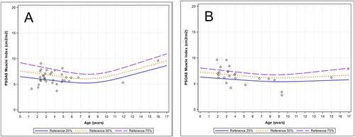

 求助内容:
求助内容: 应助结果提醒方式:


