{"title":"活体有髓神经纤维中Ranvier淋巴结的生理学研究","authors":"O. S. Sotnikov, S. V. Revenko","doi":"10.1134/S1990747822040067","DOIUrl":null,"url":null,"abstract":"<div><div><h3>\n <b>Abstract</b>—</h3><p>A fixed histological preparation cannot reveal the dynamics of morphophysiological objects being merely a basis for hypothetic physiology. At the same time, intravital microscopy of mobile and changing structures can be considered as a branch of cell physiology. The present study focused on revealing the features of this chapter of neurophysiology in relation to nerve fibers. Importantly, the preparation of living neurons isolated by the method of Tasaki inevitably produces mechanical lesions in Ranvier node structure of unknown scale and importance. These lesions can be manifested by deformation or entire elimination of the cone-shaped myelinated regions of the node and/or fiber bulb, as well as by the changes of the nodal gap. Similar alterations can emerge in the intact fiber during a long-term survival in Ringer’s solution. Electron microscopy showed that in hypotonic solutions, swelling and increase in the volume of neuroplasm in the paranodal loops were accompanied by its expansion into the axoplasmic territory in the cone during narrowing of the axon. These processes were reversible, and they probably reflected a novel form of metabolic transmembrane neuron–glial exchange of glucose, amino acids, and other low-molecular weight compounds leading to the formation of integrated cytoplasm of the nerve fiber. The loss of clear boundary of the myelinated cones of the node and/or fiber bulb depended on a large-scale exfoliation of individual main dense lines of Robertson and on flooding the series of paranodal loops. Hypertonic (2 M) solution of urea, which cannot provoke swelling of the cytoplasm but can denature the proteins, also induced similar alterations in the node of Ranvier. Consequently, the described changes in the nodes were not associated with the phenomenon of external osmotic changes, but with the influence of nonspecific physical alterations in conformation of axoplasmic proteins. The voltage clamp experiments with recording of nodal ionic currents demonstrated the correspondence of structural alterations to electrophysiological changes in sodium, potassium, and leakage conductance. The experiments with sodium channel modifier batrachotoxin revealed no structural alterations in Ranvier nodes during 1 h. The present and reviewed data indicate that the nodal changes probably result not from the structural alterations of axolemmal proteins, but from the conformational rearrangements of the axoplasmic ones.</p></div></div>","PeriodicalId":484,"journal":{"name":"Biochemistry (Moscow), Supplement Series A: Membrane and Cell Biology","volume":"16 3","pages":"224 - 235"},"PeriodicalIF":1.1000,"publicationDate":"2022-08-15","publicationTypes":"Journal Article","fieldsOfStudy":null,"isOpenAccess":false,"openAccessPdf":"","citationCount":"1","resultStr":"{\"title\":\"Physiology of Ranvier Nodes in Living Myelinated Nerve Fibers\",\"authors\":\"O. S. Sotnikov, S. V. Revenko\",\"doi\":\"10.1134/S1990747822040067\",\"DOIUrl\":null,\"url\":null,\"abstract\":\"<div><div><h3>\\n <b>Abstract</b>—</h3><p>A fixed histological preparation cannot reveal the dynamics of morphophysiological objects being merely a basis for hypothetic physiology. At the same time, intravital microscopy of mobile and changing structures can be considered as a branch of cell physiology. The present study focused on revealing the features of this chapter of neurophysiology in relation to nerve fibers. Importantly, the preparation of living neurons isolated by the method of Tasaki inevitably produces mechanical lesions in Ranvier node structure of unknown scale and importance. These lesions can be manifested by deformation or entire elimination of the cone-shaped myelinated regions of the node and/or fiber bulb, as well as by the changes of the nodal gap. Similar alterations can emerge in the intact fiber during a long-term survival in Ringer’s solution. Electron microscopy showed that in hypotonic solutions, swelling and increase in the volume of neuroplasm in the paranodal loops were accompanied by its expansion into the axoplasmic territory in the cone during narrowing of the axon. These processes were reversible, and they probably reflected a novel form of metabolic transmembrane neuron–glial exchange of glucose, amino acids, and other low-molecular weight compounds leading to the formation of integrated cytoplasm of the nerve fiber. The loss of clear boundary of the myelinated cones of the node and/or fiber bulb depended on a large-scale exfoliation of individual main dense lines of Robertson and on flooding the series of paranodal loops. Hypertonic (2 M) solution of urea, which cannot provoke swelling of the cytoplasm but can denature the proteins, also induced similar alterations in the node of Ranvier. Consequently, the described changes in the nodes were not associated with the phenomenon of external osmotic changes, but with the influence of nonspecific physical alterations in conformation of axoplasmic proteins. The voltage clamp experiments with recording of nodal ionic currents demonstrated the correspondence of structural alterations to electrophysiological changes in sodium, potassium, and leakage conductance. The experiments with sodium channel modifier batrachotoxin revealed no structural alterations in Ranvier nodes during 1 h. The present and reviewed data indicate that the nodal changes probably result not from the structural alterations of axolemmal proteins, but from the conformational rearrangements of the axoplasmic ones.</p></div></div>\",\"PeriodicalId\":484,\"journal\":{\"name\":\"Biochemistry (Moscow), Supplement Series A: Membrane and Cell Biology\",\"volume\":\"16 3\",\"pages\":\"224 - 235\"},\"PeriodicalIF\":1.1000,\"publicationDate\":\"2022-08-15\",\"publicationTypes\":\"Journal Article\",\"fieldsOfStudy\":null,\"isOpenAccess\":false,\"openAccessPdf\":\"\",\"citationCount\":\"1\",\"resultStr\":null,\"platform\":\"Semanticscholar\",\"paperid\":null,\"PeriodicalName\":\"Biochemistry (Moscow), Supplement Series A: Membrane and Cell Biology\",\"FirstCategoryId\":\"2\",\"ListUrlMain\":\"https://link.springer.com/article/10.1134/S1990747822040067\",\"RegionNum\":0,\"RegionCategory\":null,\"ArticlePicture\":[],\"TitleCN\":null,\"AbstractTextCN\":null,\"PMCID\":null,\"EPubDate\":\"\",\"PubModel\":\"\",\"JCR\":\"Q4\",\"JCRName\":\"CELL BIOLOGY\",\"Score\":null,\"Total\":0}","platform":"Semanticscholar","paperid":null,"PeriodicalName":"Biochemistry (Moscow), Supplement Series A: Membrane and Cell Biology","FirstCategoryId":"2","ListUrlMain":"https://link.springer.com/article/10.1134/S1990747822040067","RegionNum":0,"RegionCategory":null,"ArticlePicture":[],"TitleCN":null,"AbstractTextCN":null,"PMCID":null,"EPubDate":"","PubModel":"","JCR":"Q4","JCRName":"CELL BIOLOGY","Score":null,"Total":0}
Physiology of Ranvier Nodes in Living Myelinated Nerve Fibers
Abstract—
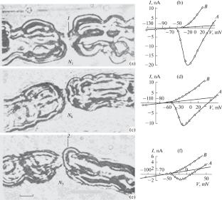
A fixed histological preparation cannot reveal the dynamics of morphophysiological objects being merely a basis for hypothetic physiology. At the same time, intravital microscopy of mobile and changing structures can be considered as a branch of cell physiology. The present study focused on revealing the features of this chapter of neurophysiology in relation to nerve fibers. Importantly, the preparation of living neurons isolated by the method of Tasaki inevitably produces mechanical lesions in Ranvier node structure of unknown scale and importance. These lesions can be manifested by deformation or entire elimination of the cone-shaped myelinated regions of the node and/or fiber bulb, as well as by the changes of the nodal gap. Similar alterations can emerge in the intact fiber during a long-term survival in Ringer’s solution. Electron microscopy showed that in hypotonic solutions, swelling and increase in the volume of neuroplasm in the paranodal loops were accompanied by its expansion into the axoplasmic territory in the cone during narrowing of the axon. These processes were reversible, and they probably reflected a novel form of metabolic transmembrane neuron–glial exchange of glucose, amino acids, and other low-molecular weight compounds leading to the formation of integrated cytoplasm of the nerve fiber. The loss of clear boundary of the myelinated cones of the node and/or fiber bulb depended on a large-scale exfoliation of individual main dense lines of Robertson and on flooding the series of paranodal loops. Hypertonic (2 M) solution of urea, which cannot provoke swelling of the cytoplasm but can denature the proteins, also induced similar alterations in the node of Ranvier. Consequently, the described changes in the nodes were not associated with the phenomenon of external osmotic changes, but with the influence of nonspecific physical alterations in conformation of axoplasmic proteins. The voltage clamp experiments with recording of nodal ionic currents demonstrated the correspondence of structural alterations to electrophysiological changes in sodium, potassium, and leakage conductance. The experiments with sodium channel modifier batrachotoxin revealed no structural alterations in Ranvier nodes during 1 h. The present and reviewed data indicate that the nodal changes probably result not from the structural alterations of axolemmal proteins, but from the conformational rearrangements of the axoplasmic ones.
期刊介绍:
Biochemistry (Moscow), Supplement Series A: Membrane and Cell Biology is an international peer reviewed journal that publishes original articles on physical, chemical, and molecular mechanisms that underlie basic properties of biological membranes and mediate membrane-related cellular functions. The primary topics of the journal are membrane structure, mechanisms of membrane transport, bioenergetics and photobiology, intracellular signaling as well as membrane aspects of cell biology, immunology, and medicine. The journal is multidisciplinary and gives preference to those articles that employ a variety of experimental approaches, basically in biophysics but also in biochemistry, cytology, and molecular biology. The journal publishes articles that strive for unveiling membrane and cellular functions through innovative theoretical models and computer simulations.
 求助内容:
求助内容: 应助结果提醒方式:


