Eunjin Kim, Jiyoung Lee, Seulgi Noh, Ohkyung Kwon, Ji Young Mun
{"title":"扫描电子显微镜阵列层析成像的双重染色方法","authors":"Eunjin Kim, Jiyoung Lee, Seulgi Noh, Ohkyung Kwon, Ji Young Mun","doi":"10.1186/s42649-020-00033-8","DOIUrl":null,"url":null,"abstract":"<p>Scanning electron microscopy (SEM) plays a central role in analyzing structures by imaging a large area of brain tissue at nanometer scales. A vast amount of data in the large area are required to study structural changes of cellular organelles in a specific cell, such as neurons, astrocytes, oligodendrocytes, and microglia among brain tissue, at sufficient resolution. Array tomography is a useful method for large-area imaging, and the osmium-thiocarbohydrazide-osmium (OTO) and ferrocyanide-reduced osmium methods are commonly used to enhance membrane contrast.</p><p>Because many samples prepared using the conventional technique without en bloc staining are considered inadequate for array tomography, we suggested an alternative technique using post-staining conventional samples and compared the advantages.</p>","PeriodicalId":470,"journal":{"name":"Applied Microscopy","volume":"50 1","pages":""},"PeriodicalIF":0.0000,"publicationDate":"2020-06-22","publicationTypes":"Journal Article","fieldsOfStudy":null,"isOpenAccess":false,"openAccessPdf":"https://sci-hub-pdf.com/10.1186/s42649-020-00033-8","citationCount":"2","resultStr":"{\"title\":\"Double staining method for array tomography using scanning electron microscopy\",\"authors\":\"Eunjin Kim, Jiyoung Lee, Seulgi Noh, Ohkyung Kwon, Ji Young Mun\",\"doi\":\"10.1186/s42649-020-00033-8\",\"DOIUrl\":null,\"url\":null,\"abstract\":\"<p>Scanning electron microscopy (SEM) plays a central role in analyzing structures by imaging a large area of brain tissue at nanometer scales. A vast amount of data in the large area are required to study structural changes of cellular organelles in a specific cell, such as neurons, astrocytes, oligodendrocytes, and microglia among brain tissue, at sufficient resolution. Array tomography is a useful method for large-area imaging, and the osmium-thiocarbohydrazide-osmium (OTO) and ferrocyanide-reduced osmium methods are commonly used to enhance membrane contrast.</p><p>Because many samples prepared using the conventional technique without en bloc staining are considered inadequate for array tomography, we suggested an alternative technique using post-staining conventional samples and compared the advantages.</p>\",\"PeriodicalId\":470,\"journal\":{\"name\":\"Applied Microscopy\",\"volume\":\"50 1\",\"pages\":\"\"},\"PeriodicalIF\":0.0000,\"publicationDate\":\"2020-06-22\",\"publicationTypes\":\"Journal Article\",\"fieldsOfStudy\":null,\"isOpenAccess\":false,\"openAccessPdf\":\"https://sci-hub-pdf.com/10.1186/s42649-020-00033-8\",\"citationCount\":\"2\",\"resultStr\":null,\"platform\":\"Semanticscholar\",\"paperid\":null,\"PeriodicalName\":\"Applied Microscopy\",\"FirstCategoryId\":\"1085\",\"ListUrlMain\":\"https://link.springer.com/article/10.1186/s42649-020-00033-8\",\"RegionNum\":0,\"RegionCategory\":null,\"ArticlePicture\":[],\"TitleCN\":null,\"AbstractTextCN\":null,\"PMCID\":null,\"EPubDate\":\"\",\"PubModel\":\"\",\"JCR\":\"Q3\",\"JCRName\":\"Immunology and Microbiology\",\"Score\":null,\"Total\":0}","platform":"Semanticscholar","paperid":null,"PeriodicalName":"Applied Microscopy","FirstCategoryId":"1085","ListUrlMain":"https://link.springer.com/article/10.1186/s42649-020-00033-8","RegionNum":0,"RegionCategory":null,"ArticlePicture":[],"TitleCN":null,"AbstractTextCN":null,"PMCID":null,"EPubDate":"","PubModel":"","JCR":"Q3","JCRName":"Immunology and Microbiology","Score":null,"Total":0}
Double staining method for array tomography using scanning electron microscopy
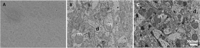
Scanning electron microscopy (SEM) plays a central role in analyzing structures by imaging a large area of brain tissue at nanometer scales. A vast amount of data in the large area are required to study structural changes of cellular organelles in a specific cell, such as neurons, astrocytes, oligodendrocytes, and microglia among brain tissue, at sufficient resolution. Array tomography is a useful method for large-area imaging, and the osmium-thiocarbohydrazide-osmium (OTO) and ferrocyanide-reduced osmium methods are commonly used to enhance membrane contrast.
Because many samples prepared using the conventional technique without en bloc staining are considered inadequate for array tomography, we suggested an alternative technique using post-staining conventional samples and compared the advantages.
Applied MicroscopyImmunology and Microbiology-Applied Microbiology and Biotechnology
CiteScore
3.40
自引率
0.00%
发文量
10
审稿时长
10 weeks
期刊介绍:
Applied Microscopy is a peer-reviewed journal sponsored by the Korean Society of Microscopy. The journal covers all the interdisciplinary fields of technological developments in new microscopy methods and instrumentation and their applications to biological or materials science for determining structure and chemistry. ISSN: 22875123, 22874445.
 求助内容:
求助内容: 应助结果提醒方式:


