A. M. Surin, L. R. Gorbacheva, I. G. Savinkova, R. R. Sharipov, V. G. Pinelis
{"title":"谷氨酸解除培养大鼠海马神经元Ca2+稳态时线粒体基质和细胞质的pH变化","authors":"A. M. Surin, L. R. Gorbacheva, I. G. Savinkova, R. R. Sharipov, V. G. Pinelis","doi":"10.1134/S1990747822040079","DOIUrl":null,"url":null,"abstract":"<p>The effect of high concentrations of glutamate (Glu) on primary cultures of neurons from the rat brain led to a strong depolarization of mitochondria, which developed synchronously with a secondary increase in the intracellular free Ca<sup>2+</sup> concentration (delayed calcium deregulation, DCD). Simultaneously with measurements of the intracellular free Ca<sup>2+</sup> concentration ([Ca<sup>2+</sup>]<sub>i</sub>), pH was measured in the mitochondrial matrix (pH<sub>m</sub>) and cytosol (pH<sub>c</sub>) of neurons when exposed to a toxic dose of Glu (100 µM). For this purpose, pH-sensitive green fluorescent protein mtYFP in mitochondria and pH-sensitive red fluorescent protein mKate in cytosol were expressed in primary cultures from the hippocampus of newborn rats. The resulting neuronal culture was loaded with the Ca<sup>2+</sup> indicator Fura-FF; [Ca<sup>2+</sup>]<sub>i</sub>, pH<sub>m</sub> and pH<sub>c</sub> were simultaneously measured in those neurons that expressed both mtYFP and mKate. It was found that during the first phase of the [Ca<sup>2+</sup>]<sub>i</sub> response to Glu, when partial depolarization of mitochondria was observed, there was an increase in the pH gradient between the mitochondrial matrix and the cytosol (ΔpH), which compensated for the decrease in the electrical component of the mitochondrial potential (∆Ψ<sub>m</sub>), thereby maintaining the constancy of the electrochemical potential of mitochondria. The development of DCD led to an abrupt decrease in ∆Ψ<sub>m</sub> and ΔpH in the soma of neurons; however, a complete collapse of ΔpH was not observed. This may mean that DCD was not caused by a nonspecific megapore in the inner mitochondrial membrane (mPTP), as is commonly believed. Alternatively, part of the mitochondria in the soma of neurons could retain the barrier properties of the inner membrane and did not form mPTP even with the development of DCD and reaching a high [Ca<sup>2+</sup>]<sub>i</sub> plateau.</p>","PeriodicalId":484,"journal":{"name":"Biochemistry (Moscow), Supplement Series A: Membrane and Cell Biology","volume":"16 3","pages":"236 - 245"},"PeriodicalIF":1.1000,"publicationDate":"2022-08-15","publicationTypes":"Journal Article","fieldsOfStudy":null,"isOpenAccess":false,"openAccessPdf":"","citationCount":"0","resultStr":"{\"title\":\"pH Changes in the Mitochondrial Matrix and Cytosol under Glutamate Deregulation of Ca2+ Homeostasis in Cultured Rat Hippocampal Neurons\",\"authors\":\"A. M. Surin, L. R. Gorbacheva, I. G. Savinkova, R. R. Sharipov, V. G. Pinelis\",\"doi\":\"10.1134/S1990747822040079\",\"DOIUrl\":null,\"url\":null,\"abstract\":\"<p>The effect of high concentrations of glutamate (Glu) on primary cultures of neurons from the rat brain led to a strong depolarization of mitochondria, which developed synchronously with a secondary increase in the intracellular free Ca<sup>2+</sup> concentration (delayed calcium deregulation, DCD). Simultaneously with measurements of the intracellular free Ca<sup>2+</sup> concentration ([Ca<sup>2+</sup>]<sub>i</sub>), pH was measured in the mitochondrial matrix (pH<sub>m</sub>) and cytosol (pH<sub>c</sub>) of neurons when exposed to a toxic dose of Glu (100 µM). For this purpose, pH-sensitive green fluorescent protein mtYFP in mitochondria and pH-sensitive red fluorescent protein mKate in cytosol were expressed in primary cultures from the hippocampus of newborn rats. The resulting neuronal culture was loaded with the Ca<sup>2+</sup> indicator Fura-FF; [Ca<sup>2+</sup>]<sub>i</sub>, pH<sub>m</sub> and pH<sub>c</sub> were simultaneously measured in those neurons that expressed both mtYFP and mKate. It was found that during the first phase of the [Ca<sup>2+</sup>]<sub>i</sub> response to Glu, when partial depolarization of mitochondria was observed, there was an increase in the pH gradient between the mitochondrial matrix and the cytosol (ΔpH), which compensated for the decrease in the electrical component of the mitochondrial potential (∆Ψ<sub>m</sub>), thereby maintaining the constancy of the electrochemical potential of mitochondria. The development of DCD led to an abrupt decrease in ∆Ψ<sub>m</sub> and ΔpH in the soma of neurons; however, a complete collapse of ΔpH was not observed. This may mean that DCD was not caused by a nonspecific megapore in the inner mitochondrial membrane (mPTP), as is commonly believed. Alternatively, part of the mitochondria in the soma of neurons could retain the barrier properties of the inner membrane and did not form mPTP even with the development of DCD and reaching a high [Ca<sup>2+</sup>]<sub>i</sub> plateau.</p>\",\"PeriodicalId\":484,\"journal\":{\"name\":\"Biochemistry (Moscow), Supplement Series A: Membrane and Cell Biology\",\"volume\":\"16 3\",\"pages\":\"236 - 245\"},\"PeriodicalIF\":1.1000,\"publicationDate\":\"2022-08-15\",\"publicationTypes\":\"Journal Article\",\"fieldsOfStudy\":null,\"isOpenAccess\":false,\"openAccessPdf\":\"\",\"citationCount\":\"0\",\"resultStr\":null,\"platform\":\"Semanticscholar\",\"paperid\":null,\"PeriodicalName\":\"Biochemistry (Moscow), Supplement Series A: Membrane and Cell Biology\",\"FirstCategoryId\":\"2\",\"ListUrlMain\":\"https://link.springer.com/article/10.1134/S1990747822040079\",\"RegionNum\":0,\"RegionCategory\":null,\"ArticlePicture\":[],\"TitleCN\":null,\"AbstractTextCN\":null,\"PMCID\":null,\"EPubDate\":\"\",\"PubModel\":\"\",\"JCR\":\"Q4\",\"JCRName\":\"CELL BIOLOGY\",\"Score\":null,\"Total\":0}","platform":"Semanticscholar","paperid":null,"PeriodicalName":"Biochemistry (Moscow), Supplement Series A: Membrane and Cell Biology","FirstCategoryId":"2","ListUrlMain":"https://link.springer.com/article/10.1134/S1990747822040079","RegionNum":0,"RegionCategory":null,"ArticlePicture":[],"TitleCN":null,"AbstractTextCN":null,"PMCID":null,"EPubDate":"","PubModel":"","JCR":"Q4","JCRName":"CELL BIOLOGY","Score":null,"Total":0}
pH Changes in the Mitochondrial Matrix and Cytosol under Glutamate Deregulation of Ca2+ Homeostasis in Cultured Rat Hippocampal Neurons
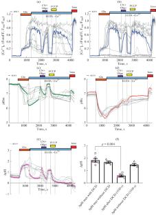
The effect of high concentrations of glutamate (Glu) on primary cultures of neurons from the rat brain led to a strong depolarization of mitochondria, which developed synchronously with a secondary increase in the intracellular free Ca2+ concentration (delayed calcium deregulation, DCD). Simultaneously with measurements of the intracellular free Ca2+ concentration ([Ca2+]i), pH was measured in the mitochondrial matrix (pHm) and cytosol (pHc) of neurons when exposed to a toxic dose of Glu (100 µM). For this purpose, pH-sensitive green fluorescent protein mtYFP in mitochondria and pH-sensitive red fluorescent protein mKate in cytosol were expressed in primary cultures from the hippocampus of newborn rats. The resulting neuronal culture was loaded with the Ca2+ indicator Fura-FF; [Ca2+]i, pHm and pHc were simultaneously measured in those neurons that expressed both mtYFP and mKate. It was found that during the first phase of the [Ca2+]i response to Glu, when partial depolarization of mitochondria was observed, there was an increase in the pH gradient between the mitochondrial matrix and the cytosol (ΔpH), which compensated for the decrease in the electrical component of the mitochondrial potential (∆Ψm), thereby maintaining the constancy of the electrochemical potential of mitochondria. The development of DCD led to an abrupt decrease in ∆Ψm and ΔpH in the soma of neurons; however, a complete collapse of ΔpH was not observed. This may mean that DCD was not caused by a nonspecific megapore in the inner mitochondrial membrane (mPTP), as is commonly believed. Alternatively, part of the mitochondria in the soma of neurons could retain the barrier properties of the inner membrane and did not form mPTP even with the development of DCD and reaching a high [Ca2+]i plateau.
期刊介绍:
Biochemistry (Moscow), Supplement Series A: Membrane and Cell Biology is an international peer reviewed journal that publishes original articles on physical, chemical, and molecular mechanisms that underlie basic properties of biological membranes and mediate membrane-related cellular functions. The primary topics of the journal are membrane structure, mechanisms of membrane transport, bioenergetics and photobiology, intracellular signaling as well as membrane aspects of cell biology, immunology, and medicine. The journal is multidisciplinary and gives preference to those articles that employ a variety of experimental approaches, basically in biophysics but also in biochemistry, cytology, and molecular biology. The journal publishes articles that strive for unveiling membrane and cellular functions through innovative theoretical models and computer simulations.
 求助内容:
求助内容: 应助结果提醒方式:


