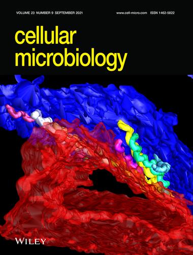
封面图片:询问钩端螺旋体在极化肾近端小管上皮细胞中迁移过程中顶端连接复合体的分解(细胞微生物学,2021年9月)

IF 2.6 2区 生物学 Q3 CELL BIOLOGY
Isabel Sebastián, Nobuhiko Okura, Bruno M. Humbel, Jun Xu, Idam Hermawan, Chiaki Matsuura, Malgorzata Hall, Chitoshi Takayama, Tetsu Yamashiro, Shuichi Nakamura, Claudia Toma
{"title":"封面图片:询问钩端螺旋体在极化肾近端小管上皮细胞中迁移过程中顶端连接复合体的分解(细胞微生物学,2021年9月)","authors":"Isabel Sebastián,&nbsp;Nobuhiko Okura,&nbsp;Bruno M. Humbel,&nbsp;Jun Xu,&nbsp;Idam Hermawan,&nbsp;Chiaki Matsuura,&nbsp;Malgorzata Hall,&nbsp;Chitoshi Takayama,&nbsp;Tetsu Yamashiro,&nbsp;Shuichi Nakamura,&nbsp;Claudia Toma","doi":"10.1111/cmi.13382","DOIUrl":null,"url":null,"abstract":"<p>Focused ion beam-scanning electron microscopy image of renal proximal tubule epithelial cells infected for 24 hrs with <i>Leptospira interrogans</i>. Leptospires localized in the gap between two adjacent cells (red and blue). For further details, readers are referred to the article by Sebastián et al. on p. e13343 of this issue.\n\n <figure>\n <div><picture>\n <source></source></picture><p></p>\n </div>\n </figure></p>","PeriodicalId":9844,"journal":{"name":"Cellular Microbiology","volume":"23 9","pages":""},"PeriodicalIF":2.6000,"publicationDate":"2021-08-17","publicationTypes":"Journal Article","fieldsOfStudy":null,"isOpenAccess":false,"openAccessPdf":"https://sci-hub-pdf.com/10.1111/cmi.13382","citationCount":"0","resultStr":"{\"title\":\"Cover Image: Disassembly of the apical junctional complex during the transmigration of Leptospira interrogans across polarized renal proximal tubule epithelial cells (Cellular Microbiology 09/2021)\",\"authors\":\"Isabel Sebastián,&nbsp;Nobuhiko Okura,&nbsp;Bruno M. Humbel,&nbsp;Jun Xu,&nbsp;Idam Hermawan,&nbsp;Chiaki Matsuura,&nbsp;Malgorzata Hall,&nbsp;Chitoshi Takayama,&nbsp;Tetsu Yamashiro,&nbsp;Shuichi Nakamura,&nbsp;Claudia Toma\",\"doi\":\"10.1111/cmi.13382\",\"DOIUrl\":null,\"url\":null,\"abstract\":\"<p>Focused ion beam-scanning electron microscopy image of renal proximal tubule epithelial cells infected for 24 hrs with <i>Leptospira interrogans</i>. Leptospires localized in the gap between two adjacent cells (red and blue). For further details, readers are referred to the article by Sebastián et al. on p. e13343 of this issue.\\n\\n <figure>\\n <div><picture>\\n <source></source></picture><p></p>\\n </div>\\n </figure></p>\",\"PeriodicalId\":9844,\"journal\":{\"name\":\"Cellular Microbiology\",\"volume\":\"23 9\",\"pages\":\"\"},\"PeriodicalIF\":2.6000,\"publicationDate\":\"2021-08-17\",\"publicationTypes\":\"Journal Article\",\"fieldsOfStudy\":null,\"isOpenAccess\":false,\"openAccessPdf\":\"https://sci-hub-pdf.com/10.1111/cmi.13382\",\"citationCount\":\"0\",\"resultStr\":null,\"platform\":\"Semanticscholar\",\"paperid\":null,\"PeriodicalName\":\"Cellular Microbiology\",\"FirstCategoryId\":\"99\",\"ListUrlMain\":\"https://onlinelibrary.wiley.com/doi/10.1111/cmi.13382\",\"RegionNum\":2,\"RegionCategory\":\"生物学\",\"ArticlePicture\":[],\"TitleCN\":null,\"AbstractTextCN\":null,\"PMCID\":null,\"EPubDate\":\"\",\"PubModel\":\"\",\"JCR\":\"Q3\",\"JCRName\":\"CELL BIOLOGY\",\"Score\":null,\"Total\":0}","platform":"Semanticscholar","paperid":null,"PeriodicalName":"Cellular Microbiology","FirstCategoryId":"99","ListUrlMain":"https://onlinelibrary.wiley.com/doi/10.1111/cmi.13382","RegionNum":2,"RegionCategory":"生物学","ArticlePicture":[],"TitleCN":null,"AbstractTextCN":null,"PMCID":null,"EPubDate":"","PubModel":"","JCR":"Q3","JCRName":"CELL BIOLOGY","Score":null,"Total":0}
引用次数: 0

摘要

被钩端螺旋体感染24小时后肾近端小管上皮细胞的聚焦离子束扫描电镜图像。钩端螺旋体位于两个相邻细胞之间的间隙(红色和蓝色)。欲了解更多细节,请参阅Sebastián等人在本期e13343页上的文章。
本文章由计算机程序翻译,如有差异,请以英文原文为准。

Cover Image: Disassembly of the apical junctional complex during the transmigration of Leptospira interrogans across polarized renal proximal tubule epithelial cells (Cellular Microbiology 09/2021)

Cover Image: Disassembly of the apical junctional complex during the transmigration of Leptospira interrogans across polarized renal proximal tubule epithelial cells (Cellular Microbiology 09/2021)

Focused ion beam-scanning electron microscopy image of renal proximal tubule epithelial cells infected for 24 hrs with Leptospira interrogans. Leptospires localized in the gap between two adjacent cells (red and blue). For further details, readers are referred to the article by Sebastián et al. on p. e13343 of this issue.

求助全文
通过发布文献求助,成功后即可免费获取论文全文。 去求助
来源期刊
Cellular Microbiology
Cellular Microbiology 生物-微生物学
CiteScore
9.70
自引率
0.00%
发文量
26
审稿时长
3 months
期刊介绍: Cellular Microbiology aims to publish outstanding contributions to the understanding of interactions between microbes, prokaryotes and eukaryotes, and their host in the context of pathogenic or mutualistic relationships, including co-infections and microbiota. We welcome studies on single cells, animals and plants, and encourage the use of model hosts and organoid cultures. Submission on cell and molecular biological aspects of microbes, such as their intracellular organization or the establishment and maintenance of their architecture in relation to virulence and pathogenicity are also encouraged. Contributions must provide mechanistic insights supported by quantitative data obtained through imaging, cellular, biochemical, structural or genetic approaches.
×
引用
GB/T 7714-2015
复制
MLA
复制
APA
复制
导出至
BibTeX EndNote RefMan NoteFirst NoteExpress
×
提示
您的信息不完整,为了账户安全,请先补充。
现在去补充
×
提示
您因"违规操作"
具体请查看互助需知
我知道了
×
提示
确定
请完成安全验证×
copy
已复制链接
快去分享给好友吧!
我知道了
右上角分享
点击右上角分享
0
联系我们:info@booksci.cn Book学术提供免费学术资源搜索服务,方便国内外学者检索中英文文献。致力于提供最便捷和优质的服务体验。 Copyright © 2023 布克学术 All rights reserved.
京ICP备2023020795号-1
ghs 京公网安备 11010802042870号
Book学术文献互助
Book学术文献互助群
群 号:481959085
Book学术官方微信