Rieko Tanaka-Yachi, Kazuko Aizawa, Kie shimizu, Hidenori Akutsu, Kazuaki Nakamura
{"title":"低密度细胞培养通过在HepG2细胞中形成紧密连接来增强肝功能","authors":"Rieko Tanaka-Yachi, Kazuko Aizawa, Kie shimizu, Hidenori Akutsu, Kazuaki Nakamura","doi":"10.1111/boc.202200002","DOIUrl":null,"url":null,"abstract":"<div>\n \n \n <section>\n \n <h3> Background Information</h3>\n \n <p>An in vitro evaluation system using cultured hepatocytes is the most useful method in preclinical research, such as drug metabolism and toxicity test. Human hepatocytes should be used in an in vitro evaluation system because the expression of drug-metabolizing enzymes varies among animal species. HepG2 cells, a liver cancer-derived cell line, are widely used as a human hepatocyte model; however, their hepatic functions are generally weak.</p>\n </section>\n \n <section>\n \n <h3> Results</h3>\n \n <p>In this study, we showed that low-density HepG2 cell culture induces hepatic function. The morphology of HepG2 cells was altered depending on the cell density at the time of seeding. Low-density cultured HepG2 cells proliferated as tightly packed colonies. The HepG2 cell colonies in low-density culture demonstrated enhanced tight junction formation. Tight junction protein gene expression levels, such as those of zonula occludens-1 (ZO-1), junctional adhesion molecule 1 (JAM), claudin, occludin, and tricellulin, increased in low-density cultured HepG2 cells. Phases I and II metabolic enzymes, phase III transporter gene expression, and CYP3A4 activity also increased in low-density cultured HepG2 cells. Occludin and tricellulin knockdown inhibited the increased hepatic function in low-density cultures. Tricellulin knockdown reduced the expression of hepatocyte nuclear factor 6 (HNF6), CCAAT/enhancer-binding protein alpha (CEBPA), and aryl hydrocarbon receptor (AHR). In addition, the expression of nuclear receptor subfamily 1 group h member 2 (NR1H2) increased in low-density cultures, canceled by occludin and tricellulin knockdown.</p>\n </section>\n \n <section>\n \n <h3> Conclusions</h3>\n \n <p>Our results suggest that low-density HepG2 cell cultures enhance hepatic function by promoting tight junction formation and demonstrate the importance of cell density in drug evaluation using hepatocyte cell lines.</p>\n </section>\n </div>","PeriodicalId":8859,"journal":{"name":"Biology of the Cell","volume":"114 9","pages":"225-236"},"PeriodicalIF":2.4000,"publicationDate":"2022-05-23","publicationTypes":"Journal Article","fieldsOfStudy":null,"isOpenAccess":false,"openAccessPdf":"","citationCount":"0","resultStr":"{\"title\":\"Low-density cell culture enhances hepatic function through tight junction formation in HepG2 cells\",\"authors\":\"Rieko Tanaka-Yachi, Kazuko Aizawa, Kie shimizu, Hidenori Akutsu, Kazuaki Nakamura\",\"doi\":\"10.1111/boc.202200002\",\"DOIUrl\":null,\"url\":null,\"abstract\":\"<div>\\n \\n \\n <section>\\n \\n <h3> Background Information</h3>\\n \\n <p>An in vitro evaluation system using cultured hepatocytes is the most useful method in preclinical research, such as drug metabolism and toxicity test. Human hepatocytes should be used in an in vitro evaluation system because the expression of drug-metabolizing enzymes varies among animal species. HepG2 cells, a liver cancer-derived cell line, are widely used as a human hepatocyte model; however, their hepatic functions are generally weak.</p>\\n </section>\\n \\n <section>\\n \\n <h3> Results</h3>\\n \\n <p>In this study, we showed that low-density HepG2 cell culture induces hepatic function. The morphology of HepG2 cells was altered depending on the cell density at the time of seeding. Low-density cultured HepG2 cells proliferated as tightly packed colonies. The HepG2 cell colonies in low-density culture demonstrated enhanced tight junction formation. Tight junction protein gene expression levels, such as those of zonula occludens-1 (ZO-1), junctional adhesion molecule 1 (JAM), claudin, occludin, and tricellulin, increased in low-density cultured HepG2 cells. Phases I and II metabolic enzymes, phase III transporter gene expression, and CYP3A4 activity also increased in low-density cultured HepG2 cells. Occludin and tricellulin knockdown inhibited the increased hepatic function in low-density cultures. Tricellulin knockdown reduced the expression of hepatocyte nuclear factor 6 (HNF6), CCAAT/enhancer-binding protein alpha (CEBPA), and aryl hydrocarbon receptor (AHR). In addition, the expression of nuclear receptor subfamily 1 group h member 2 (NR1H2) increased in low-density cultures, canceled by occludin and tricellulin knockdown.</p>\\n </section>\\n \\n <section>\\n \\n <h3> Conclusions</h3>\\n \\n <p>Our results suggest that low-density HepG2 cell cultures enhance hepatic function by promoting tight junction formation and demonstrate the importance of cell density in drug evaluation using hepatocyte cell lines.</p>\\n </section>\\n </div>\",\"PeriodicalId\":8859,\"journal\":{\"name\":\"Biology of the Cell\",\"volume\":\"114 9\",\"pages\":\"225-236\"},\"PeriodicalIF\":2.4000,\"publicationDate\":\"2022-05-23\",\"publicationTypes\":\"Journal Article\",\"fieldsOfStudy\":null,\"isOpenAccess\":false,\"openAccessPdf\":\"\",\"citationCount\":\"0\",\"resultStr\":null,\"platform\":\"Semanticscholar\",\"paperid\":null,\"PeriodicalName\":\"Biology of the Cell\",\"FirstCategoryId\":\"99\",\"ListUrlMain\":\"https://onlinelibrary.wiley.com/doi/10.1111/boc.202200002\",\"RegionNum\":4,\"RegionCategory\":\"生物学\",\"ArticlePicture\":[],\"TitleCN\":null,\"AbstractTextCN\":null,\"PMCID\":null,\"EPubDate\":\"\",\"PubModel\":\"\",\"JCR\":\"Q4\",\"JCRName\":\"CELL BIOLOGY\",\"Score\":null,\"Total\":0}","platform":"Semanticscholar","paperid":null,"PeriodicalName":"Biology of the Cell","FirstCategoryId":"99","ListUrlMain":"https://onlinelibrary.wiley.com/doi/10.1111/boc.202200002","RegionNum":4,"RegionCategory":"生物学","ArticlePicture":[],"TitleCN":null,"AbstractTextCN":null,"PMCID":null,"EPubDate":"","PubModel":"","JCR":"Q4","JCRName":"CELL BIOLOGY","Score":null,"Total":0}
Low-density cell culture enhances hepatic function through tight junction formation in HepG2 cells
Background Information
An in vitro evaluation system using cultured hepatocytes is the most useful method in preclinical research, such as drug metabolism and toxicity test. Human hepatocytes should be used in an in vitro evaluation system because the expression of drug-metabolizing enzymes varies among animal species. HepG2 cells, a liver cancer-derived cell line, are widely used as a human hepatocyte model; however, their hepatic functions are generally weak.
Results
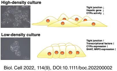
In this study, we showed that low-density HepG2 cell culture induces hepatic function. The morphology of HepG2 cells was altered depending on the cell density at the time of seeding. Low-density cultured HepG2 cells proliferated as tightly packed colonies. The HepG2 cell colonies in low-density culture demonstrated enhanced tight junction formation. Tight junction protein gene expression levels, such as those of zonula occludens-1 (ZO-1), junctional adhesion molecule 1 (JAM), claudin, occludin, and tricellulin, increased in low-density cultured HepG2 cells. Phases I and II metabolic enzymes, phase III transporter gene expression, and CYP3A4 activity also increased in low-density cultured HepG2 cells. Occludin and tricellulin knockdown inhibited the increased hepatic function in low-density cultures. Tricellulin knockdown reduced the expression of hepatocyte nuclear factor 6 (HNF6), CCAAT/enhancer-binding protein alpha (CEBPA), and aryl hydrocarbon receptor (AHR). In addition, the expression of nuclear receptor subfamily 1 group h member 2 (NR1H2) increased in low-density cultures, canceled by occludin and tricellulin knockdown.
Conclusions
Our results suggest that low-density HepG2 cell cultures enhance hepatic function by promoting tight junction formation and demonstrate the importance of cell density in drug evaluation using hepatocyte cell lines.
期刊介绍:
The journal publishes original research articles and reviews on all aspects of cellular, molecular and structural biology, developmental biology, cell physiology and evolution. It will publish articles or reviews contributing to the understanding of the elementary biochemical and biophysical principles of live matter organization from the molecular, cellular and tissues scales and organisms.
This includes contributions directed towards understanding biochemical and biophysical mechanisms, structure-function relationships with respect to basic cell and tissue functions, development, development/evolution relationship, morphogenesis, stem cell biology, cell biology of disease, plant cell biology, as well as contributions directed toward understanding integrated processes at the organelles, cell and tissue levels. Contributions using approaches such as high resolution imaging, live imaging, quantitative cell biology and integrated biology; as well as those using innovative genetic and epigenetic technologies, ex-vivo tissue engineering, cellular, tissue and integrated functional analysis, and quantitative biology and modeling to demonstrate original biological principles are encouraged.
 求助内容:
求助内容: 应助结果提醒方式:


