3D打印技术在复杂肾结石经皮肾镜取石术中的应用

Guobin Zhao, Xin Liu, Yu-hong Tang, H. Su, Lidong Tang, C. Feng, Chaoyang Li, Yu Wang, Haibin Ling
{"title":"3D打印技术在复杂肾结石经皮肾镜取石术中的应用","authors":"Guobin Zhao, Xin Liu, Yu-hong Tang, H. Su, Lidong Tang, C. Feng, Chaoyang Li, Yu Wang, Haibin Ling","doi":"10.3760/CMA.J.ISSN.1008-6315.2020.02.009","DOIUrl":null,"url":null,"abstract":"Objective \nTo investigate the application of 3D printing technique in percutaneous nephrolithotomy (PCNL) of patients with complicated kidney stones. \n \n \nMethods \nFrom January 2015 to December 2017, 60 patients with complicated kidney stones were enrolled in the First Affiliated Hospital of Hebei North University for prospective study, and PCNL was proposed.All the patients were randomly divided into 3D print group (30 cases) and conventional image inspection group (30 cases, control group). Before operation, CT urography (CTU) was used in both groups.In 3D printing group, digital imaging and communications in medicine (DICOM) files of CT were extracted for 3D image postprocessing, and thermoplastic materials were used to print 3D model.In the 3D printing group, the digital imaging and communications in medicine (DICOM) files of CT were extracted for 3D image post-processing, and the 3D model was printed with thermoplastic materials.According to the comprehensive planning of 3D kidney model, a virtual safe and reliable percutaneous renal access was established for each patient, and PCNL was executed.The patients in the two groups were compared before, during and after operation.Preoperative: age, sex, body mass index, blood creatinine, stone size and CT value.During the operation: (1) the target renal calices location time; (2) the conformity between the preoperative planning and the actual operation; (3) the operation completion time.After operation: (1) stone removal rate; (2) hemoglobin reduction level; (3) postoperative recovery. \n \n \nResults \nAll the 60 patients successfully completed the operation, 30 patients successfully printed out the 3D model, which can accurately express the relationship between the stone and the adjacent anatomical structure, the internal renal artery and the collecting system.Positioning time of 3D printing group in target renal calices((2.9 ± 1.5) min vs.(5.8 ± 1.7) min, P=0.023), coincidence between simulated and actual puncture calices((89.5 ± 3.5)% vs.(60.2 ± 5.7)%, P=0.005), postoperative stone removal rate ((89.9 ± 4.5)% vs.(75.9 ± 5.2)%, P=0.009), and hemoglobin levels((1.4 ± 0.5) g/L vs.(2.9 ± 1.4) g/L, P=0.032) were superior to the control group, and the difference was statistically significant.But there was no significant difference between the two groups (all P>0.05). \n \n \nConclusion \nThe 3D printed kidney model truly restores the anatomical details around the kidneys and stones, providing a stereoscopic and intuitive way to perform surgery, so it maybe has a significance guidance for percutaneous nephrolithotomy. \n \n \nKey words: \nKidney stones; Percutaneous nephrolithotomy; Kidney model; Surgery simulation","PeriodicalId":10365,"journal":{"name":"中国综合临床","volume":null,"pages":null},"PeriodicalIF":0.0000,"publicationDate":"2020-03-01","publicationTypes":"Journal Article","fieldsOfStudy":null,"isOpenAccess":false,"openAccessPdf":"","citationCount":"0","resultStr":"{\"title\":\"Application of 3D printing technique in percutaneous nephrolithotomy of patients with complicated kidney stones\",\"authors\":\"Guobin Zhao, Xin Liu, Yu-hong Tang, H. Su, Lidong Tang, C. Feng, Chaoyang Li, Yu Wang, Haibin Ling\",\"doi\":\"10.3760/CMA.J.ISSN.1008-6315.2020.02.009\",\"DOIUrl\":null,\"url\":null,\"abstract\":\"Objective \\nTo investigate the application of 3D printing technique in percutaneous nephrolithotomy (PCNL) of patients with complicated kidney stones. \\n \\n \\nMethods \\nFrom January 2015 to December 2017, 60 patients with complicated kidney stones were enrolled in the First Affiliated Hospital of Hebei North University for prospective study, and PCNL was proposed.All the patients were randomly divided into 3D print group (30 cases) and conventional image inspection group (30 cases, control group). Before operation, CT urography (CTU) was used in both groups.In 3D printing group, digital imaging and communications in medicine (DICOM) files of CT were extracted for 3D image postprocessing, and thermoplastic materials were used to print 3D model.In the 3D printing group, the digital imaging and communications in medicine (DICOM) files of CT were extracted for 3D image post-processing, and the 3D model was printed with thermoplastic materials.According to the comprehensive planning of 3D kidney model, a virtual safe and reliable percutaneous renal access was established for each patient, and PCNL was executed.The patients in the two groups were compared before, during and after operation.Preoperative: age, sex, body mass index, blood creatinine, stone size and CT value.During the operation: (1) the target renal calices location time; (2) the conformity between the preoperative planning and the actual operation; (3) the operation completion time.After operation: (1) stone removal rate; (2) hemoglobin reduction level; (3) postoperative recovery. \\n \\n \\nResults \\nAll the 60 patients successfully completed the operation, 30 patients successfully printed out the 3D model, which can accurately express the relationship between the stone and the adjacent anatomical structure, the internal renal artery and the collecting system.Positioning time of 3D printing group in target renal calices((2.9 ± 1.5) min vs.(5.8 ± 1.7) min, P=0.023), coincidence between simulated and actual puncture calices((89.5 ± 3.5)% vs.(60.2 ± 5.7)%, P=0.005), postoperative stone removal rate ((89.9 ± 4.5)% vs.(75.9 ± 5.2)%, P=0.009), and hemoglobin levels((1.4 ± 0.5) g/L vs.(2.9 ± 1.4) g/L, P=0.032) were superior to the control group, and the difference was statistically significant.But there was no significant difference between the two groups (all P>0.05). \\n \\n \\nConclusion \\nThe 3D printed kidney model truly restores the anatomical details around the kidneys and stones, providing a stereoscopic and intuitive way to perform surgery, so it maybe has a significance guidance for percutaneous nephrolithotomy. \\n \\n \\nKey words: \\nKidney stones; Percutaneous nephrolithotomy; Kidney model; Surgery simulation\",\"PeriodicalId\":10365,\"journal\":{\"name\":\"中国综合临床\",\"volume\":null,\"pages\":null},\"PeriodicalIF\":0.0000,\"publicationDate\":\"2020-03-01\",\"publicationTypes\":\"Journal Article\",\"fieldsOfStudy\":null,\"isOpenAccess\":false,\"openAccessPdf\":\"\",\"citationCount\":\"0\",\"resultStr\":null,\"platform\":\"Semanticscholar\",\"paperid\":null,\"PeriodicalName\":\"中国综合临床\",\"FirstCategoryId\":\"3\",\"ListUrlMain\":\"https://doi.org/10.3760/CMA.J.ISSN.1008-6315.2020.02.009\",\"RegionNum\":0,\"RegionCategory\":null,\"ArticlePicture\":[],\"TitleCN\":null,\"AbstractTextCN\":null,\"PMCID\":null,\"EPubDate\":\"\",\"PubModel\":\"\",\"JCR\":\"\",\"JCRName\":\"\",\"Score\":null,\"Total\":0}","platform":"Semanticscholar","paperid":null,"PeriodicalName":"中国综合临床","FirstCategoryId":"3","ListUrlMain":"https://doi.org/10.3760/CMA.J.ISSN.1008-6315.2020.02.009","RegionNum":0,"RegionCategory":null,"ArticlePicture":[],"TitleCN":null,"AbstractTextCN":null,"PMCID":null,"EPubDate":"","PubModel":"","JCR":"","JCRName":"","Score":null,"Total":0}
引用次数: 0

摘要

目的探讨三维打印技术在复杂性肾结石经皮肾取石术(PCNL)中的应用。方法2015年1月至2017年12月,在河北北方大学第一附属医院对60例复杂性肾结石患者进行前瞻性研究,并提出PCNL。将所有患者随机分为3D打印组(30例)和常规图像检查组(对照组30例)。术前两组均行CT尿路造影(CTU)检查。在3D打印组中,提取CT的数字成像和医学通信(DICOM)文件进行3D图像后处理,并使用热塑性材料打印3D模型。在3D打印组中,提取CT的数字成像和医学通信(DICOM)文件进行3D图像后处理,并用热塑性材料打印3D模型。根据三维肾脏模型的综合规划,为每位患者建立虚拟安全可靠的经皮肾穿刺入路,并执行PCNL。比较两组患者术前、术中、术后的疗效。术前:年龄、性别、体重指数、血肌酐、结石大小、CT值。术中:(1)靶肾盏定位时间;(2) 术前计划与实际操作的一致性;(3) 操作完成时间。术后:(1)结石清除率;(2) 血红蛋白降低水平;(3) 术后恢复。结果60例患者全部成功完成手术,30例患者成功打印出三维模型,能够准确表达结石与邻近解剖结构、肾内动脉和收集系统之间的关系。3D打印组在目标肾盏中的定位时间((2.9±1.5)min vs.(5.8±1.7)min,P=0.023),模拟与实际穿刺盏之间的一致性((89.5±3.5)%vs.(60.2±5.7)%,P=0.005),术后结石清除率((89.9±4.5)%vs.(75.9±5.2)%,P=0.009),血红蛋白水平((1.4±0.5)g/L vs.(2.9±1.4)g/L,P=0.032)均优于对照组,差异有统计学意义。结论3D打印的肾脏模型真实地还原了肾脏和结石周围的解剖细节,为手术提供了一种立体直观的方法,对经皮肾镜取石术有一定的指导意义。关键词:肾结石;经皮肾取石术;肾脏模型;手术模拟
本文章由计算机程序翻译,如有差异,请以英文原文为准。
Application of 3D printing technique in percutaneous nephrolithotomy of patients with complicated kidney stones
Objective To investigate the application of 3D printing technique in percutaneous nephrolithotomy (PCNL) of patients with complicated kidney stones. Methods From January 2015 to December 2017, 60 patients with complicated kidney stones were enrolled in the First Affiliated Hospital of Hebei North University for prospective study, and PCNL was proposed.All the patients were randomly divided into 3D print group (30 cases) and conventional image inspection group (30 cases, control group). Before operation, CT urography (CTU) was used in both groups.In 3D printing group, digital imaging and communications in medicine (DICOM) files of CT were extracted for 3D image postprocessing, and thermoplastic materials were used to print 3D model.In the 3D printing group, the digital imaging and communications in medicine (DICOM) files of CT were extracted for 3D image post-processing, and the 3D model was printed with thermoplastic materials.According to the comprehensive planning of 3D kidney model, a virtual safe and reliable percutaneous renal access was established for each patient, and PCNL was executed.The patients in the two groups were compared before, during and after operation.Preoperative: age, sex, body mass index, blood creatinine, stone size and CT value.During the operation: (1) the target renal calices location time; (2) the conformity between the preoperative planning and the actual operation; (3) the operation completion time.After operation: (1) stone removal rate; (2) hemoglobin reduction level; (3) postoperative recovery. Results All the 60 patients successfully completed the operation, 30 patients successfully printed out the 3D model, which can accurately express the relationship between the stone and the adjacent anatomical structure, the internal renal artery and the collecting system.Positioning time of 3D printing group in target renal calices((2.9 ± 1.5) min vs.(5.8 ± 1.7) min, P=0.023), coincidence between simulated and actual puncture calices((89.5 ± 3.5)% vs.(60.2 ± 5.7)%, P=0.005), postoperative stone removal rate ((89.9 ± 4.5)% vs.(75.9 ± 5.2)%, P=0.009), and hemoglobin levels((1.4 ± 0.5) g/L vs.(2.9 ± 1.4) g/L, P=0.032) were superior to the control group, and the difference was statistically significant.But there was no significant difference between the two groups (all P>0.05). Conclusion The 3D printed kidney model truly restores the anatomical details around the kidneys and stones, providing a stereoscopic and intuitive way to perform surgery, so it maybe has a significance guidance for percutaneous nephrolithotomy. Key words: Kidney stones; Percutaneous nephrolithotomy; Kidney model; Surgery simulation
求助全文
通过发布文献求助,成功后即可免费获取论文全文。 去求助
来源期刊
CiteScore
0.10
自引率
0.00%
发文量
16855
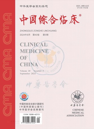
期刊介绍: Clinical Medicine of China is an academic journal organized by the Chinese Medical Association (CMA), which mainly publishes original research papers, reviews and commentaries in the field. Clinical Medicine of China is a source journal of Peking University (2000 and 2004 editions), a core journal of Chinese science and technology, an academic journal of RCCSE China Core (Extended Edition), and has been published in Chemical Abstracts of the United States (CA), Abstracts Journal of Russia (AJ), Chinese Core Journals (Selection) Database, Chinese Science and Technology Materials Directory, Wanfang Database, China Academic Journal Database, JST Japan Science and Technology Agency Database (Japanese) (2018) and other databases.
×
引用
GB/T 7714-2015
复制
MLA
复制
APA
复制
导出至
BibTeX EndNote RefMan NoteFirst NoteExpress
×
提示
您的信息不完整,为了账户安全,请先补充。
现在去补充
×
提示
您因"违规操作"
具体请查看互助需知
我知道了
×
提示
确定
请完成安全验证×
copy
已复制链接
快去分享给好友吧!
我知道了
右上角分享
点击右上角分享
0
联系我们:info@booksci.cn Book学术提供免费学术资源搜索服务,方便国内外学者检索中英文文献。致力于提供最便捷和优质的服务体验。 Copyright © 2023 布克学术 All rights reserved.
京ICP备2023020795号-1
ghs 京公网安备 11010802042870号
Book学术文献互助
Book学术文献互助群
群 号:481959085
Book学术官方微信