{"title":"利用共聚焦全玻片成像扫描仪对荧光原位杂交(FISH)进行自动三维评分","authors":"Ziv Frankenstein, Naohiro Uraoka, Umut Aypar, Ruth Aryeequaye, Mamta Rao, Meera Hameed, Yanming Zhang, Yukako Yagi","doi":"10.1186/s42649-021-00053-y","DOIUrl":null,"url":null,"abstract":"<p>Fluorescence in situ hybridization (FISH) is a technique to visualize specific DNA/RNA sequences within the cell nuclei and provide the presence, location and structural integrity of genes on chromosomes. A confocal Whole Slide Imaging (WSI) scanner technology has superior depth resolution compared to wide-field fluorescence imaging. Confocal WSI has the ability to perform serial optical sections with specimen imaging, which is critical for 3D tissue reconstruction for volumetric spatial analysis. The standard clinical manual scoring for FISH is labor-intensive, time-consuming and subjective. Application of multi-gene FISH analysis alongside 3D imaging, significantly increase the level of complexity required for an accurate 3D analysis. Therefore, the purpose of this study is to establish automated 3D FISH scoring for z-stack images from confocal WSI scanner. The algorithm and the application we developed, SHIMARIS PAFQ, successfully employs 3D calculations for clear individual cell nuclei segmentation, gene signals detection and distribution of break-apart probes signal patterns, including standard break-apart, and variant patterns due to truncation, and deletion, etc. The analysis was accurate and precise when compared with ground truth clinical manual counting and scoring reported in ten lymphoma and solid tumors cases. The algorithm and the application we developed, SHIMARIS PAFQ, is objective and more efficient than the conventional procedure. It enables the automated counting of more nuclei, precisely detecting additional abnormal signal variations in nuclei patterns and analyzes gigabyte multi-layer stacking imaging data of tissue samples from patients. Currently, we are developing a deep learning algorithm for automated tumor area detection to be integrated with SHIMARIS PAFQ.</p>","PeriodicalId":470,"journal":{"name":"Applied Microscopy","volume":"51 1","pages":""},"PeriodicalIF":0.0000,"publicationDate":"2021-04-09","publicationTypes":"Journal Article","fieldsOfStudy":null,"isOpenAccess":false,"openAccessPdf":"https://sci-hub-pdf.com/10.1186/s42649-021-00053-y","citationCount":"2","resultStr":"{\"title\":\"Automated 3D scoring of fluorescence in situ hybridization (FISH) using a confocal whole slide imaging scanner\",\"authors\":\"Ziv Frankenstein, Naohiro Uraoka, Umut Aypar, Ruth Aryeequaye, Mamta Rao, Meera Hameed, Yanming Zhang, Yukako Yagi\",\"doi\":\"10.1186/s42649-021-00053-y\",\"DOIUrl\":null,\"url\":null,\"abstract\":\"<p>Fluorescence in situ hybridization (FISH) is a technique to visualize specific DNA/RNA sequences within the cell nuclei and provide the presence, location and structural integrity of genes on chromosomes. A confocal Whole Slide Imaging (WSI) scanner technology has superior depth resolution compared to wide-field fluorescence imaging. Confocal WSI has the ability to perform serial optical sections with specimen imaging, which is critical for 3D tissue reconstruction for volumetric spatial analysis. The standard clinical manual scoring for FISH is labor-intensive, time-consuming and subjective. Application of multi-gene FISH analysis alongside 3D imaging, significantly increase the level of complexity required for an accurate 3D analysis. Therefore, the purpose of this study is to establish automated 3D FISH scoring for z-stack images from confocal WSI scanner. The algorithm and the application we developed, SHIMARIS PAFQ, successfully employs 3D calculations for clear individual cell nuclei segmentation, gene signals detection and distribution of break-apart probes signal patterns, including standard break-apart, and variant patterns due to truncation, and deletion, etc. The analysis was accurate and precise when compared with ground truth clinical manual counting and scoring reported in ten lymphoma and solid tumors cases. The algorithm and the application we developed, SHIMARIS PAFQ, is objective and more efficient than the conventional procedure. It enables the automated counting of more nuclei, precisely detecting additional abnormal signal variations in nuclei patterns and analyzes gigabyte multi-layer stacking imaging data of tissue samples from patients. Currently, we are developing a deep learning algorithm for automated tumor area detection to be integrated with SHIMARIS PAFQ.</p>\",\"PeriodicalId\":470,\"journal\":{\"name\":\"Applied Microscopy\",\"volume\":\"51 1\",\"pages\":\"\"},\"PeriodicalIF\":0.0000,\"publicationDate\":\"2021-04-09\",\"publicationTypes\":\"Journal Article\",\"fieldsOfStudy\":null,\"isOpenAccess\":false,\"openAccessPdf\":\"https://sci-hub-pdf.com/10.1186/s42649-021-00053-y\",\"citationCount\":\"2\",\"resultStr\":null,\"platform\":\"Semanticscholar\",\"paperid\":null,\"PeriodicalName\":\"Applied Microscopy\",\"FirstCategoryId\":\"1085\",\"ListUrlMain\":\"https://link.springer.com/article/10.1186/s42649-021-00053-y\",\"RegionNum\":0,\"RegionCategory\":null,\"ArticlePicture\":[],\"TitleCN\":null,\"AbstractTextCN\":null,\"PMCID\":null,\"EPubDate\":\"\",\"PubModel\":\"\",\"JCR\":\"Q3\",\"JCRName\":\"Immunology and Microbiology\",\"Score\":null,\"Total\":0}","platform":"Semanticscholar","paperid":null,"PeriodicalName":"Applied Microscopy","FirstCategoryId":"1085","ListUrlMain":"https://link.springer.com/article/10.1186/s42649-021-00053-y","RegionNum":0,"RegionCategory":null,"ArticlePicture":[],"TitleCN":null,"AbstractTextCN":null,"PMCID":null,"EPubDate":"","PubModel":"","JCR":"Q3","JCRName":"Immunology and Microbiology","Score":null,"Total":0}
Automated 3D scoring of fluorescence in situ hybridization (FISH) using a confocal whole slide imaging scanner
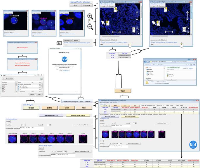
Fluorescence in situ hybridization (FISH) is a technique to visualize specific DNA/RNA sequences within the cell nuclei and provide the presence, location and structural integrity of genes on chromosomes. A confocal Whole Slide Imaging (WSI) scanner technology has superior depth resolution compared to wide-field fluorescence imaging. Confocal WSI has the ability to perform serial optical sections with specimen imaging, which is critical for 3D tissue reconstruction for volumetric spatial analysis. The standard clinical manual scoring for FISH is labor-intensive, time-consuming and subjective. Application of multi-gene FISH analysis alongside 3D imaging, significantly increase the level of complexity required for an accurate 3D analysis. Therefore, the purpose of this study is to establish automated 3D FISH scoring for z-stack images from confocal WSI scanner. The algorithm and the application we developed, SHIMARIS PAFQ, successfully employs 3D calculations for clear individual cell nuclei segmentation, gene signals detection and distribution of break-apart probes signal patterns, including standard break-apart, and variant patterns due to truncation, and deletion, etc. The analysis was accurate and precise when compared with ground truth clinical manual counting and scoring reported in ten lymphoma and solid tumors cases. The algorithm and the application we developed, SHIMARIS PAFQ, is objective and more efficient than the conventional procedure. It enables the automated counting of more nuclei, precisely detecting additional abnormal signal variations in nuclei patterns and analyzes gigabyte multi-layer stacking imaging data of tissue samples from patients. Currently, we are developing a deep learning algorithm for automated tumor area detection to be integrated with SHIMARIS PAFQ.
Applied MicroscopyImmunology and Microbiology-Applied Microbiology and Biotechnology
CiteScore
3.40
自引率
0.00%
发文量
10
审稿时长
10 weeks
期刊介绍:
Applied Microscopy is a peer-reviewed journal sponsored by the Korean Society of Microscopy. The journal covers all the interdisciplinary fields of technological developments in new microscopy methods and instrumentation and their applications to biological or materials science for determining structure and chemistry. ISSN: 22875123, 22874445.

 求助内容:
求助内容: 应助结果提醒方式:


