{"title":"原发性骨内脑膜瘤:罕见的高增殖能力恶性肿瘤","authors":"Yoshiki Sato, Koichi Mitsuya, Yuko Kakuda, Takuma Oishi, Shoichi Deguchi, Takashi Sugino, Yasuto Akiyama, Takeshi Nagashima, Kenichi Urakami, Yuji Shimoda, Keiichi Ohshima, Nakamasa Hayashi, Ken Yamaguchi","doi":"10.1055/a-2161-7710","DOIUrl":null,"url":null,"abstract":"<p><p>Primary intraosseous meningioma (PIM) is a rare tumor that arises in the skull. Histopathologically, it is generally described as a slow-growing, benign lesion. However, on rare occasions, PIM presents as a malignancy with high proliferative ability, which requires maximal resection, adjuvant radiotherapy, and subsequent careful follow-up. Because of the rarity of such cases, they present a diagnostic challenge with unusual pathological findings. Herein, we report a case of a primary intraosseous anaplastic meningioma with extensive invasion inside and outside the skull, along with the results of whole-genome analysis. Histopathological diagnosis was a World Health Organization grade 3 anaplastic meningioma. In the literature, only two cases of anaplastic PIM have been reported, so its characteristics and treatment are poorly understood. Our patient was successfully treated with tumor resection, followed by intensity-modulated radiation therapy. Follow-up imaging studies revealed no recurrence or distant metastasis, including to lung, liver, and bone, at 8 months after the surgery.</p>","PeriodicalId":44256,"journal":{"name":"Journal of Neurological Surgery Reports","volume":"84 1","pages":"e103-e108"},"PeriodicalIF":0.7000,"publicationDate":"2023-09-27","publicationTypes":"Journal Article","fieldsOfStudy":null,"isOpenAccess":false,"openAccessPdf":"https://www.ncbi.nlm.nih.gov/pmc/articles/PMC10611535/pdf/","citationCount":"0","resultStr":"{\"title\":\"A Primary Intraosseous Meningioma: A Rare Case of Malignancy with High Proliferative Ability.\",\"authors\":\"Yoshiki Sato, Koichi Mitsuya, Yuko Kakuda, Takuma Oishi, Shoichi Deguchi, Takashi Sugino, Yasuto Akiyama, Takeshi Nagashima, Kenichi Urakami, Yuji Shimoda, Keiichi Ohshima, Nakamasa Hayashi, Ken Yamaguchi\",\"doi\":\"10.1055/a-2161-7710\",\"DOIUrl\":null,\"url\":null,\"abstract\":\"<p><p>Primary intraosseous meningioma (PIM) is a rare tumor that arises in the skull. Histopathologically, it is generally described as a slow-growing, benign lesion. However, on rare occasions, PIM presents as a malignancy with high proliferative ability, which requires maximal resection, adjuvant radiotherapy, and subsequent careful follow-up. Because of the rarity of such cases, they present a diagnostic challenge with unusual pathological findings. Herein, we report a case of a primary intraosseous anaplastic meningioma with extensive invasion inside and outside the skull, along with the results of whole-genome analysis. Histopathological diagnosis was a World Health Organization grade 3 anaplastic meningioma. In the literature, only two cases of anaplastic PIM have been reported, so its characteristics and treatment are poorly understood. Our patient was successfully treated with tumor resection, followed by intensity-modulated radiation therapy. Follow-up imaging studies revealed no recurrence or distant metastasis, including to lung, liver, and bone, at 8 months after the surgery.</p>\",\"PeriodicalId\":44256,\"journal\":{\"name\":\"Journal of Neurological Surgery Reports\",\"volume\":\"84 1\",\"pages\":\"e103-e108\"},\"PeriodicalIF\":0.7000,\"publicationDate\":\"2023-09-27\",\"publicationTypes\":\"Journal Article\",\"fieldsOfStudy\":null,\"isOpenAccess\":false,\"openAccessPdf\":\"https://www.ncbi.nlm.nih.gov/pmc/articles/PMC10611535/pdf/\",\"citationCount\":\"0\",\"resultStr\":null,\"platform\":\"Semanticscholar\",\"paperid\":null,\"PeriodicalName\":\"Journal of Neurological Surgery Reports\",\"FirstCategoryId\":\"1085\",\"ListUrlMain\":\"https://doi.org/10.1055/a-2161-7710\",\"RegionNum\":0,\"RegionCategory\":null,\"ArticlePicture\":[],\"TitleCN\":null,\"AbstractTextCN\":null,\"PMCID\":null,\"EPubDate\":\"2023/7/1 0:00:00\",\"PubModel\":\"eCollection\",\"JCR\":\"Q4\",\"JCRName\":\"CLINICAL NEUROLOGY\",\"Score\":null,\"Total\":0}","platform":"Semanticscholar","paperid":null,"PeriodicalName":"Journal of Neurological Surgery Reports","FirstCategoryId":"1085","ListUrlMain":"https://doi.org/10.1055/a-2161-7710","RegionNum":0,"RegionCategory":null,"ArticlePicture":[],"TitleCN":null,"AbstractTextCN":null,"PMCID":null,"EPubDate":"2023/7/1 0:00:00","PubModel":"eCollection","JCR":"Q4","JCRName":"CLINICAL NEUROLOGY","Score":null,"Total":0}
A Primary Intraosseous Meningioma: A Rare Case of Malignancy with High Proliferative Ability.
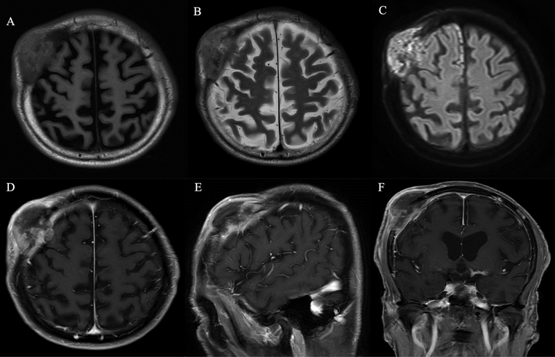
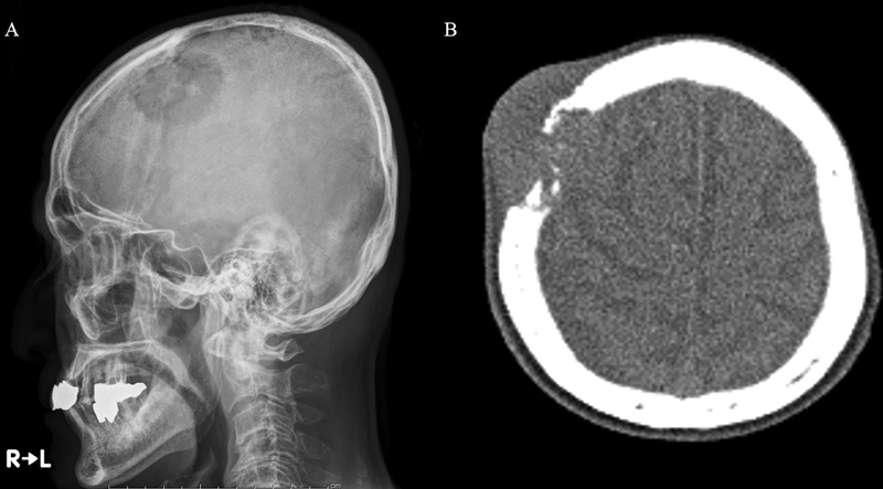
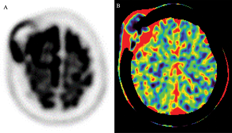
Primary intraosseous meningioma (PIM) is a rare tumor that arises in the skull. Histopathologically, it is generally described as a slow-growing, benign lesion. However, on rare occasions, PIM presents as a malignancy with high proliferative ability, which requires maximal resection, adjuvant radiotherapy, and subsequent careful follow-up. Because of the rarity of such cases, they present a diagnostic challenge with unusual pathological findings. Herein, we report a case of a primary intraosseous anaplastic meningioma with extensive invasion inside and outside the skull, along with the results of whole-genome analysis. Histopathological diagnosis was a World Health Organization grade 3 anaplastic meningioma. In the literature, only two cases of anaplastic PIM have been reported, so its characteristics and treatment are poorly understood. Our patient was successfully treated with tumor resection, followed by intensity-modulated radiation therapy. Follow-up imaging studies revealed no recurrence or distant metastasis, including to lung, liver, and bone, at 8 months after the surgery.

 求助内容:
求助内容: 应助结果提醒方式:


