{"title":"关节软骨层的超短回声时间MRI T2*标测与组织学早期退化有关。","authors":"Rui Imamura, Atsushi Teramoto, Yasutaka Murahashi, Yohei Okada, Shinichiro Okimura, Yoshihiro Akatsuka, Kota Watanabe, Toshihiko Yamashita","doi":"10.1177/19476035231205685","DOIUrl":null,"url":null,"abstract":"<p><strong>Objective: </strong>Ultra-short TE (UTE) sequences on MRI are a technique that improves the visualization of tissues with short T2 relaxation time, such as deep cartilage layers. In addition, T2* relaxation time calculated from the UTE has the potential to evaluate water molecules bound to the cartilage matrix. This study was performed to determine if there is an association between UTE-T2* relaxation time by cartilage layer and histological degeneration in knee osteoarthritis (OA).</p><p><strong>Design: </strong>Seven knees that had undergone total knee arthroplasty (TKA) were included in the study, and the lateral tibial cartilage, which had the least degeneration of the resected bones, was used as the sample. The T2* relaxation time of 4 patients with no abnormal findings on MRI was the reference relaxation time. Histological degeneration of TKA samples was assessed by the Mankin score and graded as the early OA group (≤3 points) and the advanced OA group (≥4 points). The association between T2* relaxation time and Mankin grade in each cartilage layer was compared. The effect of angiogenesis to the tidemark on T2* relaxation time was also compared.</p><p><strong>Results: </strong>T2* relaxation time of the cartilage layer was significantly longer in early OA than that in the control group. In the deep cartilage layer, the mean T2* relaxation time for angiogenesis (-) was 15.7 ms, whereas it was significantly shorter for angiogenesis (+) at 8.2 ms.</p><p><strong>Conclusions: </strong>The UTE-T2* relaxation time was associated with histological cartilage degeneration, suggesting a potential application in monitoring early cartilage degeneration.</p>","PeriodicalId":9626,"journal":{"name":"CARTILAGE","volume":" ","pages":"118-124"},"PeriodicalIF":2.7000,"publicationDate":"2025-03-01","publicationTypes":"Journal Article","fieldsOfStudy":null,"isOpenAccess":false,"openAccessPdf":"https://www.ncbi.nlm.nih.gov/pmc/articles/PMC11744601/pdf/","citationCount":"0","resultStr":"{\"title\":\"Ultra-Short Echo Time-MRI T2* Mapping of Articular Cartilage Layers Is Associated with Histological Early Degeneration.\",\"authors\":\"Rui Imamura, Atsushi Teramoto, Yasutaka Murahashi, Yohei Okada, Shinichiro Okimura, Yoshihiro Akatsuka, Kota Watanabe, Toshihiko Yamashita\",\"doi\":\"10.1177/19476035231205685\",\"DOIUrl\":null,\"url\":null,\"abstract\":\"<p><strong>Objective: </strong>Ultra-short TE (UTE) sequences on MRI are a technique that improves the visualization of tissues with short T2 relaxation time, such as deep cartilage layers. In addition, T2* relaxation time calculated from the UTE has the potential to evaluate water molecules bound to the cartilage matrix. This study was performed to determine if there is an association between UTE-T2* relaxation time by cartilage layer and histological degeneration in knee osteoarthritis (OA).</p><p><strong>Design: </strong>Seven knees that had undergone total knee arthroplasty (TKA) were included in the study, and the lateral tibial cartilage, which had the least degeneration of the resected bones, was used as the sample. The T2* relaxation time of 4 patients with no abnormal findings on MRI was the reference relaxation time. Histological degeneration of TKA samples was assessed by the Mankin score and graded as the early OA group (≤3 points) and the advanced OA group (≥4 points). The association between T2* relaxation time and Mankin grade in each cartilage layer was compared. The effect of angiogenesis to the tidemark on T2* relaxation time was also compared.</p><p><strong>Results: </strong>T2* relaxation time of the cartilage layer was significantly longer in early OA than that in the control group. In the deep cartilage layer, the mean T2* relaxation time for angiogenesis (-) was 15.7 ms, whereas it was significantly shorter for angiogenesis (+) at 8.2 ms.</p><p><strong>Conclusions: </strong>The UTE-T2* relaxation time was associated with histological cartilage degeneration, suggesting a potential application in monitoring early cartilage degeneration.</p>\",\"PeriodicalId\":9626,\"journal\":{\"name\":\"CARTILAGE\",\"volume\":\" \",\"pages\":\"118-124\"},\"PeriodicalIF\":2.7000,\"publicationDate\":\"2025-03-01\",\"publicationTypes\":\"Journal Article\",\"fieldsOfStudy\":null,\"isOpenAccess\":false,\"openAccessPdf\":\"https://www.ncbi.nlm.nih.gov/pmc/articles/PMC11744601/pdf/\",\"citationCount\":\"0\",\"resultStr\":null,\"platform\":\"Semanticscholar\",\"paperid\":null,\"PeriodicalName\":\"CARTILAGE\",\"FirstCategoryId\":\"3\",\"ListUrlMain\":\"https://doi.org/10.1177/19476035231205685\",\"RegionNum\":4,\"RegionCategory\":\"医学\",\"ArticlePicture\":[],\"TitleCN\":null,\"AbstractTextCN\":null,\"PMCID\":null,\"EPubDate\":\"2023/10/16 0:00:00\",\"PubModel\":\"Epub\",\"JCR\":\"Q1\",\"JCRName\":\"ORTHOPEDICS\",\"Score\":null,\"Total\":0}","platform":"Semanticscholar","paperid":null,"PeriodicalName":"CARTILAGE","FirstCategoryId":"3","ListUrlMain":"https://doi.org/10.1177/19476035231205685","RegionNum":4,"RegionCategory":"医学","ArticlePicture":[],"TitleCN":null,"AbstractTextCN":null,"PMCID":null,"EPubDate":"2023/10/16 0:00:00","PubModel":"Epub","JCR":"Q1","JCRName":"ORTHOPEDICS","Score":null,"Total":0}
Ultra-Short Echo Time-MRI T2* Mapping of Articular Cartilage Layers Is Associated with Histological Early Degeneration.
Objective: Ultra-short TE (UTE) sequences on MRI are a technique that improves the visualization of tissues with short T2 relaxation time, such as deep cartilage layers. In addition, T2* relaxation time calculated from the UTE has the potential to evaluate water molecules bound to the cartilage matrix. This study was performed to determine if there is an association between UTE-T2* relaxation time by cartilage layer and histological degeneration in knee osteoarthritis (OA).
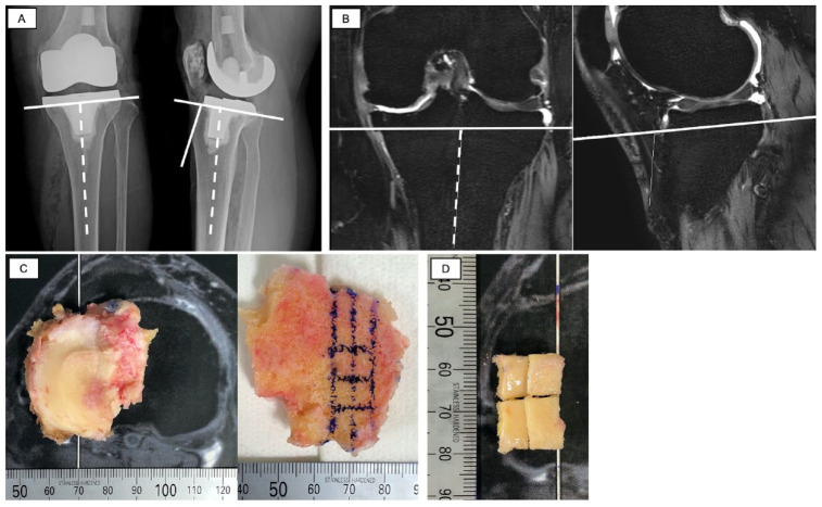
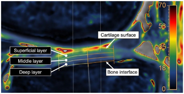
Design: Seven knees that had undergone total knee arthroplasty (TKA) were included in the study, and the lateral tibial cartilage, which had the least degeneration of the resected bones, was used as the sample. The T2* relaxation time of 4 patients with no abnormal findings on MRI was the reference relaxation time. Histological degeneration of TKA samples was assessed by the Mankin score and graded as the early OA group (≤3 points) and the advanced OA group (≥4 points). The association between T2* relaxation time and Mankin grade in each cartilage layer was compared. The effect of angiogenesis to the tidemark on T2* relaxation time was also compared.
Results: T2* relaxation time of the cartilage layer was significantly longer in early OA than that in the control group. In the deep cartilage layer, the mean T2* relaxation time for angiogenesis (-) was 15.7 ms, whereas it was significantly shorter for angiogenesis (+) at 8.2 ms.
Conclusions: The UTE-T2* relaxation time was associated with histological cartilage degeneration, suggesting a potential application in monitoring early cartilage degeneration.
期刊介绍:
CARTILAGE publishes articles related to the musculoskeletal system with particular attention to cartilage repair, development, function, degeneration, transplantation, and rehabilitation. The journal is a forum for the exchange of ideas for the many types of researchers and clinicians involved in cartilage biology and repair. A primary objective of CARTILAGE is to foster the cross-fertilization of the findings between clinical and basic sciences throughout the various disciplines involved in cartilage repair.
The journal publishes full length original manuscripts on all types of cartilage including articular, nasal, auricular, tracheal/bronchial, and intervertebral disc fibrocartilage. Manuscripts on clinical and laboratory research are welcome. Review articles, editorials, and letters are also encouraged. The ICRS envisages CARTILAGE as a forum for the exchange of knowledge among clinicians, scientists, patients, and researchers.
The International Cartilage Repair Society (ICRS) is dedicated to promotion, encouragement, and distribution of fundamental and applied research of cartilage in order to permit a better knowledge of function and dysfunction of articular cartilage and its repair.
 求助内容:
求助内容: 应助结果提醒方式:


