Margarete M Karg, May Moorefield, Emma Hoffmann, Hannah Philipose, Drenushe Krasniqi, Cindy Hoppe, Daisy Y Shu, Shintaro Shirahama, Bruce R Ksander, Magali Saint-Geniez
{"title":"小胶质细胞通过支持视网膜色素上皮的健康来保护衰老视网膜的视觉功能损失。","authors":"Margarete M Karg, May Moorefield, Emma Hoffmann, Hannah Philipose, Drenushe Krasniqi, Cindy Hoppe, Daisy Y Shu, Shintaro Shirahama, Bruce R Ksander, Magali Saint-Geniez","doi":"10.1186/s12979-023-00358-4","DOIUrl":null,"url":null,"abstract":"<p><strong>Background: </strong>Increased age is a risk factor for the development and progression of retinal diseases including age-related macular degeneration (AMD). Understanding the changes that occur in the eye due to aging is important in enhancing our understanding of AMD pathogenesis and the development of novel AMD therapies. Microglia, the resident brain and retinal immune cells are associated with both maintaining homeostasis and protection of neurons and loss of microglia homeostasis could be a significant player in age related neurodegeneration. One important characteristic of retinal aging is the migration of microglia from the inner to outer retina where they reside in the subretinal space (SRS) in contact with the retinal pigment epithelial (RPE) cells. The role of aged subretinal microglia is unknown. Here, we depleted microglia in aged C57/BL6 mice fed for 6 weeks with a chow containing PLX5622, a small molecule inhibitor of colony-stimulating factor-1 receptor (Csf1r) required for microglial survival.</p><p><strong>Results: </strong>The subretinal P2RY12 + microglia in aged mice displayed a highly amoeboid and activated morphology and were filled with autofluorescence droplets reminiscent of lipofuscin. TEM indicates that subretinal microglia actively phagocytize shed photoreceptor outer segments, one of the main functions of retinal pigmented epithelial cells. PLX5622 treatment depleted up to 90% of the retinal microglia and was associated with significant loss in visual function. Mice on the microglia depletion diet showed reduced contrast sensitivity and significantly lower electroretinogram for the c-wave, a measurement of RPE functionality, compared to age-matched controls. The loss of c-wave coincided with a loss of RPE cells and increased RPE swelling in the absence of microglia.</p><p><strong>Conclusions: </strong>We conclude that microglia preserve visual function in aged mice and support RPE cell function, by phagocytosing shed photoreceptor outer segments and lipids, therefore compensating for the known age-related decline of RPE phagocytosis.</p>","PeriodicalId":51289,"journal":{"name":"Immunity & Ageing","volume":null,"pages":null},"PeriodicalIF":5.2000,"publicationDate":"2023-10-14","publicationTypes":"Journal Article","fieldsOfStudy":null,"isOpenAccess":false,"openAccessPdf":"https://www.ncbi.nlm.nih.gov/pmc/articles/PMC10576380/pdf/","citationCount":"0","resultStr":"{\"title\":\"Microglia preserve visual function loss in the aging retina by supporting retinal pigment epithelial health.\",\"authors\":\"Margarete M Karg, May Moorefield, Emma Hoffmann, Hannah Philipose, Drenushe Krasniqi, Cindy Hoppe, Daisy Y Shu, Shintaro Shirahama, Bruce R Ksander, Magali Saint-Geniez\",\"doi\":\"10.1186/s12979-023-00358-4\",\"DOIUrl\":null,\"url\":null,\"abstract\":\"<p><strong>Background: </strong>Increased age is a risk factor for the development and progression of retinal diseases including age-related macular degeneration (AMD). Understanding the changes that occur in the eye due to aging is important in enhancing our understanding of AMD pathogenesis and the development of novel AMD therapies. Microglia, the resident brain and retinal immune cells are associated with both maintaining homeostasis and protection of neurons and loss of microglia homeostasis could be a significant player in age related neurodegeneration. One important characteristic of retinal aging is the migration of microglia from the inner to outer retina where they reside in the subretinal space (SRS) in contact with the retinal pigment epithelial (RPE) cells. The role of aged subretinal microglia is unknown. Here, we depleted microglia in aged C57/BL6 mice fed for 6 weeks with a chow containing PLX5622, a small molecule inhibitor of colony-stimulating factor-1 receptor (Csf1r) required for microglial survival.</p><p><strong>Results: </strong>The subretinal P2RY12 + microglia in aged mice displayed a highly amoeboid and activated morphology and were filled with autofluorescence droplets reminiscent of lipofuscin. TEM indicates that subretinal microglia actively phagocytize shed photoreceptor outer segments, one of the main functions of retinal pigmented epithelial cells. PLX5622 treatment depleted up to 90% of the retinal microglia and was associated with significant loss in visual function. Mice on the microglia depletion diet showed reduced contrast sensitivity and significantly lower electroretinogram for the c-wave, a measurement of RPE functionality, compared to age-matched controls. The loss of c-wave coincided with a loss of RPE cells and increased RPE swelling in the absence of microglia.</p><p><strong>Conclusions: </strong>We conclude that microglia preserve visual function in aged mice and support RPE cell function, by phagocytosing shed photoreceptor outer segments and lipids, therefore compensating for the known age-related decline of RPE phagocytosis.</p>\",\"PeriodicalId\":51289,\"journal\":{\"name\":\"Immunity & Ageing\",\"volume\":null,\"pages\":null},\"PeriodicalIF\":5.2000,\"publicationDate\":\"2023-10-14\",\"publicationTypes\":\"Journal Article\",\"fieldsOfStudy\":null,\"isOpenAccess\":false,\"openAccessPdf\":\"https://www.ncbi.nlm.nih.gov/pmc/articles/PMC10576380/pdf/\",\"citationCount\":\"0\",\"resultStr\":null,\"platform\":\"Semanticscholar\",\"paperid\":null,\"PeriodicalName\":\"Immunity & Ageing\",\"FirstCategoryId\":\"3\",\"ListUrlMain\":\"https://doi.org/10.1186/s12979-023-00358-4\",\"RegionNum\":2,\"RegionCategory\":\"医学\",\"ArticlePicture\":[],\"TitleCN\":null,\"AbstractTextCN\":null,\"PMCID\":null,\"EPubDate\":\"\",\"PubModel\":\"\",\"JCR\":\"Q1\",\"JCRName\":\"GERIATRICS & GERONTOLOGY\",\"Score\":null,\"Total\":0}","platform":"Semanticscholar","paperid":null,"PeriodicalName":"Immunity & Ageing","FirstCategoryId":"3","ListUrlMain":"https://doi.org/10.1186/s12979-023-00358-4","RegionNum":2,"RegionCategory":"医学","ArticlePicture":[],"TitleCN":null,"AbstractTextCN":null,"PMCID":null,"EPubDate":"","PubModel":"","JCR":"Q1","JCRName":"GERIATRICS & GERONTOLOGY","Score":null,"Total":0}
Microglia preserve visual function loss in the aging retina by supporting retinal pigment epithelial health.
Background: Increased age is a risk factor for the development and progression of retinal diseases including age-related macular degeneration (AMD). Understanding the changes that occur in the eye due to aging is important in enhancing our understanding of AMD pathogenesis and the development of novel AMD therapies. Microglia, the resident brain and retinal immune cells are associated with both maintaining homeostasis and protection of neurons and loss of microglia homeostasis could be a significant player in age related neurodegeneration. One important characteristic of retinal aging is the migration of microglia from the inner to outer retina where they reside in the subretinal space (SRS) in contact with the retinal pigment epithelial (RPE) cells. The role of aged subretinal microglia is unknown. Here, we depleted microglia in aged C57/BL6 mice fed for 6 weeks with a chow containing PLX5622, a small molecule inhibitor of colony-stimulating factor-1 receptor (Csf1r) required for microglial survival.
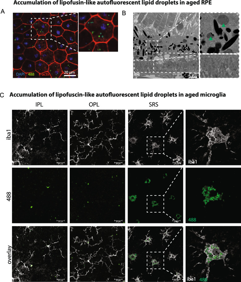
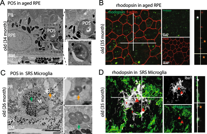
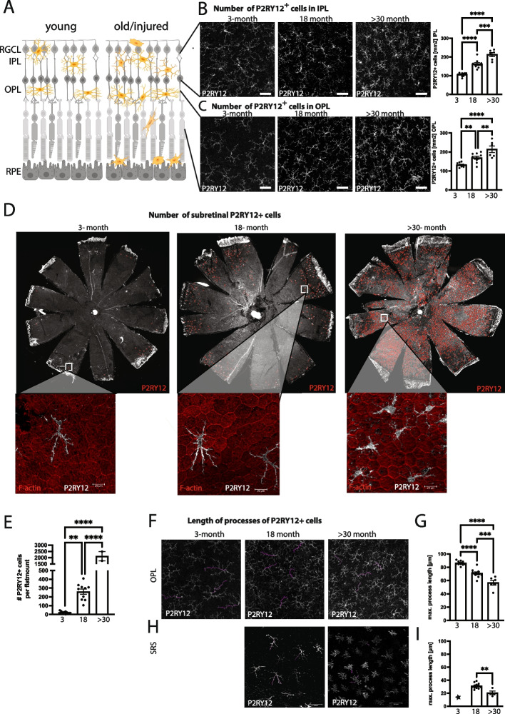
Results: The subretinal P2RY12 + microglia in aged mice displayed a highly amoeboid and activated morphology and were filled with autofluorescence droplets reminiscent of lipofuscin. TEM indicates that subretinal microglia actively phagocytize shed photoreceptor outer segments, one of the main functions of retinal pigmented epithelial cells. PLX5622 treatment depleted up to 90% of the retinal microglia and was associated with significant loss in visual function. Mice on the microglia depletion diet showed reduced contrast sensitivity and significantly lower electroretinogram for the c-wave, a measurement of RPE functionality, compared to age-matched controls. The loss of c-wave coincided with a loss of RPE cells and increased RPE swelling in the absence of microglia.
Conclusions: We conclude that microglia preserve visual function in aged mice and support RPE cell function, by phagocytosing shed photoreceptor outer segments and lipids, therefore compensating for the known age-related decline of RPE phagocytosis.
期刊介绍:
Immunity & Ageing is a specialist open access journal that was first published in 2004. The journal focuses on the impact of ageing on immune systems, the influence of aged immune systems on organismal well-being and longevity, age-associated diseases with immune etiology, and potential immune interventions to increase health span. All articles published in Immunity & Ageing are indexed in the following databases: Biological Abstracts, BIOSIS, CAS, Citebase, DOAJ, Embase, Google Scholar, Journal Citation Reports/Science Edition, OAIster, PubMed, PubMed Central, Science Citation Index Expanded, SCImago, Scopus, SOCOLAR, and Zetoc.
 求助内容:
求助内容: 应助结果提醒方式:


