Ştefan Ţălu , Robert S. Matos , Henrique Duarte da Fonseca Filho , Daniela Predoi , Simona Liliana Iconaru , Carmen Steluţa Ciobanu , Liliana Ghegoiu
{"title":"基于壳聚糖基质的掺镁羟基磷灰石复合层锚定癌症细胞的形态学和分形特征。","authors":"Ştefan Ţălu , Robert S. Matos , Henrique Duarte da Fonseca Filho , Daniela Predoi , Simona Liliana Iconaru , Carmen Steluţa Ciobanu , Liliana Ghegoiu","doi":"10.1016/j.micron.2023.103548","DOIUrl":null,"url":null,"abstract":"<div><p>In the present study, we report the development and characterization of composite layers (by spin coating) based on magnesium-doped hydroxyapatite in a chitosan matrix, containing human osteosarcoma MG63 cells anchored. Studies regarding the biocompatibility of the composite layers were performed with the aid of a MTT (3–4,5-Dimethylthiazol 2,5-diphenyltetrazolium bromide) assay. The data determined that the composite layers did not inhibit the growth and adhesion of MG63 cells to their surfaces exhibiting good biocompatibility properties. Furthermore, the attachment and development of MG63 cells on the surface of MgHApCh composite layers were investigated using atomic force microscopy (AFM). AFM topographical maps emphasized that the HApCh and 8MgHApCh composite layers surface promoted the attachment and proliferation of MG63 cells on their surface. Meanwhile, in the case of 30MgHApCh layers incubated for 48 h, a slight modification of the morphological features of the MG63 cells. In addition, the effects of the composite layers against <em>Candida albicans</em> ATCC 10231 were also evaluated. The data results from the <em>in vitro</em> antifungal assay depicted that the composite layers successfully inhibited the growth of the fungal cells onto their surface. Morphological and fractal analyses unveil cancer cell surfaces on Mg-containing composite layers with intricate 3D patterns, driven by high-frequency components. Their remarkable complexity and roughness arises from a strong multifractal nature, supporting more effective vertical growth compared to Si and HApCh surfaces. The cell viability reduced of uncoated Si surface is highlighted by its less intense 3D pattern growth. Our results show that the uncoated Si surface promotes lower viability of MG63 cancer cells, with less rough and complex 3D spatial patterns.</p></div>","PeriodicalId":18501,"journal":{"name":"Micron","volume":null,"pages":null},"PeriodicalIF":2.5000,"publicationDate":"2023-10-04","publicationTypes":"Journal Article","fieldsOfStudy":null,"isOpenAccess":false,"openAccessPdf":"","citationCount":"0","resultStr":"{\"title\":\"Morphological and fractal features of cancer cells anchored on composite layers based on magnesium-doped hydroxyapatite loaded in chitosan matrix\",\"authors\":\"Ştefan Ţălu , Robert S. Matos , Henrique Duarte da Fonseca Filho , Daniela Predoi , Simona Liliana Iconaru , Carmen Steluţa Ciobanu , Liliana Ghegoiu\",\"doi\":\"10.1016/j.micron.2023.103548\",\"DOIUrl\":null,\"url\":null,\"abstract\":\"<div><p>In the present study, we report the development and characterization of composite layers (by spin coating) based on magnesium-doped hydroxyapatite in a chitosan matrix, containing human osteosarcoma MG63 cells anchored. Studies regarding the biocompatibility of the composite layers were performed with the aid of a MTT (3–4,5-Dimethylthiazol 2,5-diphenyltetrazolium bromide) assay. The data determined that the composite layers did not inhibit the growth and adhesion of MG63 cells to their surfaces exhibiting good biocompatibility properties. Furthermore, the attachment and development of MG63 cells on the surface of MgHApCh composite layers were investigated using atomic force microscopy (AFM). AFM topographical maps emphasized that the HApCh and 8MgHApCh composite layers surface promoted the attachment and proliferation of MG63 cells on their surface. Meanwhile, in the case of 30MgHApCh layers incubated for 48 h, a slight modification of the morphological features of the MG63 cells. In addition, the effects of the composite layers against <em>Candida albicans</em> ATCC 10231 were also evaluated. The data results from the <em>in vitro</em> antifungal assay depicted that the composite layers successfully inhibited the growth of the fungal cells onto their surface. Morphological and fractal analyses unveil cancer cell surfaces on Mg-containing composite layers with intricate 3D patterns, driven by high-frequency components. Their remarkable complexity and roughness arises from a strong multifractal nature, supporting more effective vertical growth compared to Si and HApCh surfaces. The cell viability reduced of uncoated Si surface is highlighted by its less intense 3D pattern growth. Our results show that the uncoated Si surface promotes lower viability of MG63 cancer cells, with less rough and complex 3D spatial patterns.</p></div>\",\"PeriodicalId\":18501,\"journal\":{\"name\":\"Micron\",\"volume\":null,\"pages\":null},\"PeriodicalIF\":2.5000,\"publicationDate\":\"2023-10-04\",\"publicationTypes\":\"Journal Article\",\"fieldsOfStudy\":null,\"isOpenAccess\":false,\"openAccessPdf\":\"\",\"citationCount\":\"0\",\"resultStr\":null,\"platform\":\"Semanticscholar\",\"paperid\":null,\"PeriodicalName\":\"Micron\",\"FirstCategoryId\":\"5\",\"ListUrlMain\":\"https://www.sciencedirect.com/science/article/pii/S0968432823001464\",\"RegionNum\":3,\"RegionCategory\":\"工程技术\",\"ArticlePicture\":[],\"TitleCN\":null,\"AbstractTextCN\":null,\"PMCID\":null,\"EPubDate\":\"\",\"PubModel\":\"\",\"JCR\":\"Q1\",\"JCRName\":\"MICROSCOPY\",\"Score\":null,\"Total\":0}","platform":"Semanticscholar","paperid":null,"PeriodicalName":"Micron","FirstCategoryId":"5","ListUrlMain":"https://www.sciencedirect.com/science/article/pii/S0968432823001464","RegionNum":3,"RegionCategory":"工程技术","ArticlePicture":[],"TitleCN":null,"AbstractTextCN":null,"PMCID":null,"EPubDate":"","PubModel":"","JCR":"Q1","JCRName":"MICROSCOPY","Score":null,"Total":0}
Morphological and fractal features of cancer cells anchored on composite layers based on magnesium-doped hydroxyapatite loaded in chitosan matrix
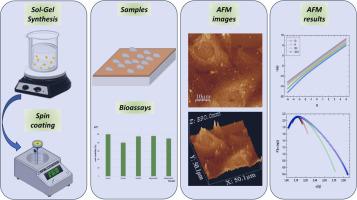
In the present study, we report the development and characterization of composite layers (by spin coating) based on magnesium-doped hydroxyapatite in a chitosan matrix, containing human osteosarcoma MG63 cells anchored. Studies regarding the biocompatibility of the composite layers were performed with the aid of a MTT (3–4,5-Dimethylthiazol 2,5-diphenyltetrazolium bromide) assay. The data determined that the composite layers did not inhibit the growth and adhesion of MG63 cells to their surfaces exhibiting good biocompatibility properties. Furthermore, the attachment and development of MG63 cells on the surface of MgHApCh composite layers were investigated using atomic force microscopy (AFM). AFM topographical maps emphasized that the HApCh and 8MgHApCh composite layers surface promoted the attachment and proliferation of MG63 cells on their surface. Meanwhile, in the case of 30MgHApCh layers incubated for 48 h, a slight modification of the morphological features of the MG63 cells. In addition, the effects of the composite layers against Candida albicans ATCC 10231 were also evaluated. The data results from the in vitro antifungal assay depicted that the composite layers successfully inhibited the growth of the fungal cells onto their surface. Morphological and fractal analyses unveil cancer cell surfaces on Mg-containing composite layers with intricate 3D patterns, driven by high-frequency components. Their remarkable complexity and roughness arises from a strong multifractal nature, supporting more effective vertical growth compared to Si and HApCh surfaces. The cell viability reduced of uncoated Si surface is highlighted by its less intense 3D pattern growth. Our results show that the uncoated Si surface promotes lower viability of MG63 cancer cells, with less rough and complex 3D spatial patterns.
期刊介绍:
Micron is an interdisciplinary forum for all work that involves new applications of microscopy or where advanced microscopy plays a central role. The journal will publish on the design, methods, application, practice or theory of microscopy and microanalysis, including reports on optical, electron-beam, X-ray microtomography, and scanning-probe systems. It also aims at the regular publication of review papers, short communications, as well as thematic issues on contemporary developments in microscopy and microanalysis. The journal embraces original research in which microscopy has contributed significantly to knowledge in biology, life science, nanoscience and nanotechnology, materials science and engineering.
 求助内容:
求助内容: 应助结果提醒方式:


