{"title":"滇南小耳猪不同年龄组脑结构分析","authors":"Yi-Fan Liu, Chang-Le Fang, Yu Pi, Teng-Fei Ke, Ji-Xiang Chu, Lin-Na Tang, Lan-Chun Zhang, Somjit Wanchana, Cheng-De Liao","doi":"10.1002/ibra.12058","DOIUrl":null,"url":null,"abstract":"<p>The objective of the study is to investigate the brain development and atrophy of Diannan small-ear pigs in different ages using magnetic resonance imaging (MRI). A total of 12 Diannan small-ear pigs were included and divided into the young group, adult group, and middle-and-old age (M&O) group according to their age. The brain structure of pigs was scanned using MRI, and the brain data obtained were statistically analyzed by signal conversion and image reconstruction. Compared with the young group, the signals of most brain structures in the adult group and M&O group were significantly decreased (<i>p</i> < 0.05). Compared with the adult group, the signal intensity of the right caudate nucleus and the right lateral ventricle in the M&O group was significantly increased, while the signal intensity of other regions was almost significantly decreased (<i>p</i> < 0.05). Compared with the young group, both adult and M&O groups had some degree of brain atrophy. Brain atrophy in the precuneus and the inferior temporal gyrus was more predominant in the M&O group in comparison with the adult group. The present study demonstrated that the brain signal of Diannan small-ear pigs gradually diminished with age, while the degree of brain atrophy was the opposite, providing the basic data on the brain of Diannan small-ear pigs.</p>","PeriodicalId":94030,"journal":{"name":"Ibrain","volume":"8 3","pages":"314-323"},"PeriodicalIF":0.0000,"publicationDate":"2022-08-13","publicationTypes":"Journal Article","fieldsOfStudy":null,"isOpenAccess":false,"openAccessPdf":"https://www.ncbi.nlm.nih.gov/pmc/articles/PMC10528979/pdf/","citationCount":"0","resultStr":"{\"title\":\"Brain structure analysis of different age groups of Diannan small-ear pigs\",\"authors\":\"Yi-Fan Liu, Chang-Le Fang, Yu Pi, Teng-Fei Ke, Ji-Xiang Chu, Lin-Na Tang, Lan-Chun Zhang, Somjit Wanchana, Cheng-De Liao\",\"doi\":\"10.1002/ibra.12058\",\"DOIUrl\":null,\"url\":null,\"abstract\":\"<p>The objective of the study is to investigate the brain development and atrophy of Diannan small-ear pigs in different ages using magnetic resonance imaging (MRI). A total of 12 Diannan small-ear pigs were included and divided into the young group, adult group, and middle-and-old age (M&O) group according to their age. The brain structure of pigs was scanned using MRI, and the brain data obtained were statistically analyzed by signal conversion and image reconstruction. Compared with the young group, the signals of most brain structures in the adult group and M&O group were significantly decreased (<i>p</i> < 0.05). Compared with the adult group, the signal intensity of the right caudate nucleus and the right lateral ventricle in the M&O group was significantly increased, while the signal intensity of other regions was almost significantly decreased (<i>p</i> < 0.05). Compared with the young group, both adult and M&O groups had some degree of brain atrophy. Brain atrophy in the precuneus and the inferior temporal gyrus was more predominant in the M&O group in comparison with the adult group. The present study demonstrated that the brain signal of Diannan small-ear pigs gradually diminished with age, while the degree of brain atrophy was the opposite, providing the basic data on the brain of Diannan small-ear pigs.</p>\",\"PeriodicalId\":94030,\"journal\":{\"name\":\"Ibrain\",\"volume\":\"8 3\",\"pages\":\"314-323\"},\"PeriodicalIF\":0.0000,\"publicationDate\":\"2022-08-13\",\"publicationTypes\":\"Journal Article\",\"fieldsOfStudy\":null,\"isOpenAccess\":false,\"openAccessPdf\":\"https://www.ncbi.nlm.nih.gov/pmc/articles/PMC10528979/pdf/\",\"citationCount\":\"0\",\"resultStr\":null,\"platform\":\"Semanticscholar\",\"paperid\":null,\"PeriodicalName\":\"Ibrain\",\"FirstCategoryId\":\"1085\",\"ListUrlMain\":\"https://onlinelibrary.wiley.com/doi/10.1002/ibra.12058\",\"RegionNum\":0,\"RegionCategory\":null,\"ArticlePicture\":[],\"TitleCN\":null,\"AbstractTextCN\":null,\"PMCID\":null,\"EPubDate\":\"\",\"PubModel\":\"\",\"JCR\":\"\",\"JCRName\":\"\",\"Score\":null,\"Total\":0}","platform":"Semanticscholar","paperid":null,"PeriodicalName":"Ibrain","FirstCategoryId":"1085","ListUrlMain":"https://onlinelibrary.wiley.com/doi/10.1002/ibra.12058","RegionNum":0,"RegionCategory":null,"ArticlePicture":[],"TitleCN":null,"AbstractTextCN":null,"PMCID":null,"EPubDate":"","PubModel":"","JCR":"","JCRName":"","Score":null,"Total":0}
Brain structure analysis of different age groups of Diannan small-ear pigs
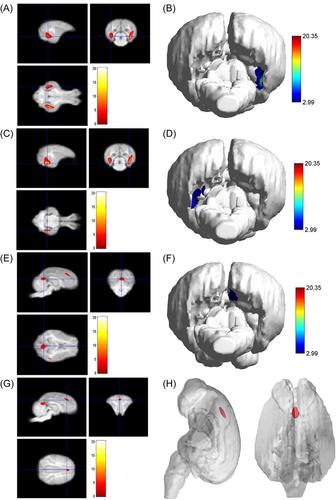
The objective of the study is to investigate the brain development and atrophy of Diannan small-ear pigs in different ages using magnetic resonance imaging (MRI). A total of 12 Diannan small-ear pigs were included and divided into the young group, adult group, and middle-and-old age (M&O) group according to their age. The brain structure of pigs was scanned using MRI, and the brain data obtained were statistically analyzed by signal conversion and image reconstruction. Compared with the young group, the signals of most brain structures in the adult group and M&O group were significantly decreased (p < 0.05). Compared with the adult group, the signal intensity of the right caudate nucleus and the right lateral ventricle in the M&O group was significantly increased, while the signal intensity of other regions was almost significantly decreased (p < 0.05). Compared with the young group, both adult and M&O groups had some degree of brain atrophy. Brain atrophy in the precuneus and the inferior temporal gyrus was more predominant in the M&O group in comparison with the adult group. The present study demonstrated that the brain signal of Diannan small-ear pigs gradually diminished with age, while the degree of brain atrophy was the opposite, providing the basic data on the brain of Diannan small-ear pigs.

 求助内容:
求助内容: 应助结果提醒方式:


