{"title":"桥本病的组织学改变:一个病例系列的超微结构研究。","authors":"Eleni Avramidou, Antonios Gkantaras, Iasonas Dermitzakis, Konstantinos Sapalidis, Maria Eleni Manthou, Paschalis Theotokis","doi":"10.3390/medicines10090051","DOIUrl":null,"url":null,"abstract":"<p><strong>Background: </strong>Hashimoto's thyroiditis (HT) is an autoimmune disease exhibiting stromal fibrosis and follicular cell destruction due to lymphoplasmacytic infiltration. Besides deprecated analyses, histopathological approaches have not employed the use of electron microscopy adequately toward delineating subcellular-level interactions.</p><p><strong>Methods: </strong>Biopsies for ultrastructural investigations were obtained from the thyroids of five patients with HT after a thyroidectomy. Transmission electron microscopy (TEM) was utilized to study representative tissue specimens.</p><p><strong>Results: </strong>Examination indicated interstitial extravasated blood cells and a plethora of plasma cells, based on their subcellular identity landmarks. These antibody-secreting cells were profoundly spotted near follicular cells, fibroblasts, and cell debris entrenched in collagenous areas. Pathological changes persistently affected subcellular components of the thyrocytes, including the nucleus, endoplasmic reticulum (ER), Golgi apparatus, mitochondria, lysosomes, and other intracellular vesicles. Interestingly, significant endothelial destruction was observed, specifically in the larger blood vessels, while the smaller vessels appeared comparatively unaffected.</p><p><strong>Conclusions: </strong>Our TEM findings highlight the immune-related alterations occurring within the thyroid stroma. The impaired vasculature component and remodeling have not been described ultrastructurally before; thus, further exploration is needed with regards to angiogenesis in HT in order to achieve successful prognostic, diagnostic, and treatment-monitoring strategies.</p>","PeriodicalId":74162,"journal":{"name":"Medicines (Basel, Switzerland)","volume":"10 9","pages":""},"PeriodicalIF":0.0000,"publicationDate":"2023-09-02","publicationTypes":"Journal Article","fieldsOfStudy":null,"isOpenAccess":false,"openAccessPdf":"https://www.ncbi.nlm.nih.gov/pmc/articles/PMC10534781/pdf/","citationCount":"0","resultStr":"{\"title\":\"Histological Alterations in Hashimoto's Disease: A Case-Series Ultrastructural Study.\",\"authors\":\"Eleni Avramidou, Antonios Gkantaras, Iasonas Dermitzakis, Konstantinos Sapalidis, Maria Eleni Manthou, Paschalis Theotokis\",\"doi\":\"10.3390/medicines10090051\",\"DOIUrl\":null,\"url\":null,\"abstract\":\"<p><strong>Background: </strong>Hashimoto's thyroiditis (HT) is an autoimmune disease exhibiting stromal fibrosis and follicular cell destruction due to lymphoplasmacytic infiltration. Besides deprecated analyses, histopathological approaches have not employed the use of electron microscopy adequately toward delineating subcellular-level interactions.</p><p><strong>Methods: </strong>Biopsies for ultrastructural investigations were obtained from the thyroids of five patients with HT after a thyroidectomy. Transmission electron microscopy (TEM) was utilized to study representative tissue specimens.</p><p><strong>Results: </strong>Examination indicated interstitial extravasated blood cells and a plethora of plasma cells, based on their subcellular identity landmarks. These antibody-secreting cells were profoundly spotted near follicular cells, fibroblasts, and cell debris entrenched in collagenous areas. Pathological changes persistently affected subcellular components of the thyrocytes, including the nucleus, endoplasmic reticulum (ER), Golgi apparatus, mitochondria, lysosomes, and other intracellular vesicles. Interestingly, significant endothelial destruction was observed, specifically in the larger blood vessels, while the smaller vessels appeared comparatively unaffected.</p><p><strong>Conclusions: </strong>Our TEM findings highlight the immune-related alterations occurring within the thyroid stroma. The impaired vasculature component and remodeling have not been described ultrastructurally before; thus, further exploration is needed with regards to angiogenesis in HT in order to achieve successful prognostic, diagnostic, and treatment-monitoring strategies.</p>\",\"PeriodicalId\":74162,\"journal\":{\"name\":\"Medicines (Basel, Switzerland)\",\"volume\":\"10 9\",\"pages\":\"\"},\"PeriodicalIF\":0.0000,\"publicationDate\":\"2023-09-02\",\"publicationTypes\":\"Journal Article\",\"fieldsOfStudy\":null,\"isOpenAccess\":false,\"openAccessPdf\":\"https://www.ncbi.nlm.nih.gov/pmc/articles/PMC10534781/pdf/\",\"citationCount\":\"0\",\"resultStr\":null,\"platform\":\"Semanticscholar\",\"paperid\":null,\"PeriodicalName\":\"Medicines (Basel, Switzerland)\",\"FirstCategoryId\":\"1085\",\"ListUrlMain\":\"https://doi.org/10.3390/medicines10090051\",\"RegionNum\":0,\"RegionCategory\":null,\"ArticlePicture\":[],\"TitleCN\":null,\"AbstractTextCN\":null,\"PMCID\":null,\"EPubDate\":\"\",\"PubModel\":\"\",\"JCR\":\"\",\"JCRName\":\"\",\"Score\":null,\"Total\":0}","platform":"Semanticscholar","paperid":null,"PeriodicalName":"Medicines (Basel, Switzerland)","FirstCategoryId":"1085","ListUrlMain":"https://doi.org/10.3390/medicines10090051","RegionNum":0,"RegionCategory":null,"ArticlePicture":[],"TitleCN":null,"AbstractTextCN":null,"PMCID":null,"EPubDate":"","PubModel":"","JCR":"","JCRName":"","Score":null,"Total":0}
Histological Alterations in Hashimoto's Disease: A Case-Series Ultrastructural Study.
Background: Hashimoto's thyroiditis (HT) is an autoimmune disease exhibiting stromal fibrosis and follicular cell destruction due to lymphoplasmacytic infiltration. Besides deprecated analyses, histopathological approaches have not employed the use of electron microscopy adequately toward delineating subcellular-level interactions.
Methods: Biopsies for ultrastructural investigations were obtained from the thyroids of five patients with HT after a thyroidectomy. Transmission electron microscopy (TEM) was utilized to study representative tissue specimens.
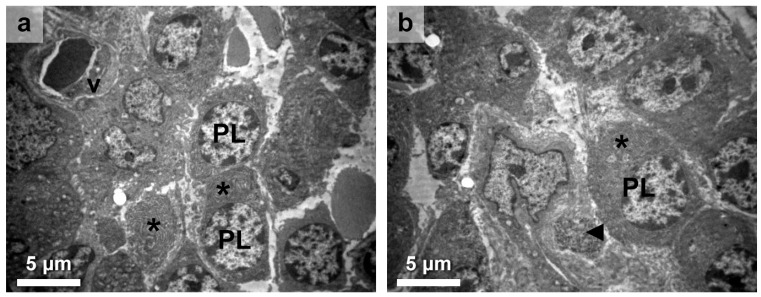
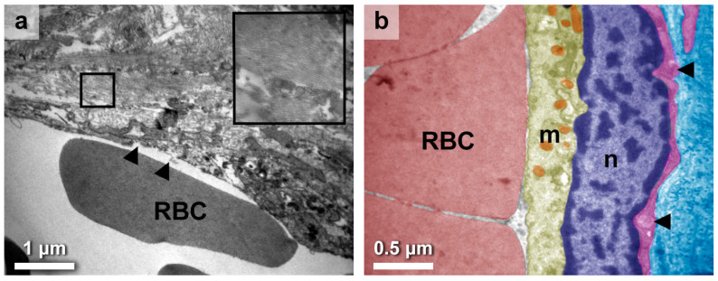
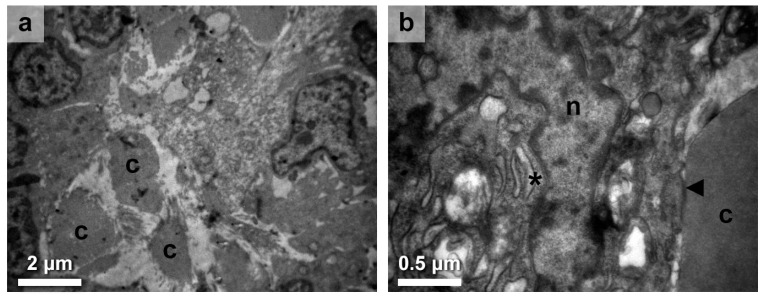
Results: Examination indicated interstitial extravasated blood cells and a plethora of plasma cells, based on their subcellular identity landmarks. These antibody-secreting cells were profoundly spotted near follicular cells, fibroblasts, and cell debris entrenched in collagenous areas. Pathological changes persistently affected subcellular components of the thyrocytes, including the nucleus, endoplasmic reticulum (ER), Golgi apparatus, mitochondria, lysosomes, and other intracellular vesicles. Interestingly, significant endothelial destruction was observed, specifically in the larger blood vessels, while the smaller vessels appeared comparatively unaffected.
Conclusions: Our TEM findings highlight the immune-related alterations occurring within the thyroid stroma. The impaired vasculature component and remodeling have not been described ultrastructurally before; thus, further exploration is needed with regards to angiogenesis in HT in order to achieve successful prognostic, diagnostic, and treatment-monitoring strategies.

 求助内容:
求助内容: 应助结果提醒方式:


