{"title":"用锥形束计算机断层扫描技术评估伊朗南部亚群中恒牙和犬牙的牙根扩张。","authors":"Safoora Sahebi, Alireza Razavian, Neshat Maddahi, Bahar Asheghi, Maryam Zangooei Booshehri","doi":"10.30476/dentjods.2022.95451.1874","DOIUrl":null,"url":null,"abstract":"<p><strong>Statement of the problem: </strong>Developmental anomalies such as dilacerations can affect the eruption pattern of permanent anterior teeth. They are characterized by a curvature in the crown and roots of the teeth relative to their normal axis. This anomaly can cause some complexities in routine dental treatments such as root canal treatment, orthodontics, and surgery.</p><p><strong>Purpose: </strong>The purpose of this study was to assess the prevalence of dilaceration in maxillary and mandibular anterior and canine teeth in Shiraz, Iran using cone-beam computed tomography (CBCT).</p><p><strong>Materials and method: </strong>In this retrospective study, a total of 1537 encompassed 400 CBCT images collected from 4 private radiology clinics in Shiraz were assessed. Each tooth was radiographically examined in order to diagnose root dilacerations considering their location (apical, middle, coronal), position in the jaw (maxillary or mandibular), direction (mesial, distal buccal and palatal/lingual), and severity of dilaceration (mild, moderate, and severe). The obtained data were analyzed by Chi-square statistical test and Fisher's exact test.</p><p><strong>Results: </strong>In this study, out of 1537 studied teeth, 5.98% had dilaceration. The maxillary and mandibular canine teeth (9.8% and 9.7%, respectively) were significantly the most common teeth involved in this anomaly (<i>p</i>< 0.001). The distal direction with mild severity in the apical third of the root was also the most common result obtained from this study (p<0.001). In addition, there was no statistically significant relationship between gender and type of jaw regarding the prevalence of dilaceration in the studied dental groups (p=0.670 and p=0.231, respectively).</p><p><strong>Conclusion: </strong>In the current research, it was demonstrated through CBCT records that the prevalence of dilaceration in maxillary and mandibular anterior and canine teeth is relatively uncommon. The most prevalent dilaceration was found to be distal direction with mild severity in the apical third of the root.</p>","PeriodicalId":73702,"journal":{"name":"Journal of dentistry (Shiraz, Iran)","volume":"24 3","pages":"320-327"},"PeriodicalIF":0.0000,"publicationDate":"2023-09-01","publicationTypes":"Journal Article","fieldsOfStudy":null,"isOpenAccess":false,"openAccessPdf":"https://www.ncbi.nlm.nih.gov/pmc/articles/PMC10506143/pdf/","citationCount":"0","resultStr":"{\"title\":\"Evaluation of Root Dilaceration in Permanent Anterior and Canine Teeth in the Southern Subpopulation of Iran Using Cone-Beam Computed Tomography.\",\"authors\":\"Safoora Sahebi, Alireza Razavian, Neshat Maddahi, Bahar Asheghi, Maryam Zangooei Booshehri\",\"doi\":\"10.30476/dentjods.2022.95451.1874\",\"DOIUrl\":null,\"url\":null,\"abstract\":\"<p><strong>Statement of the problem: </strong>Developmental anomalies such as dilacerations can affect the eruption pattern of permanent anterior teeth. They are characterized by a curvature in the crown and roots of the teeth relative to their normal axis. This anomaly can cause some complexities in routine dental treatments such as root canal treatment, orthodontics, and surgery.</p><p><strong>Purpose: </strong>The purpose of this study was to assess the prevalence of dilaceration in maxillary and mandibular anterior and canine teeth in Shiraz, Iran using cone-beam computed tomography (CBCT).</p><p><strong>Materials and method: </strong>In this retrospective study, a total of 1537 encompassed 400 CBCT images collected from 4 private radiology clinics in Shiraz were assessed. Each tooth was radiographically examined in order to diagnose root dilacerations considering their location (apical, middle, coronal), position in the jaw (maxillary or mandibular), direction (mesial, distal buccal and palatal/lingual), and severity of dilaceration (mild, moderate, and severe). The obtained data were analyzed by Chi-square statistical test and Fisher's exact test.</p><p><strong>Results: </strong>In this study, out of 1537 studied teeth, 5.98% had dilaceration. The maxillary and mandibular canine teeth (9.8% and 9.7%, respectively) were significantly the most common teeth involved in this anomaly (<i>p</i>< 0.001). The distal direction with mild severity in the apical third of the root was also the most common result obtained from this study (p<0.001). In addition, there was no statistically significant relationship between gender and type of jaw regarding the prevalence of dilaceration in the studied dental groups (p=0.670 and p=0.231, respectively).</p><p><strong>Conclusion: </strong>In the current research, it was demonstrated through CBCT records that the prevalence of dilaceration in maxillary and mandibular anterior and canine teeth is relatively uncommon. The most prevalent dilaceration was found to be distal direction with mild severity in the apical third of the root.</p>\",\"PeriodicalId\":73702,\"journal\":{\"name\":\"Journal of dentistry (Shiraz, Iran)\",\"volume\":\"24 3\",\"pages\":\"320-327\"},\"PeriodicalIF\":0.0000,\"publicationDate\":\"2023-09-01\",\"publicationTypes\":\"Journal Article\",\"fieldsOfStudy\":null,\"isOpenAccess\":false,\"openAccessPdf\":\"https://www.ncbi.nlm.nih.gov/pmc/articles/PMC10506143/pdf/\",\"citationCount\":\"0\",\"resultStr\":null,\"platform\":\"Semanticscholar\",\"paperid\":null,\"PeriodicalName\":\"Journal of dentistry (Shiraz, Iran)\",\"FirstCategoryId\":\"1085\",\"ListUrlMain\":\"https://doi.org/10.30476/dentjods.2022.95451.1874\",\"RegionNum\":0,\"RegionCategory\":null,\"ArticlePicture\":[],\"TitleCN\":null,\"AbstractTextCN\":null,\"PMCID\":null,\"EPubDate\":\"\",\"PubModel\":\"\",\"JCR\":\"\",\"JCRName\":\"\",\"Score\":null,\"Total\":0}","platform":"Semanticscholar","paperid":null,"PeriodicalName":"Journal of dentistry (Shiraz, Iran)","FirstCategoryId":"1085","ListUrlMain":"https://doi.org/10.30476/dentjods.2022.95451.1874","RegionNum":0,"RegionCategory":null,"ArticlePicture":[],"TitleCN":null,"AbstractTextCN":null,"PMCID":null,"EPubDate":"","PubModel":"","JCR":"","JCRName":"","Score":null,"Total":0}
Evaluation of Root Dilaceration in Permanent Anterior and Canine Teeth in the Southern Subpopulation of Iran Using Cone-Beam Computed Tomography.
Statement of the problem: Developmental anomalies such as dilacerations can affect the eruption pattern of permanent anterior teeth. They are characterized by a curvature in the crown and roots of the teeth relative to their normal axis. This anomaly can cause some complexities in routine dental treatments such as root canal treatment, orthodontics, and surgery.
Purpose: The purpose of this study was to assess the prevalence of dilaceration in maxillary and mandibular anterior and canine teeth in Shiraz, Iran using cone-beam computed tomography (CBCT).
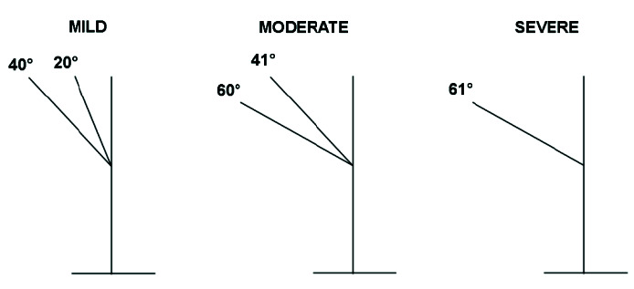
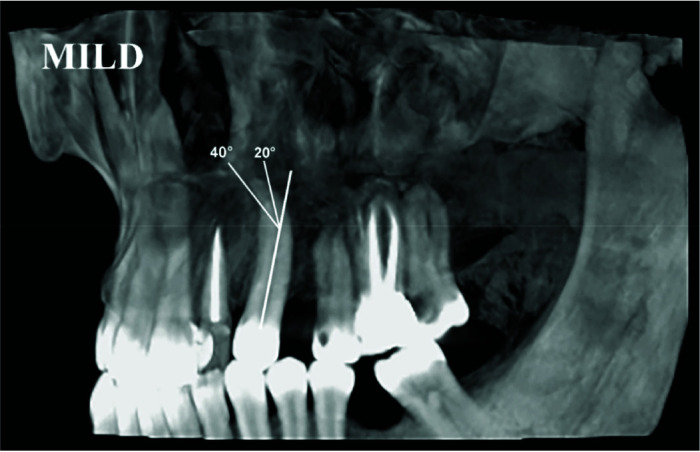
Materials and method: In this retrospective study, a total of 1537 encompassed 400 CBCT images collected from 4 private radiology clinics in Shiraz were assessed. Each tooth was radiographically examined in order to diagnose root dilacerations considering their location (apical, middle, coronal), position in the jaw (maxillary or mandibular), direction (mesial, distal buccal and palatal/lingual), and severity of dilaceration (mild, moderate, and severe). The obtained data were analyzed by Chi-square statistical test and Fisher's exact test.
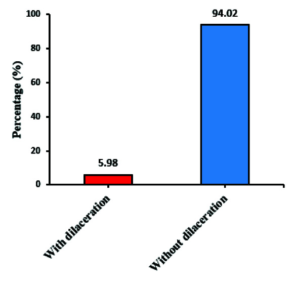
Results: In this study, out of 1537 studied teeth, 5.98% had dilaceration. The maxillary and mandibular canine teeth (9.8% and 9.7%, respectively) were significantly the most common teeth involved in this anomaly (p< 0.001). The distal direction with mild severity in the apical third of the root was also the most common result obtained from this study (p<0.001). In addition, there was no statistically significant relationship between gender and type of jaw regarding the prevalence of dilaceration in the studied dental groups (p=0.670 and p=0.231, respectively).
Conclusion: In the current research, it was demonstrated through CBCT records that the prevalence of dilaceration in maxillary and mandibular anterior and canine teeth is relatively uncommon. The most prevalent dilaceration was found to be distal direction with mild severity in the apical third of the root.

 求助内容:
求助内容: 应助结果提醒方式:


