Andrzej Grzybowski, Divya Parthasarathy Rao, Piotr Brona, Kalpa Negiloni, Tomasz Krzywicki, Florian M Savoy
{"title":"糖尿病视网膜病变图像自动评估软件的诊断准确性:IDx DR和MediosAI。","authors":"Andrzej Grzybowski, Divya Parthasarathy Rao, Piotr Brona, Kalpa Negiloni, Tomasz Krzywicki, Florian M Savoy","doi":"10.1159/000534098","DOIUrl":null,"url":null,"abstract":"<p><strong>Introduction: </strong>Numerous studies have demonstrated the use of artificial intelligence (AI) for early detection of referable diabetic retinopathy (RDR). A direct comparison of these multiple automated diabetic retinopathy (DR) image assessment softwares (ARIAs) is, however, challenging. We retrospectively compared the performance of two modern ARIAs, IDx-DR and Medios AI.</p><p><strong>Methods: </strong>In this retrospective-comparative study, retinal images with sufficient image quality were run on both ARIAs. They were captured in 811 consecutive patients with diabetes visiting diabetic clinics in Poland. For each patient, four non-mydriatic images, 45° field of view, i.e., two sets of one optic disc and one macula-centered image using Topcon NW400 were captured. Images were manually graded for severity of DR as no DR, any DR (mild non-proliferative diabetic retinopathy [NPDR] or more severe disease), RDR (moderate NPDR or more severe disease and/or clinically significant diabetic macular edema [CSDME]), or sight-threatening DR (severe NPDR or more severe disease and/or CSDME) by certified graders. The ARIA output was compared to manual consensus image grading (reference standard).</p><p><strong>Results: </strong>On 807 patients, based on consensus grading, there was no evidence of DR in 543 patients (67%). Any DR was seen in 264 (33%) patients, of which 174 (22%) were RDR and 41 (5%) were sight-threatening DR. The sensitivity of detecting RDR against reference standard grading was 95% (95% CI: 91, 98%) and the specificity was 80% (95% CI: 77, 83%) for Medios AI. They were 99% (95% CI: 96, 100%) and 68% (95% CI: 64, 72%) for IDx-DR, respectively.</p><p><strong>Conclusion: </strong>Both the ARIAs achieved satisfactory accuracy, with few false negatives. Although false-positive results generate additional costs and workload, missed cases raise the most concern whenever automated screening is debated.</p>","PeriodicalId":19662,"journal":{"name":"Ophthalmic Research","volume":" ","pages":"1286-1292"},"PeriodicalIF":2.0000,"publicationDate":"2023-01-01","publicationTypes":"Journal Article","fieldsOfStudy":null,"isOpenAccess":false,"openAccessPdf":"https://www.ncbi.nlm.nih.gov/pmc/articles/PMC10619585/pdf/","citationCount":"0","resultStr":"{\"title\":\"Diagnostic Accuracy of Automated Diabetic Retinopathy Image Assessment Softwares: IDx-DR and Medios Artificial Intelligence.\",\"authors\":\"Andrzej Grzybowski, Divya Parthasarathy Rao, Piotr Brona, Kalpa Negiloni, Tomasz Krzywicki, Florian M Savoy\",\"doi\":\"10.1159/000534098\",\"DOIUrl\":null,\"url\":null,\"abstract\":\"<p><strong>Introduction: </strong>Numerous studies have demonstrated the use of artificial intelligence (AI) for early detection of referable diabetic retinopathy (RDR). A direct comparison of these multiple automated diabetic retinopathy (DR) image assessment softwares (ARIAs) is, however, challenging. We retrospectively compared the performance of two modern ARIAs, IDx-DR and Medios AI.</p><p><strong>Methods: </strong>In this retrospective-comparative study, retinal images with sufficient image quality were run on both ARIAs. They were captured in 811 consecutive patients with diabetes visiting diabetic clinics in Poland. For each patient, four non-mydriatic images, 45° field of view, i.e., two sets of one optic disc and one macula-centered image using Topcon NW400 were captured. Images were manually graded for severity of DR as no DR, any DR (mild non-proliferative diabetic retinopathy [NPDR] or more severe disease), RDR (moderate NPDR or more severe disease and/or clinically significant diabetic macular edema [CSDME]), or sight-threatening DR (severe NPDR or more severe disease and/or CSDME) by certified graders. The ARIA output was compared to manual consensus image grading (reference standard).</p><p><strong>Results: </strong>On 807 patients, based on consensus grading, there was no evidence of DR in 543 patients (67%). Any DR was seen in 264 (33%) patients, of which 174 (22%) were RDR and 41 (5%) were sight-threatening DR. The sensitivity of detecting RDR against reference standard grading was 95% (95% CI: 91, 98%) and the specificity was 80% (95% CI: 77, 83%) for Medios AI. They were 99% (95% CI: 96, 100%) and 68% (95% CI: 64, 72%) for IDx-DR, respectively.</p><p><strong>Conclusion: </strong>Both the ARIAs achieved satisfactory accuracy, with few false negatives. Although false-positive results generate additional costs and workload, missed cases raise the most concern whenever automated screening is debated.</p>\",\"PeriodicalId\":19662,\"journal\":{\"name\":\"Ophthalmic Research\",\"volume\":\" \",\"pages\":\"1286-1292\"},\"PeriodicalIF\":2.0000,\"publicationDate\":\"2023-01-01\",\"publicationTypes\":\"Journal Article\",\"fieldsOfStudy\":null,\"isOpenAccess\":false,\"openAccessPdf\":\"https://www.ncbi.nlm.nih.gov/pmc/articles/PMC10619585/pdf/\",\"citationCount\":\"0\",\"resultStr\":null,\"platform\":\"Semanticscholar\",\"paperid\":null,\"PeriodicalName\":\"Ophthalmic Research\",\"FirstCategoryId\":\"3\",\"ListUrlMain\":\"https://doi.org/10.1159/000534098\",\"RegionNum\":4,\"RegionCategory\":\"医学\",\"ArticlePicture\":[],\"TitleCN\":null,\"AbstractTextCN\":null,\"PMCID\":null,\"EPubDate\":\"2023/9/27 0:00:00\",\"PubModel\":\"Epub\",\"JCR\":\"Q2\",\"JCRName\":\"OPHTHALMOLOGY\",\"Score\":null,\"Total\":0}","platform":"Semanticscholar","paperid":null,"PeriodicalName":"Ophthalmic Research","FirstCategoryId":"3","ListUrlMain":"https://doi.org/10.1159/000534098","RegionNum":4,"RegionCategory":"医学","ArticlePicture":[],"TitleCN":null,"AbstractTextCN":null,"PMCID":null,"EPubDate":"2023/9/27 0:00:00","PubModel":"Epub","JCR":"Q2","JCRName":"OPHTHALMOLOGY","Score":null,"Total":0}
Diagnostic Accuracy of Automated Diabetic Retinopathy Image Assessment Softwares: IDx-DR and Medios Artificial Intelligence.
Introduction: Numerous studies have demonstrated the use of artificial intelligence (AI) for early detection of referable diabetic retinopathy (RDR). A direct comparison of these multiple automated diabetic retinopathy (DR) image assessment softwares (ARIAs) is, however, challenging. We retrospectively compared the performance of two modern ARIAs, IDx-DR and Medios AI.
Methods: In this retrospective-comparative study, retinal images with sufficient image quality were run on both ARIAs. They were captured in 811 consecutive patients with diabetes visiting diabetic clinics in Poland. For each patient, four non-mydriatic images, 45° field of view, i.e., two sets of one optic disc and one macula-centered image using Topcon NW400 were captured. Images were manually graded for severity of DR as no DR, any DR (mild non-proliferative diabetic retinopathy [NPDR] or more severe disease), RDR (moderate NPDR or more severe disease and/or clinically significant diabetic macular edema [CSDME]), or sight-threatening DR (severe NPDR or more severe disease and/or CSDME) by certified graders. The ARIA output was compared to manual consensus image grading (reference standard).
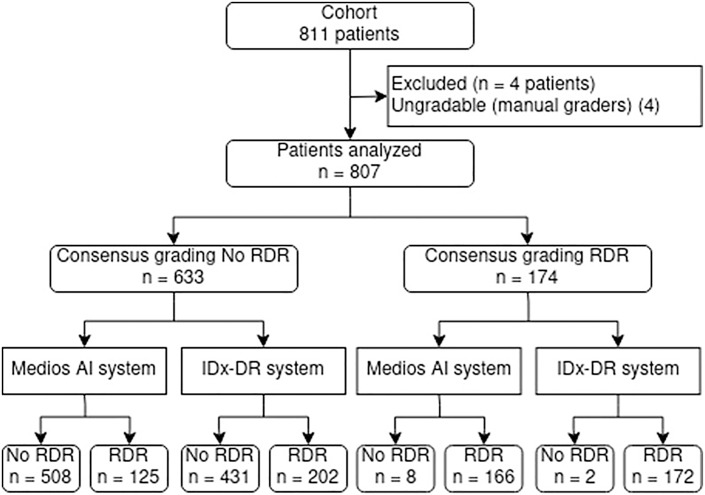
Results: On 807 patients, based on consensus grading, there was no evidence of DR in 543 patients (67%). Any DR was seen in 264 (33%) patients, of which 174 (22%) were RDR and 41 (5%) were sight-threatening DR. The sensitivity of detecting RDR against reference standard grading was 95% (95% CI: 91, 98%) and the specificity was 80% (95% CI: 77, 83%) for Medios AI. They were 99% (95% CI: 96, 100%) and 68% (95% CI: 64, 72%) for IDx-DR, respectively.
Conclusion: Both the ARIAs achieved satisfactory accuracy, with few false negatives. Although false-positive results generate additional costs and workload, missed cases raise the most concern whenever automated screening is debated.
期刊介绍:
''Ophthalmic Research'' features original papers and reviews reporting on translational and clinical studies. Authors from throughout the world cover research topics on every field in connection with physical, physiologic, pharmacological, biochemical and molecular biological aspects of ophthalmology. This journal also aims to provide a record of international clinical research for both researchers and clinicians in ophthalmology. Finally, the transfer of information from fundamental research to clinical research and clinical practice is particularly welcome.

 求助内容:
求助内容: 应助结果提醒方式:


