{"title":"MR定量成像在非酒精性脂肪肝中的临床应用。","authors":"Yushi Tsujita, Keitaro Sofue, Eisuke Ueshima, Yoshiko Ueno, Masatoshi Hori, Takamichi Murakami","doi":"10.2463/mrms.rev.2021-0152","DOIUrl":null,"url":null,"abstract":"<p><p>Viral hepatitis was previously the most common cause of chronic liver disease. However, in recent years, nonalcoholic fatty liver disease (NAFLD) cases have been increasing, especially in developed countries. NAFLD is histologically characterized by fat, fibrosis, and inflammation in the liver, eventually leading to cirrhosis and hepatocellular carcinoma. Although biopsy is the gold standard for the assessment of the liver parenchyma, quantitative evaluation methods, such as ultrasound, CT, and MRI, have been reported to have good diagnostic performances. The quantification of liver fat, fibrosis, and inflammation is expected to be clinically useful in terms of the prognosis, early intervention, and treatment response for the management of NAFLD. The aim of this review was to discuss the basics and prospects of MRI-based tissue quantifications of the liver, mainly focusing on proton density fat fraction for the quantification of fat deposition, MR elastography for the quantification of fibrosis, and multifrequency MR elastography for the evaluation of inflammation.</p>","PeriodicalId":94126,"journal":{"name":"Magnetic resonance in medical sciences : MRMS : an official journal of Japan Society of Magnetic Resonance in Medicine","volume":"22 4","pages":"435-445"},"PeriodicalIF":0.0000,"publicationDate":"2023-10-01","publicationTypes":"Journal Article","fieldsOfStudy":null,"isOpenAccess":false,"openAccessPdf":"https://ftp.ncbi.nlm.nih.gov/pub/pmc/oa_pdf/bc/79/mrms-22-435.PMC10552668.pdf","citationCount":"0","resultStr":"{\"title\":\"Clinical Application of Quantitative MR Imaging in Nonalcoholic Fatty Liver Disease.\",\"authors\":\"Yushi Tsujita, Keitaro Sofue, Eisuke Ueshima, Yoshiko Ueno, Masatoshi Hori, Takamichi Murakami\",\"doi\":\"10.2463/mrms.rev.2021-0152\",\"DOIUrl\":null,\"url\":null,\"abstract\":\"<p><p>Viral hepatitis was previously the most common cause of chronic liver disease. However, in recent years, nonalcoholic fatty liver disease (NAFLD) cases have been increasing, especially in developed countries. NAFLD is histologically characterized by fat, fibrosis, and inflammation in the liver, eventually leading to cirrhosis and hepatocellular carcinoma. Although biopsy is the gold standard for the assessment of the liver parenchyma, quantitative evaluation methods, such as ultrasound, CT, and MRI, have been reported to have good diagnostic performances. The quantification of liver fat, fibrosis, and inflammation is expected to be clinically useful in terms of the prognosis, early intervention, and treatment response for the management of NAFLD. The aim of this review was to discuss the basics and prospects of MRI-based tissue quantifications of the liver, mainly focusing on proton density fat fraction for the quantification of fat deposition, MR elastography for the quantification of fibrosis, and multifrequency MR elastography for the evaluation of inflammation.</p>\",\"PeriodicalId\":94126,\"journal\":{\"name\":\"Magnetic resonance in medical sciences : MRMS : an official journal of Japan Society of Magnetic Resonance in Medicine\",\"volume\":\"22 4\",\"pages\":\"435-445\"},\"PeriodicalIF\":0.0000,\"publicationDate\":\"2023-10-01\",\"publicationTypes\":\"Journal Article\",\"fieldsOfStudy\":null,\"isOpenAccess\":false,\"openAccessPdf\":\"https://ftp.ncbi.nlm.nih.gov/pub/pmc/oa_pdf/bc/79/mrms-22-435.PMC10552668.pdf\",\"citationCount\":\"0\",\"resultStr\":null,\"platform\":\"Semanticscholar\",\"paperid\":null,\"PeriodicalName\":\"Magnetic resonance in medical sciences : MRMS : an official journal of Japan Society of Magnetic Resonance in Medicine\",\"FirstCategoryId\":\"1085\",\"ListUrlMain\":\"https://doi.org/10.2463/mrms.rev.2021-0152\",\"RegionNum\":0,\"RegionCategory\":null,\"ArticlePicture\":[],\"TitleCN\":null,\"AbstractTextCN\":null,\"PMCID\":null,\"EPubDate\":\"2022/5/18 0:00:00\",\"PubModel\":\"Epub\",\"JCR\":\"\",\"JCRName\":\"\",\"Score\":null,\"Total\":0}","platform":"Semanticscholar","paperid":null,"PeriodicalName":"Magnetic resonance in medical sciences : MRMS : an official journal of Japan Society of Magnetic Resonance in Medicine","FirstCategoryId":"1085","ListUrlMain":"https://doi.org/10.2463/mrms.rev.2021-0152","RegionNum":0,"RegionCategory":null,"ArticlePicture":[],"TitleCN":null,"AbstractTextCN":null,"PMCID":null,"EPubDate":"2022/5/18 0:00:00","PubModel":"Epub","JCR":"","JCRName":"","Score":null,"Total":0}
Clinical Application of Quantitative MR Imaging in Nonalcoholic Fatty Liver Disease.
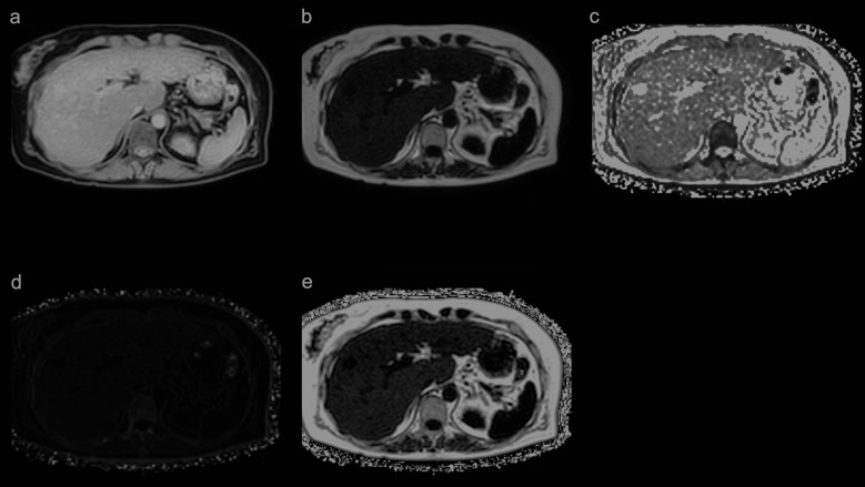
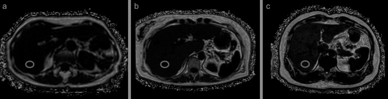
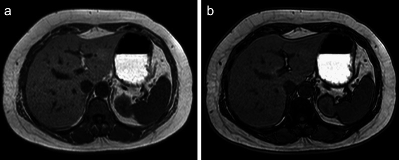
Viral hepatitis was previously the most common cause of chronic liver disease. However, in recent years, nonalcoholic fatty liver disease (NAFLD) cases have been increasing, especially in developed countries. NAFLD is histologically characterized by fat, fibrosis, and inflammation in the liver, eventually leading to cirrhosis and hepatocellular carcinoma. Although biopsy is the gold standard for the assessment of the liver parenchyma, quantitative evaluation methods, such as ultrasound, CT, and MRI, have been reported to have good diagnostic performances. The quantification of liver fat, fibrosis, and inflammation is expected to be clinically useful in terms of the prognosis, early intervention, and treatment response for the management of NAFLD. The aim of this review was to discuss the basics and prospects of MRI-based tissue quantifications of the liver, mainly focusing on proton density fat fraction for the quantification of fat deposition, MR elastography for the quantification of fibrosis, and multifrequency MR elastography for the evaluation of inflammation.

 求助内容:
求助内容: 应助结果提醒方式:


