{"title":"一例口腔牙源性角化囊肿:多模态成像特征,包括计算机断层扫描、扩散加权磁共振成像和超声检查。","authors":"Yasuhito Tezuka, Takahiro Oneyama, Yoriaki Kanri, Shuji Toya, Yasuo Okada, Ichiro Ogura","doi":"10.1007/s11282-023-00712-8","DOIUrl":null,"url":null,"abstract":"<p><p>Odontogenic keratocyst (OKC) is a relatively common non-inflammatory jaw lesion. OKC is known to occur most often in the mandibular angle and mandibular ramus, but rarely outside the bone. In this report, we describe characteristic multimodality imaging of OKC in the buccal space, especially diffusion-weighted MR imaging (DWI) with apparent diffusion coefficient (ADC) mapping, extra-oral and intra-oral ultrasonography. On clinical examination, an approximately 20 mm in diameter mass with elastic hardness was found the left side of the buccal area. Contrast-enhanced CT showed areas of internal non-contrast lesions in the left buccal space. On T1-weighted image, the mass showed multilocular high signal intensity, and homogeneous internal. T2-weighted images revealed high signal at the marginal part and slightly median signal in the internal part. STIR images revealed a heterogeneous high signal in the interior. Furthermore, DWI and ADC map showed high signal and moderate-to-low signal intensity, respectively. ADC value of the lesion was 1.55 × 10<sup>-3</sup> mm<sup>2</sup> s<sup>-1</sup>. On extra-oral ultrasonography, the tumor showed clear boundary, hypoechoic, homogeneous internal architecture and vascular signals, and heterogeneous hard of the lesion. On intra-oral ultrasonography also showed clear boundary, hypoechoic, homogeneous internal architecture, heterogeneous hard of the tumor, and back echo enhance. The histopathologic diagnosis based on a full excisional specimen was odontogenic keratocyst. This case suggests that multimodality imaging, especially MR imaging with ADC and DWI, and extra and intra-oral ultrasonography with color Doppler imaging and elastography, could be effective for evaluating buccal lesions.</p>","PeriodicalId":56103,"journal":{"name":"Oral Radiology","volume":" ","pages":"304-309"},"PeriodicalIF":1.6000,"publicationDate":"2024-04-01","publicationTypes":"Journal Article","fieldsOfStudy":null,"isOpenAccess":false,"openAccessPdf":"","citationCount":"0","resultStr":"{\"title\":\"A case of odontogenic keratocyst in the buccal space: characterization by multimodality imaging including computed tomography, diffusion-weighted magnetic resonance imaging, and ultrasonography.\",\"authors\":\"Yasuhito Tezuka, Takahiro Oneyama, Yoriaki Kanri, Shuji Toya, Yasuo Okada, Ichiro Ogura\",\"doi\":\"10.1007/s11282-023-00712-8\",\"DOIUrl\":null,\"url\":null,\"abstract\":\"<p><p>Odontogenic keratocyst (OKC) is a relatively common non-inflammatory jaw lesion. OKC is known to occur most often in the mandibular angle and mandibular ramus, but rarely outside the bone. In this report, we describe characteristic multimodality imaging of OKC in the buccal space, especially diffusion-weighted MR imaging (DWI) with apparent diffusion coefficient (ADC) mapping, extra-oral and intra-oral ultrasonography. On clinical examination, an approximately 20 mm in diameter mass with elastic hardness was found the left side of the buccal area. Contrast-enhanced CT showed areas of internal non-contrast lesions in the left buccal space. On T1-weighted image, the mass showed multilocular high signal intensity, and homogeneous internal. T2-weighted images revealed high signal at the marginal part and slightly median signal in the internal part. STIR images revealed a heterogeneous high signal in the interior. Furthermore, DWI and ADC map showed high signal and moderate-to-low signal intensity, respectively. ADC value of the lesion was 1.55 × 10<sup>-3</sup> mm<sup>2</sup> s<sup>-1</sup>. On extra-oral ultrasonography, the tumor showed clear boundary, hypoechoic, homogeneous internal architecture and vascular signals, and heterogeneous hard of the lesion. On intra-oral ultrasonography also showed clear boundary, hypoechoic, homogeneous internal architecture, heterogeneous hard of the tumor, and back echo enhance. The histopathologic diagnosis based on a full excisional specimen was odontogenic keratocyst. This case suggests that multimodality imaging, especially MR imaging with ADC and DWI, and extra and intra-oral ultrasonography with color Doppler imaging and elastography, could be effective for evaluating buccal lesions.</p>\",\"PeriodicalId\":56103,\"journal\":{\"name\":\"Oral Radiology\",\"volume\":\" \",\"pages\":\"304-309\"},\"PeriodicalIF\":1.6000,\"publicationDate\":\"2024-04-01\",\"publicationTypes\":\"Journal Article\",\"fieldsOfStudy\":null,\"isOpenAccess\":false,\"openAccessPdf\":\"\",\"citationCount\":\"0\",\"resultStr\":null,\"platform\":\"Semanticscholar\",\"paperid\":null,\"PeriodicalName\":\"Oral Radiology\",\"FirstCategoryId\":\"3\",\"ListUrlMain\":\"https://doi.org/10.1007/s11282-023-00712-8\",\"RegionNum\":3,\"RegionCategory\":\"医学\",\"ArticlePicture\":[],\"TitleCN\":null,\"AbstractTextCN\":null,\"PMCID\":null,\"EPubDate\":\"2023/9/19 0:00:00\",\"PubModel\":\"Epub\",\"JCR\":\"Q3\",\"JCRName\":\"DENTISTRY, ORAL SURGERY & MEDICINE\",\"Score\":null,\"Total\":0}","platform":"Semanticscholar","paperid":null,"PeriodicalName":"Oral Radiology","FirstCategoryId":"3","ListUrlMain":"https://doi.org/10.1007/s11282-023-00712-8","RegionNum":3,"RegionCategory":"医学","ArticlePicture":[],"TitleCN":null,"AbstractTextCN":null,"PMCID":null,"EPubDate":"2023/9/19 0:00:00","PubModel":"Epub","JCR":"Q3","JCRName":"DENTISTRY, ORAL SURGERY & MEDICINE","Score":null,"Total":0}
A case of odontogenic keratocyst in the buccal space: characterization by multimodality imaging including computed tomography, diffusion-weighted magnetic resonance imaging, and ultrasonography.
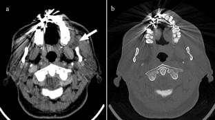
Odontogenic keratocyst (OKC) is a relatively common non-inflammatory jaw lesion. OKC is known to occur most often in the mandibular angle and mandibular ramus, but rarely outside the bone. In this report, we describe characteristic multimodality imaging of OKC in the buccal space, especially diffusion-weighted MR imaging (DWI) with apparent diffusion coefficient (ADC) mapping, extra-oral and intra-oral ultrasonography. On clinical examination, an approximately 20 mm in diameter mass with elastic hardness was found the left side of the buccal area. Contrast-enhanced CT showed areas of internal non-contrast lesions in the left buccal space. On T1-weighted image, the mass showed multilocular high signal intensity, and homogeneous internal. T2-weighted images revealed high signal at the marginal part and slightly median signal in the internal part. STIR images revealed a heterogeneous high signal in the interior. Furthermore, DWI and ADC map showed high signal and moderate-to-low signal intensity, respectively. ADC value of the lesion was 1.55 × 10⁻³ mm2 s-1. On extra-oral ultrasonography, the tumor showed clear boundary, hypoechoic, homogeneous internal architecture and vascular signals, and heterogeneous hard of the lesion. On intra-oral ultrasonography also showed clear boundary, hypoechoic, homogeneous internal architecture, heterogeneous hard of the tumor, and back echo enhance. The histopathologic diagnosis based on a full excisional specimen was odontogenic keratocyst. This case suggests that multimodality imaging, especially MR imaging with ADC and DWI, and extra and intra-oral ultrasonography with color Doppler imaging and elastography, could be effective for evaluating buccal lesions.
期刊介绍:
As the official English-language journal of the Japanese Society for Oral and Maxillofacial Radiology and the Asian Academy of Oral and Maxillofacial Radiology, Oral Radiology is intended to be a forum for international collaboration in head and neck diagnostic imaging and all related fields. Oral Radiology features cutting-edge research papers, review articles, case reports, and technical notes from both the clinical and experimental fields. As membership in the Society is not a prerequisite, contributions are welcome from researchers and clinicians worldwide.

 求助内容:
求助内容: 应助结果提醒方式:


