{"title":"健康的年轻男性和女性志愿者在排尿开始时的脑干激活是否不同?高清晰度7特斯拉磁共振成像研究。","authors":"Bradley Schott, Darshil Choksi, Khue Tran, Christof Karmonik, Betsy Salazar, Timothy Boone, Rose Khavari","doi":"10.5213/inj.2346104.052","DOIUrl":null,"url":null,"abstract":"<p><strong>Purpose: </strong>Assessing brainstem function in humans through typical neuroimaging modalities has been challenging. Our objective was to evaluate brain and brainstem activation patterns during initiation of voiding in healthy males and females utilizing a 7 Tesla magnetic resonance imaging (MRI) scanner and a noninvasive brain-bladder functional MRI (fMRI) protocol.</p><p><strong>Methods: </strong>Twenty healthy adult volunteers (10 males and 10 females) with no history of urinary symptoms were recruited. Each volunteer underwent a clinic uroflow and postvoid residual assessment and was asked to consume water prior to entering the scanner. Anatomical and diffusion tensor images were obtained first, followed by a blood oxygenation level dependent (BOLD) resting-state fMRI (rs-fMRI) during the empty bladder. Subjects indicated when they felt the urge to void, and a full bladder rs-fMRI was obtained. Once completed, the subjects began 5 voiding cycles, where the first 7.5 seconds of each voiding cycle was identified as \"initiation of voiding.\" BOLD activation maps were generated, and regions of interests with a t-value greater than 2.1 were deemed statistically significant.</p><p><strong>Results: </strong>We present 5 distinct regions within the periaqueductal gray (PAG) and pontine micturition center (PMC) with statistically significant activation associated with an initiation of voiding in both men and women, 3 within the PAG and 2 within the PMC. Several additional areas in the brain also demonstrated activation as well. When comparing males to females, there was an overall lower BOLD activation seen in females throughout all regions, with the exception of the caudate lobe.</p><p><strong>Conclusion: </strong>Our study effectively defines regions within the PAG and PMC involved in initiation of voiding in healthy volunteers. To our knowledge, this is the first study investigating differences between male and female brainstem activation utilizing an ultra-high definition 7T MRI.</p>","PeriodicalId":14466,"journal":{"name":"International Neurourology Journal","volume":"27 3","pages":"174-181"},"PeriodicalIF":2.1000,"publicationDate":"2023-09-01","publicationTypes":"Journal Article","fieldsOfStudy":null,"isOpenAccess":false,"openAccessPdf":"https://ftp.ncbi.nlm.nih.gov/pub/pmc/oa_pdf/ba/6d/inj-2346104-052.PMC10556429.pdf","citationCount":"0","resultStr":"{\"title\":\"Is the Brainstem Activation Different Between Healthy Young Male and Female Volunteers at Initiation of Voiding? A High Definition 7-Tesla Magnetic Resonance Imaging Study.\",\"authors\":\"Bradley Schott, Darshil Choksi, Khue Tran, Christof Karmonik, Betsy Salazar, Timothy Boone, Rose Khavari\",\"doi\":\"10.5213/inj.2346104.052\",\"DOIUrl\":null,\"url\":null,\"abstract\":\"<p><strong>Purpose: </strong>Assessing brainstem function in humans through typical neuroimaging modalities has been challenging. Our objective was to evaluate brain and brainstem activation patterns during initiation of voiding in healthy males and females utilizing a 7 Tesla magnetic resonance imaging (MRI) scanner and a noninvasive brain-bladder functional MRI (fMRI) protocol.</p><p><strong>Methods: </strong>Twenty healthy adult volunteers (10 males and 10 females) with no history of urinary symptoms were recruited. Each volunteer underwent a clinic uroflow and postvoid residual assessment and was asked to consume water prior to entering the scanner. Anatomical and diffusion tensor images were obtained first, followed by a blood oxygenation level dependent (BOLD) resting-state fMRI (rs-fMRI) during the empty bladder. Subjects indicated when they felt the urge to void, and a full bladder rs-fMRI was obtained. Once completed, the subjects began 5 voiding cycles, where the first 7.5 seconds of each voiding cycle was identified as \\\"initiation of voiding.\\\" BOLD activation maps were generated, and regions of interests with a t-value greater than 2.1 were deemed statistically significant.</p><p><strong>Results: </strong>We present 5 distinct regions within the periaqueductal gray (PAG) and pontine micturition center (PMC) with statistically significant activation associated with an initiation of voiding in both men and women, 3 within the PAG and 2 within the PMC. Several additional areas in the brain also demonstrated activation as well. When comparing males to females, there was an overall lower BOLD activation seen in females throughout all regions, with the exception of the caudate lobe.</p><p><strong>Conclusion: </strong>Our study effectively defines regions within the PAG and PMC involved in initiation of voiding in healthy volunteers. To our knowledge, this is the first study investigating differences between male and female brainstem activation utilizing an ultra-high definition 7T MRI.</p>\",\"PeriodicalId\":14466,\"journal\":{\"name\":\"International Neurourology Journal\",\"volume\":\"27 3\",\"pages\":\"174-181\"},\"PeriodicalIF\":2.1000,\"publicationDate\":\"2023-09-01\",\"publicationTypes\":\"Journal Article\",\"fieldsOfStudy\":null,\"isOpenAccess\":false,\"openAccessPdf\":\"https://ftp.ncbi.nlm.nih.gov/pub/pmc/oa_pdf/ba/6d/inj-2346104-052.PMC10556429.pdf\",\"citationCount\":\"0\",\"resultStr\":null,\"platform\":\"Semanticscholar\",\"paperid\":null,\"PeriodicalName\":\"International Neurourology Journal\",\"FirstCategoryId\":\"3\",\"ListUrlMain\":\"https://doi.org/10.5213/inj.2346104.052\",\"RegionNum\":3,\"RegionCategory\":\"医学\",\"ArticlePicture\":[],\"TitleCN\":null,\"AbstractTextCN\":null,\"PMCID\":null,\"EPubDate\":\"2023/9/30 0:00:00\",\"PubModel\":\"Epub\",\"JCR\":\"Q3\",\"JCRName\":\"UROLOGY & NEPHROLOGY\",\"Score\":null,\"Total\":0}","platform":"Semanticscholar","paperid":null,"PeriodicalName":"International Neurourology Journal","FirstCategoryId":"3","ListUrlMain":"https://doi.org/10.5213/inj.2346104.052","RegionNum":3,"RegionCategory":"医学","ArticlePicture":[],"TitleCN":null,"AbstractTextCN":null,"PMCID":null,"EPubDate":"2023/9/30 0:00:00","PubModel":"Epub","JCR":"Q3","JCRName":"UROLOGY & NEPHROLOGY","Score":null,"Total":0}
Is the Brainstem Activation Different Between Healthy Young Male and Female Volunteers at Initiation of Voiding? A High Definition 7-Tesla Magnetic Resonance Imaging Study.
Purpose: Assessing brainstem function in humans through typical neuroimaging modalities has been challenging. Our objective was to evaluate brain and brainstem activation patterns during initiation of voiding in healthy males and females utilizing a 7 Tesla magnetic resonance imaging (MRI) scanner and a noninvasive brain-bladder functional MRI (fMRI) protocol.
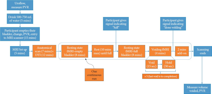
Methods: Twenty healthy adult volunteers (10 males and 10 females) with no history of urinary symptoms were recruited. Each volunteer underwent a clinic uroflow and postvoid residual assessment and was asked to consume water prior to entering the scanner. Anatomical and diffusion tensor images were obtained first, followed by a blood oxygenation level dependent (BOLD) resting-state fMRI (rs-fMRI) during the empty bladder. Subjects indicated when they felt the urge to void, and a full bladder rs-fMRI was obtained. Once completed, the subjects began 5 voiding cycles, where the first 7.5 seconds of each voiding cycle was identified as "initiation of voiding." BOLD activation maps were generated, and regions of interests with a t-value greater than 2.1 were deemed statistically significant.
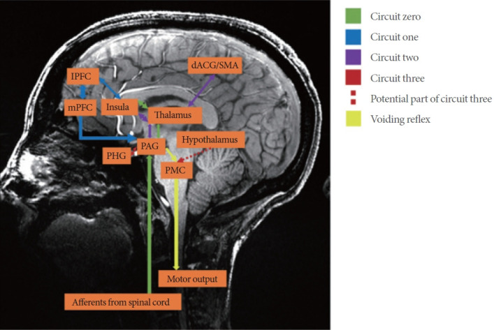
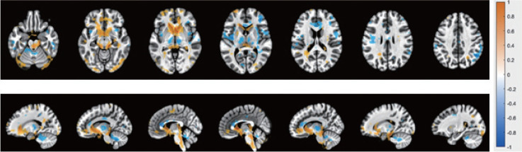
Results: We present 5 distinct regions within the periaqueductal gray (PAG) and pontine micturition center (PMC) with statistically significant activation associated with an initiation of voiding in both men and women, 3 within the PAG and 2 within the PMC. Several additional areas in the brain also demonstrated activation as well. When comparing males to females, there was an overall lower BOLD activation seen in females throughout all regions, with the exception of the caudate lobe.
Conclusion: Our study effectively defines regions within the PAG and PMC involved in initiation of voiding in healthy volunteers. To our knowledge, this is the first study investigating differences between male and female brainstem activation utilizing an ultra-high definition 7T MRI.
期刊介绍:
The International Neurourology Journal (Int Neurourol J, INJ) is a quarterly international journal that publishes high-quality research papers that provide the most significant and promising achievements in the fields of clinical neurourology and fundamental science. Specifically, fundamental science includes the most influential research papers from all fields of science and technology, revolutionizing what physicians and researchers practicing the art of neurourology worldwide know. Thus, we welcome valuable basic research articles to introduce cutting-edge translational research of fundamental sciences to clinical neurourology. In the editorials, urologists will present their perspectives on these articles. The original mission statement of the INJ was published on October 12, 1997.
INJ provides authors a fast review of their work and makes a decision in an average of three to four weeks of receiving submissions. If accepted, articles are posted online in fully citable form. Supplementary issues will be published interim to quarterlies, as necessary, to fully allow berth to accept and publish relevant articles.
 求助内容:
求助内容: 应助结果提醒方式:


